
 EasySep™小鼠TIL(CD45)正选试剂盒


搜索结果: 'methocult media formulations for human hematopoietic cells serum containing'
-

产品类型:
产品号#:
05850
05857
05870
05875
85850
85857
85870
85875
产品名:
mTeSR™1
mTeSR™1
-
产品类型:
产品号#:
05412
05455
产品名:
MesenCult™ 脂肪分化试剂盒 (人)
MesenCult™-ACF软骨细胞分化试剂盒
-
-
产品类型:
产品号#:
18756
18756RF
产品名:
EasySep™小鼠SCA1正选试剂盒
RoboSep™ 小鼠SCA1正选试剂盒含滤芯吸头
-
产品类型:
产品号#:
05850
05857
05870
05875
85850
85857
85870
85875
产品名:
mTeSR™1
mTeSR™1
-
产品类型:
产品号#:
05230
100-0483
100-0484
85850
85857
产品名:
STEMdiff™ 三胚层分化试剂盒
Hausser Scientificᵀᴹ 明线血球计数板
ReLeSR™
mTeSR™1
mTeSR™1
-
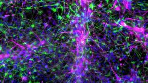 实验方案How to Co-Culture Astrocytes and NGN2 mRNA-Driven Induced Forebrain Neurons Derived from Human Pluripotent Stem Cells
产品类型:
研究方向:
疾病建模,神经科学,药物发现和毒性检测
产品号#:
产品名:
-
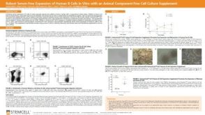 科学海报Robust Serum-Free Expansion of Human B Cells In Vitro with an Animal Component-Free Cell Culture Supplement
产品类型:
Conference:
PEGS 2020
产品号#:
产品名:
发布日期: 09/01/2020 -
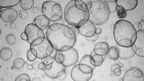
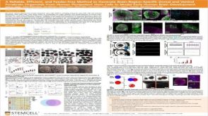 科学海报A Reliable, Efficient, and Feeder-Free Method to Generate Brain-Region-Specific Dorsal and Ventral Forebrain Organoids From Human Pluripotent Stem Cells to Model Early Human Brain Development
产品类型:
Conference:
CSHL 3D Brain 2019
产品号#:
产品名:
发布日期: 12/20/2019 -
产品类型:
产品号#:
09500
产品名:
BIT 9500血清替代物

 沪公网安备31010102008431号