Cai S et al. (APR 2011)
Clinical cancer research : an official journal of the American Association for Cancer Research 17 8 2195--206
Humanized bone marrow mouse model as a preclinical tool to assess therapy-mediated hematotoxicity.
PURPOSE: Preclinical in vivo studies can help guide the selection of agents and regimens for clinical testing. However,one of the challenges in screening anticancer therapies is the assessment of off-target human toxicity. There is a need for in vivo models that can simulate efficacy and toxicities of promising therapeutic regimens. For example,hematopoietic cells of human origin are particularly sensitive to a variety of chemotherapeutic regimens,but in vivo models to assess potential toxicities have not been developed. In this study,a xenograft model containing humanized bone marrow is utilized as an in vivo assay to monitor hematotoxicity. EXPERIMENTAL DESIGN: A proof-of-concept,temozolomide-based regimen was developed that inhibits tumor xenograft growth. This regimen was selected for testing because it has been previously shown to cause myelosuppression in mice and humans. The dose-intensive regimen was administered to NOD.Cg-Prkdc(scid)IL2rg(tm1Wjl)/Sz (NOD/SCID/γchain(null)),reconstituted with human hematopoietic cells,and the impact of treatment on human hematopoiesis was evaluated. RESULTS: The dose-intensive regimen resulted in significant decreases in growth of human glioblastoma xenografts. When this regimen was administered to mice containing humanized bone marrow,flow cytometric analyses indicated that the human bone marrow cells were significantly more sensitive to treatment than the murine bone marrow cells and that the regimen was highly toxic to human-derived hematopoietic cells of all lineages (progenitor,lymphoid,and myeloid). CONCLUSIONS: The humanized bone marrow xenograft model described has the potential to be used as a platform for monitoring the impact of anticancer therapies on human hematopoiesis and could lead to subsequent refinement of therapies prior to clinical evaluation.
View Publication
产品类型:
产品号#:
03434
03444
04434
04444
84434
84444
产品名:
MethoCult™ GF M3434
MethoCult™ GF M3434
MethoCult™ H4434 Classic
MethoCult™ H4434 Classic
(Sep 2024)
Nature Communications 15
Sequence variants influencing the regulation of serum IgG subclass levels
Immunoglobulin G (IgG) is the main isotype of antibody in human blood. IgG consists of four subclasses (IgG1 to IgG4),encoded by separate constant region genes within the Ig heavy chain locus (IGH). Here,we report a genome-wide association study on blood IgG subclass levels. Across 4334 adults and 4571 individuals under 18 years,we discover ten new and identify four known variants at five loci influencing IgG subclass levels. These variants also affect the risk of asthma,autoimmune diseases,and blood traits. Seven variants map to the IGH locus,three to the Fcγ receptor (FCGR) locus,and two to the human leukocyte antigen (HLA) region,affecting the levels of all IgG subclasses. The most significant associations are observed between the G1m (f),G2m(n) and G3m(b*) allotypes,and IgG1,IgG2 and IgG3,respectively. Additionally,we describe selective associations with IgG4 at 16p11.2 (ITGAX) and 17q21.1 (IKZF3,ZPBP2,GSDMB,ORMDL3). Interestingly,the latter coincides with a highly pleiotropic signal where the allele associated with lower IgG4 levels protects against childhood asthma but predisposes to inflammatory bowel disease. Our results provide insight into the regulation of antibody-mediated immunity that can potentially be useful in the development of antibody based therapeutics. Immunoglobulin G (IgG) is the main isotype of antibody in human blood. Here the authors describe 14 genetic variants that affect IgG levels in blood. The data provide new insight into the regulation of humoral immunity that could be useful in the development of antibody-based therapeutics.
View Publication
产品类型:
产品号#:
18000
产品名:
EasySep™磁极
Garidou L et al. (SEP 2009)
Journal of virology 83 17 8905--15
Therapeutic memory T cells require costimulation for effective clearance of a persistent viral infection.
Persistent viral infections are a major health concern worldwide. During persistent infection,overwhelming viral replication and the rapid loss of antiviral T-cell function can prevent immune-mediated clearance of the infection,and therapies to reanimate the immune response and purge persistent viruses have been largely unsuccessful. Adoptive immunotherapy using memory T cells is a highly successful therapeutic approach to eradicate a persistent viral infection. Understanding precisely how therapeutically administered memory T cells achieve clearance should improve our ability to terminate states of viral persistence in humans. Mice persistently infected from birth with lymphocytic choriomeningitis virus are tolerant to the pathogen at the T-cell level and thus provide an excellent model to evaluate immunotherapeutic regimens. Previously,we demonstrated that adoptively transferred memory T cells require recipient dendritic cells to effectively purge an established persistent viral infection. However,the mechanisms that reactivate and sustain memory T-cell responses during clearance of such an infection remain unclear. Here we establish that therapeutic memory T cells require CD80 and CD86 costimulatory signals to efficiently clear an established persistent viral infection in vivo. Early blockade of costimulatory pathways with CTLA-4-Fc decreased the secondary expansion of virus-specific CD8(+) and CD4(+) memory T cells as well as their ability to produce antiviral cytokines and purge the persistent infection. Late costimulation blockade also reduced virus-specific T-cell numbers,illustrating that sustained interactions with costimulatory molecules is required for efficient T-cell expansion. These findings indicate that antiviral memory T cells require costimulation to efficiently clear a persistent viral infection and that costimulatory pathways can be targeted to modulate the magnitude of an adoptive immunotherapeutic regimen.
View Publication
产品类型:
产品号#:
18758
18758RF
18768
18768RF
产品名:
Chen G et al. (AUG 2010)
Cell stem cell 7 2 240--8
Actin-myosin contractility is responsible for the reduced viability of dissociated human embryonic stem cells.
Human ESCs are the pluripotent precursor of the three embryonic germ layers. Human ESCs exhibit basal-apical polarity,junctional complexes,integrin-dependent matrix adhesion,and E-cadherin-dependent cell-cell adhesion,all characteristics shared by the epiblast epithelium of the intact mammalian embryo. After disruption of epithelial structures,programmed cell death is commonly observed. If individualized human ESCs are prevented from reattaching and forming colonies,their viability is significantly reduced. Here,we show that actin-myosin contraction is a critical effector of the cell death response to human ESC dissociation. Inhibition of myosin heavy chain ATPase,downregulation of myosin heavy chain,and downregulation of myosin light chain all increase survival and cloning efficiency of individualized human ESCs. ROCK inhibition decreases phosphorylation of myosin light chain,suggesting that inhibition of actin-myosin contraction is also the mechanism through which ROCK inhibitors increase cloning efficiency of human ESCs.
View Publication
产品类型:
产品号#:
05850
05857
05870
05875
72402
72404
85850
85857
85870
85875
产品名:
(-)-Blebbistatin
(-)-Blebbistatin
mTeSR™1
mTeSR™1
Safinia N et al. (FEB 2016)
Oncotarget 7 7 7563--77
Successful expansion of functional and stable regulatory T cells for immunotherapy in liver transplantation.
Strategies to prevent organ transplant rejection whilst minimizing long-term immunosuppression are currently under intense investigation with regulatory T cells (Tregs) nearing clinical application. The clinical trial,ThRIL,recently commenced at King's College London,proposes to use Treg cell therapy to induce tolerance in liver transplant recipients,the success of which has the potential to revolutionize the management of these patients and enable a future of drug-free transplants. This is the first report of the manufacture of clinical grade Tregs from prospective liver transplant recipients via a CliniMACS-based GMP isolation technique and expanded using anti-CD3/CD28 beads,IL-2 and rapamycin. We report the enrichment of a pure,stable population of Tregs (textgreater95% CD4(+)CD25(+)FOXP3(+)),reaching adequate numbers for their clinical application. Our protocol proved successful in,influencing the expansion of superior functional Tregs,as compared to freshly isolated cells,whilst also preventing their conversion to Th17 cells under pro-inflammatory conditions. We conclude with the manufacture of the final Treg product in the clinical research facility (CRF),a prerequisite for the clinical application of these cells. The data presented in this manuscript together with the much-anticipated clinical results from ThRIL,will undoubtedly inform the improved management of the liver transplant recipient.
View Publication
产品类型:
产品号#:
07930
07931
07940
07955
07956
07959
07954
100-1061
07952
产品名:
CryoStor® CS10
CryoStor® CS10
CryoStor® CS10
CryoStor® CS10
CryoStor® CS10
CryoStor® CS10
CryoStor® CS10
Carpentier A et al. (MAR 2016)
Stem Cell Research 16 3 640--650
Hepatic differentiation of human pluripotent stem cells in miniaturized format suitable for high-throughput screen
The establishment of protocols to differentiate human pluripotent stem cells (hPSCs) including embryonic (ESC) and induced pluripotent (iPSC) stem cells into functional hepatocyte-like cells (HLCs) creates new opportunities to study liver metabolism,genetic diseases and infection of hepatotropic viruses (hepatitis B and C viruses) in the context of specific genetic background. While supporting efficient differentiation to HLCs,the published protocols are limited in terms of differentiation into fully mature hepatocytes and in a smaller-well format. This limitation handicaps the application of these cells to high-throughput assays. Here we describe a protocol allowing efficient and consistent hepatic differentiation of hPSCs in 384-well plates into functional hepatocyte-like cells,which remain differentiated for more than 3 weeks. This protocol affords the unique opportunity to miniaturize the hPSC-based differentiation technology and facilitates screening for molecules in modulating liver differentiation,metabolism,genetic network,and response to infection or other external stimuli.
View Publication
产品类型:
产品号#:
05110
05850
05857
05870
05875
85850
85857
85870
85875
产品名:
STEMdiff™定型内胚层检测试剂盒
mTeSR™1
mTeSR™1
Luna JI et al. (MAY 2011)
Tissue engineering. Part C,Methods 17 5 579--88
Multiscale biomimetic topography for the alignment of neonatal and embryonic stem cell-derived heart cells.
Nano- and microscale topographical cues play critical roles in the induction and maintenance of various cellular functions,including morphology,adhesion,gene regulation,and communication. Recent studies indicate that structure and function at the heart tissue level is exquisitely sensitive to mechanical cues at the nano-scale as well as at the microscale level. Although fabrication methods exist for generating topographical features for cell culture,current techniques,especially those with nanoscale resolution,are typically complex,prohibitively expensive,and not accessible to most biology laboratories. Here,we present a tunable culture platform comprised of biomimetic wrinkles that simulate the heart's complex anisotropic and multiscale architecture for facile and robust cardiac cell alignment. We demonstrate the cellular and subcellular alignment of both neonatal mouse cardiomyocytes as well as those derived from human embryonic stem cells. By mimicking the fibrillar network of the extracellular matrix,this system enables monitoring of protein localization in real time and therefore the high-resolution study of phenotypic and physiologic responses to in-vivo like topographical cues.
View Publication
产品类型:
产品号#:
05850
05857
05870
05875
85850
85857
85870
85875
产品名:
mTeSR™1
mTeSR™1
Zhang R et al. (JAN 2013)
Nature communications 4 1335
A thermoresponsive and chemically defined hydrogel for long-term culture of human embryonic stem cells
Cultures of human embryonic stem cell typically rely on protein matrices or feeder cells to support attachment and growth,while mechanical,enzymatic or chemical cell dissociation methods are used for cellular passaging. However,these methods are ill defined,thus introducing variability into the system,and may damage cells. They also exert selective pressures favouring cell aneuploidy and loss of differentiation potential. Here we report the identification of a family of chemically defined thermoresponsive synthetic hydrogels based on 2-(diethylamino)ethyl acrylate,which support long-term human embryonic stem cell growth and pluripotency over a period of 2-6 months. The hydrogels permitted gentle,reagent-free cell passaging by virtue of transient modulation of the ambient temperature from 37 to 15 °C for 30 min. These chemically defined alternatives to currently used,undefined biological substrates represent a flexible and scalable approach for improving the definition,efficacy and safety of human embryonic stem cell culture systems for research,industrial and clinical applications.
View Publication
产品类型:
产品号#:
05850
05857
05870
05875
85850
85857
85870
85875
产品名:
mTeSR™1
mTeSR™1
Choi SM et al. (JUN 2013)
Hepatology 57 6 2458--2468
Efficient drug screening and gene correction for treating liver disease using patient-specific stem cells
UNLABELLED: Patient-specific induced pluripotent stem cells (iPSCs) represent a potential source for developing novel drug and cell therapies. Although increasing numbers of disease-specific iPSCs have been generated,there has been limited progress in iPSC-based drug screening/discovery for liver diseases,and the low gene-targeting efficiency in human iPSCs warrants further improvement. Using iPSC lines from patients with alpha-1 antitrypsin (AAT) deficiency,for which there is currently no drug or gene therapy available,we established a platform to discover new drug candidates and correct disease-causing mutation with a high efficiency. A high-throughput format screening assay,based on our hepatic differentiation protocol,was implemented to facilitate automated quantification of cellular AAT accumulation using a 96-well immunofluorescence reader. To expedite the eventual application of lead compounds to patients,we conducted drug screening utilizing our established library of clinical compounds (the Johns Hopkins Drug Library) with extensive safety profiles. Through a blind large-scale drug screening,five clinical drugs were identified to reduce AAT accumulation in diverse patient iPSC-derived hepatocyte-like cells. In addition,using the recently developed transcription activator-like effector nuclease technology,we achieved high gene-targeting efficiency in AAT-deficiency patient iPSCs with 25%-33% of the clones demonstrating simultaneous targeting at both diseased alleles. The hepatocyte-like cells derived from the gene-corrected iPSCs were functional without the mutant AAT accumulation. This highly efficient and cost-effective targeting technology will broadly benefit both basic and translational applications.backslashnbackslashnCONCLUSIONS: Our results demonstrated the feasibility of effective large-scale drug screening using an iPSC-based disease model and highly robust gene targeting in human iPSCs,both of which are critical for translating the iPSC technology into novel therapies for untreatable diseases.
View Publication
产品类型:
产品号#:
05850
05857
05870
05875
85850
85857
85870
85875
产品名:
mTeSR™1
mTeSR™1
E. J. Oh et al. (Jul 2025)
Cells 14 14
Modeling Aberrant Angiogenesis in Arteriovenous Malformations Using Endothelial Cells and Organoids for Pharmacological Treatment
Arteriovenous malformations (AVMs) are congenital vascular anomalies defined by abnormal direct connections between arteries and veins due to their complex structure or endovascular approaches. Pharmacological strategies targeting the underlying molecular mechanisms are thus gaining increasing attention in an effort to determine the mechanism involved in AVM regulation. In this study,we examined 30 human tissue samples,comprising 10 vascular samples,10 human fibroblasts derived from AVM tissue,and 10 vascular samples derived from healthy individuals. The pharmacological agents thalidomide,U0126,and rapamycin were applied to the isolated endothelial cells (ECs). The pharmacological treatments reduced the proliferation of AVM ECs and downregulated miR-135b-5p,a biomarker associated with AVMs. The expression levels of angiogenesis-related genes,including VEGF,ANG2,FSTL1,and MARCKS,decreased; in comparison,CSPG4,a gene related to capillary networks,was upregulated. Following analysis of these findings,skin samples from 10 AVM patients were reprogrammed into induced pluripotent stem cells (iPSCs) to generate AVM blood vessel organoids. Treatment of these AVM blood vessel organoids with thalidomide,U0126,and rapamycin resulted in a reduction in the expression of the EC markers CD31 and α-SMA. The establishment of AVM blood vessel organoids offers a physiologically relevant in vitro model for disease characterization and drug screening. The authors of future studies should aim to refine this model using advanced techniques,such as microfluidic systems,to more efficiently replicate AVMs’ pathology and support the development of personalized therapies.
View Publication
产品类型:
产品号#:
100-0651
产品名:
STEMdiff™ 血管类器官试剂盒
(Jun 2024)
Materials Today Bio 26 4
Nanofiber-microwell cell culture system for spatially patterned differentiation of pluripotent stem cells in 3D
The intricate interplay between biochemical and physical cues dictates pluripotent stem cell (PSC) differentiation to form various tissues. While biochemical modulation has been extensively studied,the role of biophysical microenvironments in early lineage commitment remains elusive. Here,we introduce a novel 3D cell culture system combining electrospun nanofibers with microfabricated polydimethylsiloxane (PDMS) patterns. This system enables the controlled formation of semispherical human induced pluripotent stem cell (hiPSC) colonies,facilitating the investigation of local mechanical stem cell niches on mechano-responsive signaling and lineage specification. Our system unveiled spatially organized RhoA activity coupled with actin-myosin cable formation,suggesting mechano-dependent hiPSC behaviors. Nodal network analysis of RNA-seq data revealed RhoA downstream regulation of YAP signaling,DNA histone modifications,and patterned germ layer specification. Notably,altering colony morphology through controlled PDMS microwell shaping effectively modulated the spatial distribution of mechano-sensitive mediators and subsequent differentiation. This study provides a cell culture platform to decipher the role of biophysical cues in early embryogenesis,offering valuable insights for material design in tissue engineering and regenerative medicine applications. Graphical abstractImage 1
View Publication

 EasySep™小鼠TIL(CD45)正选试剂盒


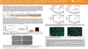 科学海报STEMdiff™ Endothelial: A New Culture Workflow for Efficient Derivation and Expansion of Endothelial Cells From Human Pluripotent Stem Cells

 沪公网安备31010102008431号