Primitive human hematopoietic cells are enriched in cord blood compared with adult bone marrow or mobilized peripheral blood as measured by the quantitative in vivo SCID-repopulating cell assay.
We have previously reported the development of in vivo functional assays for primitive human hematopoietic cells based on their ability to repopulate the bone marrow (BM) of severe combined immunodeficient (SCID) and nonobese diabetic/SCID (NOD/SCID) mice following intravenous transplantation. Accumulated data from gene marking and cell purification experiments indicate that the engrafting cells (defined as SCID-repopulating cells or SRC) are biologically distinct from and more primitive than most cells that can be assayed in vitro. Here we demonstrate through limiting dilution analysis that the NOD/SCID xenotransplant model provides a quantitative assay for SRC. Using this assay,the frequency of SRC in cord blood (CB) was found to be 1 in 9.3 x 10(5) cells. This was significantly higher than the frequency of 1 SRC in 3.0 x 10(6) adult BM cells or 1 in 6.0 x 10(6) mobilized peripheral blood (PB) cells from normal donors. Mice transplanted with limiting numbers of SRC were engrafted with both lymphoid and multilineage myeloid human cells. This functional assay is currently the only available method for quantitative analysis of human hematopoietic cells with repopulating capacity. Both CB and mobilized PB are increasingly being used as alternative sources of hematopoietic stem cells in allogeneic transplantation. Thus,the findings reported here will have important clinical as well as biologic implications.
View Publication
产品类型:
产品号#:
28600
产品名:
L-Calc™有限稀释软件
Fraga AM et al. (MAR 2011)
Cell Transplantation 20 3 431--40
Establishment of a Brazilian line of human embryonic stem cells in defined medium: implications for cell therapy in an ethnically diverse population.
Pluripotent human embryonic stem (hES) cells are an important experimental tool for basic and applied research,and a potential source of different tissues for transplantation. However,one important challenge for the clinical use of these cells is the issue of immunocompatibility,which may be dealt with by the establishment of hES cell banks to attend different populations. Here we describe the derivation and characterization of a line of hES cells from the Brazilian population,named BR-1,in commercial defined medium. In contrast to the other hES cell lines established in defined medium,BR-1 maintained a stable normal karyotype as determined by genomic array analysis after 6 months in continuous culture (passage 29). To our knowledge,this is the first reported line of hES cells derived in South America. We have determined its genomic ancestry and compared the HLA-profile of BR-1 and another 22 hES cell lines established elsewhere with those of the Brazilian population,finding they would match only 0.011% of those individuals. Our results highlight the challenges involved in hES cell banking for populations with a high degree of ethnic admixture.
View Publication
Myogenesis GRC 2017,Till and McCulloch Meeting 2017
产品号#:
05980
05982
05983
产品名:
MyoCult™-SF 扩增添加物试剂盒 (人)
MyoCult™-SF 扩增10X添加物(人)
MyoCult™-SF 贴附基质
Ortiz-Sá et al. (JAN 2009)
Leukemia 23 1 59--70
Enhanced cytotoxicity of an anti-transferrin receptor IgG3-avidin fusion protein in combination with gambogic acid against human malignant hematopoietic cells: functional relevance of iron, the receptor, and reactive oxygen species.
The human transferrin receptor (hTfR) is a target for cancer immunotherapy due to its overexpression on the surface of cancer cells. We previously developed an antibody-avidin fusion protein that targets hTfR (anti-hTfR IgG3-Av) and exhibits intrinsic cytotoxicity against certain malignant cells. Gambogic acid (GA),a drug that also binds hTfR,induces cytotoxicity in several malignant cell lines. We now report that anti-hTfR IgG3-Av and GA induce cytotoxicity in a new broader panel of hematopoietic malignant cell lines. Our results show that the effect of anti-hTfR IgG3-Av is iron-dependent whereas that of GA is iron-independent in all cells tested. In addition,we observed that GA exerts a TfR-independent cytotoxicity. We also found that GA increases the generation of reactive oxygen species that may play a role in the cytotoxicity induced by this drug. Additive cytotoxicity was observed by simultaneous combination treatment with these drugs and synergy by using anti-hTfR IgG3-Av as a chemosensitizing agent. In addition,we found a concentration of GA that is toxic to malignant hematopoietic cells but not to human hematopoietic progenitor cells. Our results suggest that these two compounds may be effective,alone or in combination,for the treatment of human hematopoietic malignancies.
View Publication
产品类型:
产品号#:
04434
04444
产品名:
MethoCult™ H4434 Classic
MethoCult™ H4434 Classic
Ma R et al. (APR 2015)
Thyroid 25 4 455--461
Human embryonic stem cells form functional thyroid follicles.
OBJECTIVE: The molecular events that lead to human thyroid cell speciation remain incompletely characterized. It has been shown that overexpression of the regulatory transcription factors Pax8 and Nkx2-1 (ttf-1) directs murine embryonic stem (mES) cells to differentiate into thyroid follicular cells by initiating a transcriptional regulatory network. Such cells subsequently organized into three-dimensional follicular structures in the presence of extracellular matrix. In the current study,human embryonic stem (hES) cells were studied with the aim of recapitulating this scenario and producing functional human thyroid cell lines. METHODS: Reporter gene tagged pEZ-lentiviral vectors were used to express human PAX8-eGFP and NKX2-1-mCherry in the H9 hES cell line followed by differentiation into thyroid cells directed by Activin A and thyrotropin (TSH). RESULTS: Both transcription factors were expressed efficiently in hES cells expressing either PAX8,NKX2-1,or in combination in the hES cells,which had low endogenous expression of these transcription factors. Further differentiation of the double transfected cells showed the expression of thyroid-specific genes,including thyroglobulin (TG),thyroid peroxidase (TPO),the sodium/iodide symporter (NIS),and the TSH receptor (TSHR) as assessed by reverse transcription polymerase chain reaction and immunostaining. Most notably,the Activin/TSH-induced differentiation approach resulted in thyroid follicle formation and abundant TG protein expression within the follicular lumens. On stimulation with TSH,these hES-derived follicles were also capable of dose-dependent cAMP generation and radioiodine uptake,indicating functional thyroid epithelial cells. CONCLUSION: The induced expression of PAX8 and NKX2-1 in hES cells was followed by differentiation into thyroid epithelial cells and their commitment to form functional three-dimensional neo-follicular structures. The data provide proof of principal that hES cells can be committed to thyroid cell speciation under appropriate conditions.
View Publication
产品类型:
产品号#:
05850
05857
05870
05875
85850
85857
85870
85875
产品名:
mTeSR™1
mTeSR™1
Yeo C et al. (SEP 2009)
Regenerative Medicine 4 5 689--696
Ficoll-Paque™ versus Lymphoprep™: a comparative study of two density gradient media for therapeutic bone marrow mononuclear cell preparations
AIMS Contradictory outcomes from recent clinical trials investigating the transplantation of autologous bone marrow mononuclear cell (BM-MNC) fraction containing stem/progenitor cells to damaged myocardium,following acute myocardial infarction,may be,in part,due to the different cell isolation protocols used. We compared total BM-MNC numbers and its cellular subsets obtained following isolation using Ficoll-Paque and Lymphoprep - two different density gradient media used in the clinical trials. MATERIALS & METHODS Bone marrow samples were taken from patients entered into the REGENERATE-IHD clinical trial after 5 days of subcutaneous granulocyte colony-stimulating factor injections. Each sample was divided equally for BM-MNC isolation using Ficoll-Paque and Lymphoprep,keeping all other procedural steps constant. Isolated fractions were characterized for hematopoietic stem cells,endothelial progenitor cells,T lymphocytes,B lymphocytes and NK cells using cell surface markers CD34(+),CD133(+)VEGFR2(+),CD45(+)CD3(+),CD45(+)CD19(+) and CD45(+)CD16(+)CD56(+),respectively. There were no significant differences in the absolute numbers and percentage cell recovery of various mononuclear cell types recovered following separation using either density gradient media. Cell viability and the proportion of various cell phenotypes investigated were similar between the two media. They were also equally efficient in excluding unwanted red blood cells,granulocytes and platelets from the final cell products. CONCLUSION We demonstrated that the composition and quantity of cell types found within therapeutic BM-MNC preparations for use in clinical trials of cardiac stem cell transplantation are not influenced by the type of density gradient media used when comparing Ficoll-Paque and Lymphoprep.
View Publication
产品类型:
产品号#:
07801
07811
07851
07861
18060
18061
产品名:
Lymphoprep™
Lymphoprep™
Lymphoprep™
Lymphoprep™
Qué et al. (JUN 2011)
Blood 117 22 5918--30
Smad4 binds Hoxa9 in the cytoplasm and protects primitive hematopoietic cells against nuclear activation by Hoxa9 and leukemia transformation.
We studied leukemic stem cells (LSCs) in a Smad4(-/-) mouse model of acute myelogenous leukemia (AML) induced either by the HOXA9 gene or by the fusion oncogene NUP98-HOXA9. Although Hoxa9-Smad4 complexes accumulate in the cytoplasm of normal hematopoietic stem cells and progenitor cells (HSPCs) transduced with these oncogenes,there is no cytoplasmic stabilization of HOXA9 in Smad4(-/-) HSPCs,and as a consequence increased levels of Hoxa9 is observed in the nucleus leading to increased immortalization in vitro. Loss of Smad4 accelerates the development of leukemia in vivo because of an increase in transformation of HSPCs. Therefore,the cytoplasmic binding of Hoxa9 by Smad4 is a mechanism to protect Hoxa9-induced transformation of normal HSPCs. Because Smad4 is a potent tumor suppressor involved in growth control,we developed a strategy to modify the subcellular distribution of Smad4. We successfully disrupted the interaction between Hoxa9 and Smad4 to activate the TGF-β pathway and apoptosis,leading to a loss of LSCs. Together,these findings reveal a major role for Smad4 in the negative regulation of leukemia initiation and maintenance induced by HOXA9/NUP98-HOXA9 and provide strong evidence that antagonizing Smad4 stabilization by these oncoproteins might be a promising novel therapeutic approach in leukemia.
View Publication
产品类型:
产品号#:
03434
03444
03236
产品名:
MethoCult™ GF M3434
MethoCult™ GF M3434
MethoCult™ SF M3236
Pijuan-Galitó et al. (NOV 2014)
Journal of Biological Chemistry 289 48 33492--33502
Serum Inter-$\$-inhibitor activates the Yes tyrosine kinase and YAP/TEAD transcriptional complex in mouse embryonic stem cells.
We have previously demonstrated that the Src family kinase Yes,the Yes-associated protein (YAP) and TEA domain TEAD2 transcription factor pathway are activated by leukemia inhibitory factor (LIF) and contribute to mouse embryonic stem (mES) cell maintenance of pluripotency and self-renewal. In addition,we have shown that fetal bovine serum (FBS) induces Yes auto-phosphorylation and activation. In the present study we confirm that serum also activates TEAD-dependent transcription in a time- and dose-dependent manner and we identify Inter-α-inhibitor (IαI) as a component in serum capable of activating the Yes/YAP/TEAD pathway by inducing Yes auto-phosphorylation,YAP nuclear localization and TEAD-dependent transcription. The cleaved heavy chain 2 (HC2) sub-component of IαI,is demonstrated to be responsible for this effect. Moreover,IαI is also shown to efficiently increase expression of TEAD-downstream target genes including well-known stem cell factors Nanog and Oct 3/4. IαI is not produced by the ES cells per se but is added to the cells via the cell culture medium containing serum or serum-derived components such as bovine serum albumin (BSA). In conclusion,we describe a novel function of IαI in activating key pluripotency pathways associated with ES cell maintenance and self-renewal.
View Publication
产品类型:
产品号#:
05850
05857
05870
05875
85850
85857
85870
85875
产品名:
mTeSR™1
mTeSR™1
Deglincerti A et al. (NOV 2016)
Nature protocols 11 11 2223--2232
Self-organization of human embryonic stem cells on micropatterns.
Fate allocation in the gastrulating embryo is spatially organized as cells differentiate into specialized cell types depending on their positions with respect to the body axes. There is a need for in vitro protocols that allow the study of spatial organization associated with this developmental transition. Although embryoid bodies and organoids can exhibit some spatial organization of differentiated cells,methods that generate embryoid bodies or organoids do not yield consistent and fully reproducible results. Here,we describe a micropatterning approach in which human embryonic stem cells are confined to disk-shaped,submillimeter colonies. After 42 h of BMP4 stimulation,cells form self-organized differentiation patterns in concentric radial domains,which express specific markers associated with the embryonic germ layers,reminiscent of gastrulating embryos. Our protocol takes 3 d; it uses commercial microfabricated slides (from CYTOO),human laminin-521 (LN-521) as extracellular matrix coating,and either conditioned or chemically defined medium (mTeSR). Differentiation patterns within individual colonies can be determined by immunofluorescence and analyzed with cellular resolution. Both the size of the micropattern and the type of medium affect the patterning outcome. The protocol is appropriate for personnel with basic stem cell culture training. This protocol describes a robust platform for quantitative analysis of the mechanisms associated with pattern formation at the onset of gastrulation.
View Publication
产品类型:
产品号#:
05850
05857
05870
05875
85850
85857
85870
85875
产品名:
mTeSR™1
mTeSR™1
Gerrits A et al. (APR 2010)
Blood 115 13 2610--8
Cellular barcoding tool for clonal analysis in the hematopoietic system.
Clonal analysis is important for many areas of hematopoietic stem cell research,including in vitro cell expansion,gene therapy,and cancer progression and treatment. A common approach to measure clonality of retrovirally transduced cells is to perform integration site analysis using Southern blotting or polymerase chain reaction-based methods. Although these methods are useful in principle,they generally provide a low-resolution,biased,and incomplete assessment of clonality. To overcome those limitations,we labeled retroviral vectors with random sequence tags or barcodes." On integration�
View Publication
产品类型:
产品号#:
09600
09650
产品名:
StemSpan™ SFEM
StemSpan™ SFEM
Kharas MG et al. (JUN 2004)
Blood 103 11 4268--75
Phosphoinositide 3-kinase signaling is essential for ABL oncogene-mediated transformation of B-lineage cells.
BCR-ABL and v-ABL are oncogenic forms of the Abl tyrosine kinase that can cause leukemias in mice and humans. ABL oncogenes trigger multiple signaling pathways whose contribution to transformation varies among cell types. Activation of phosphoinositide 3-kinase (PI3K) is essential for ABL-dependent proliferation and survival in some cell types,and global PI3K inhibitors can enhance the antileukemia effects of the Abl kinase inhibitor imatinib. Although a significant fraction of BCR-ABL-induced human leukemias are of B-cell origin,little is known about PI3K signaling mechanisms in B-lineage cells transformed by ABL oncogenes. Here we show that activation of class I(A) PI3K and downstream inactivation of FOXO transcription factors are essential for survival of murine pro/pre-B cells transformed by v-ABL or BCR-ABL. In addition,analysis of mice lacking individual PI3K genes indicates that products of the Pik3r1 gene contribute to transformation efficiency by BCR-ABL. These findings establish a role for PI3K signaling in B-lineage transformation by ABL oncogenes.
View Publication
产品类型:
产品号#:
03630
产品名:
MethoCult™ M3630
Allan LL et al. (SEP 2009)
Blood 114 12 2411--6
Apolipoprotein-mediated lipid antigen presentation in B cells provides a pathway for innate help by NKT cells.
Natural killer T (NKT) cells are innate-like lymphocytes that recognize lipid antigens and have been shown to enhance B-cell activation and antibody production. B cells typically recruit T-cell help by presenting internalized antigens recognized by their surface antigen receptor. Here,we demonstrate a highly efficient means whereby human B cells present lipid antigens to NKT cells,capturing the antigen using apolipoprotein E (apoE) and the low-density lipoprotein receptor (LDL-R). ApoE dramatically enhances B-cell presentation of alpha-galactosylceramide (alphaGalCer),an exogenous CD1d presented antigen,inducing activation of NKT cells and the subsequent activation of B cells. B cells express the LDL-R on activation,and the activation of NKT cells by B cells is completely LDL-R dependent,as shown by blocking experiments and the complete lack of presentation when using apoE2,an isoform of apoE incapable of LDL-R binding. The dependence on apoE and the LDL-R is much more pronounced in B cells than we had previously seen in dendritic cells,which can apparently use alternate pathways of lipid antigen uptake. Thus,B cells use an apolipoprotein-mediated pathway of lipid antigen presentation,which constitutes a form of innate help for B cells by NKT cells.
View Publication

 EasySep™小鼠TIL(CD45)正选试剂盒


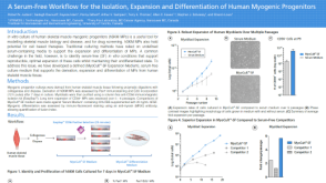 科学海报A Serum-Free Workflow for the Isolation, Expansion and Differentiation of Human Myogenic Progenitors
 沪公网安备31010102008431号