Burkhardt MF et al. (SEP 2013)
Molecular and Cellular Neuroscience 56 355--364
A cellular model for sporadic ALS using patient-derived induced pluripotent stem cells
Development of therapeutics for genetically complex neurodegenerative diseases such as sporadic amyotrophic lateral sclerosis (ALS) has largely been hampered by lack of relevant disease models. Reprogramming of sporadic ALS patients' fibroblasts into induced pluripotent stem cells (iPSC) and differentiation into affected neurons that show a disease phenotype could provide a cellular model for disease mechanism studies and drug discovery. Here we report the reprogramming to pluripotency of fibroblasts from a large cohort of healthy controls and ALS patients and their differentiation into motor neurons. We demonstrate that motor neurons derived from three sALS patients show de novo TDP-43 aggregation and that the aggregates recapitulate pathology in postmortem tissue from one of the same patients from which the iPSC were derived. We configured a high-content chemical screen using the TDP-43 aggregate endpoint both in lower motor neurons and upper motor neuron like cells and identified FDA-approved small molecule modulators including Digoxin demonstrating the feasibility of patient-derived iPSC-based disease modeling for drug screening.
View Publication
产品类型:
产品号#:
05850
05857
05870
05875
85850
85857
85870
85875
产品名:
mTeSR™1
mTeSR™1
X. Qi et al. (May 2025)
Cell Death & Disease 16 1
KLF7-regulated ITGA2 as a therapeutic target for inhibiting oral cancer stem cells
Cancer stem cells (CSCs) play crucial roles in tumor metastasis,therapy resistance,and immune evasion. Identifying and understanding the factors that regulate the stemness of tumor cells presents promising opportunities for developing effective therapeutic strategies. In this study on oral squamous cell carcinoma (OSCC),we confirmed the key role of KLF7 in maintaining the stemness of OSCC. Using chromatin immunoprecipitation sequencing and dual-luciferase assays,we identified ITGA2,a membrane receptor,as a key downstream gene regulated by KLF7 in the maintenance of stemness. Tumor sphere formation assays,flow cytometry analyses,and in vivo limiting dilution tumorigenicity evaluations demonstrated that knocking down ITGA2 significantly impaired stemness. Upon binding to its extracellular matrix (ECM) ligand,type I collagen,ITGA2 activates stemness-associated signaling pathways,including PI3K-AKT,MAPK,and Hippo. TC-I 15,a small-molecule inhibitor of the ITGA2-collagen interaction,significantly sensitizes oral squamous cell carcinoma (OSCC) to cisplatin in xenograft models. In summary,we reveal that the KLF7/ITGA2 axis is a crucial modulator of stemness in OSCC. Our findings suggest that ITGA2 is a promising therapeutic target,offering a novel anti-CSC strategy. Subject terms: Cancer stem cells,Cancer therapeutic resistance
View Publication
Reconstitution of the functional human hematopoietic microenvironment derived from human mesenchymal stem cells in the murine bone marrow compartment.
Hematopoiesis is maintained by specific interactions between both hematopoietic and nonhematopoietic cells. Whereas hematopoietic stem cells (HSCs) have been extensively studied both in vitro and in vivo,little is known about the in vivo characteristics of stem cells of the nonhematopoietic component,known as mesenchymal stem cells (MSCs). Here we have visualized and characterized human MSCs in vivo following intramedullary transplantation of enhanced green fluorescent protein-marked human MSCs (eGFP-MSCs) into the bone marrow (BM) of nonobese diabetic/severe combined immunodeficiency (NOD/SCID) mice. Between 4 to 10 weeks after transplantation,eGFP-MSCs that engrafted in murine BM integrated into the hematopoietic microenvironment (HME) of the host mouse. They differentiated into pericytes,myofibroblasts,BM stromal cells,osteocytes in bone,bone-lining osteoblasts,and endothelial cells,which constituted the functional components of the BM HME. The presence of human MSCs in murine BM resulted in an increase in functionally and phenotypically primitive human hematopoietic cells. Human MSC-derived cells that reconstituted the HME appeared to contribute to the maintenance of human hematopoiesis by actively interacting with primitive human hematopoietic cells.
View Publication
产品类型:
产品号#:
04034
04044
产品名:
MethoCult™ H4034 Optimum
MethoCult™ H4034 Optimum
Naramura M et al. (SEP 2010)
Proceedings of the National Academy of Sciences of the United States of America 107 37 16274--9
Rapidly fatal myeloproliferative disorders in mice with deletion of Casitas B-cell lymphoma (Cbl) and Cbl-b in hematopoietic stem cells.
Casitas B-cell lymphoma (Cbl)-family E3 ubiquitin ligases are negative regulators of tyrosine kinase signaling. Recent work has revealed a critical role of Cbl in the maintenance of hematopoietic stem cell (HSC) homeostasis,and mutations in CBL have been identified in myeloid malignancies. Here we show that,in contrast to Cbl or Cbl-b single-deficient mice,concurrent loss of Cbl and Cbl-b in the HSC compartment leads to an early-onset lethal myeloproliferative disease in mice. Cbl,Cbl-b double-deficient bone marrow cells are hypersensitive to cytokines,and show altered biochemical response to thrombopoietin. Thus,Cbl and Cbl-b play redundant but essential roles in HSC regulation,whose breakdown leads to hematological abnormalities that phenocopy crucial aspects of mutant Cbl-driven human myeloid malignancies.
View Publication
产品类型:
产品号#:
03234
产品名:
MethoCult™ M3234
S. H. Park et al. (may 2019)
Nucleic acids research
Highly efficient editing of the beta-globin gene in patient-derived hematopoietic stem and progenitor cells to treat sickle cell disease.
Sickle cell disease (SCD) is a monogenic disorder that affects millions worldwide. Allogeneic hematopoietic stem cell transplantation is the only available cure. Here,we demonstrate the use of CRISPR/Cas9 and a short single-stranded oligonucleotide template to correct the sickle mutation in the beta-globin gene in hematopoietic stem and progenitor cells (HSPCs) from peripheral blood or bone marrow of patients with SCD,with 24.5 ± 7.6{\%} efficiency without selection. Erythrocytes derived from gene-edited cells showed a marked reduction of sickle cells,with the level of normal hemoglobin (HbA) increased to 25.3 ± 13.9{\%}. Gene-corrected SCD HSPCs retained the ability to engraft when transplanted into non-obese diabetic (NOD)-SCID-gamma (NSG) mice with detectable levels of gene correction 16-19 weeks post-transplantation. We show that,by using a high-fidelity SpyCas9 that maintained the same level of on-target gene modification,the off-target effects including chromosomal rearrangements were significantly reduced. Taken together,our results demonstrate efficient gene correction of the sickle mutation in both peripheral blood and bone marrow-derived SCD HSPCs,a significant reduction in sickling of red blood cells,engraftment of gene-edited SCD HSPCs in vivo and the importance of reducing off-target effects; all are essential for moving genome editing based SCD treatment into clinical practice.
View Publication
产品类型:
产品号#:
04434
04444
产品名:
MethoCult™ H4434 Classic
MethoCult™ H4434 Classic
Bieback K et al. (JAN 2004)
Stem cells (Dayton,Ohio) 22 4 625--34
Critical parameters for the isolation of mesenchymal stem cells from umbilical cord blood.
Evidence has emerged that mesenchymal stem cells (MSCs) represent a promising population for supporting new clinical concepts in cellular therapy. However,attempts to isolate MSCs from umbilical cord blood (UCB) of full-term deliveries have previously either failed or been characterized by a low yield. We investigated whether cells with MSC characteristics and multi-lineage differentiation potential can be cultivated from UCB of healthy newborns and whether yields might be maximized by optimal culture conditions or by defining UCB quality criteria. Using optimized isolation and culture conditions,in up to 63% of 59 low-volume UCB units,cells showing a characteristic mesenchymal morphology and immune phenotype (MSC-like cells) were isolated. These were similar to control MSCs from adult bone marrow (BM). The frequency of MSC-like cells ranged from 0 to 2.3 clones per 1 x 10(8) mononuclear cells (MNCs). The cell clones proliferated extensively with at least 20 population doublings within eight passages. In addition,osteogenic and chondrogenic differentiation demonstrated a multi-lineage capacity comparable with BM MSCs. However,in contrast to MSCs,MSC-like cells showed a reduced sensitivity to undergo adipogenic differentiation. Crucial points to isolate MSC-like cells from UCB were a time from collection to isolation of less than 15 hours,a net volume of more than 33 ml,and an MNC count of more than 1 x 10(8) MNCs. Because MSC-like cells can be isolated at high efficacy from full-term UCB donations,we regard UCB as an additional stem cell source for experimental and potentially clinical purposes.
View Publication
产品类型:
产品号#:
05401
05402
05411
产品名:
MesenCult™ MSC 基础培养基(人)
MesenCult™ MSC 刺激补充剂(人)
MesenCult™ 增殖试剂盒(人)
Meng G et al. (APR 2016)
Methods in molecular biology (Clifton,N.J.)
An Effective and Reliable Xeno-free Cryopreservation Protocol for Single Human Pluripotent Stem Cells.
Efficient cryopreservation of human pluripotent stem cells (hPSCs) in chemically defined,xeno-free conditions is highly desirable for medical research and clinical applications such as cell-based therapies. Here we present a simple and effective slow freezing-rapid thawing protocol for the cryopreservation of feeder-free,single hPSCs. This cryopreservation protocol involves the supplementation of 10 % dimethyl sulfoxide (DMSO) and 10 $$M Rho-associated kinase inhibitor Y-27632 into two types of xeno-free,defined media supplements (Knockout Serum Replacement and TeSR2). High post-thaw cell recovery (˜90 %) and cell expansion (˜70 %) can be achieved using this protocol. The cryopreserved single cells retain the morphological characteristics of hPSCs and differentiation capabilities of pluripotent stem cells.
View Publication
Molecular basis for an attenuated cytoplasmic dsRNA response in human embryonic stem cells
The introduction of double stranded RNA (dsRNA) into the cytoplasm of mammalian cells usually leads to a potent antiviral response resulting in the rapid induction of interferon beta (IFNβ). This response can be mediated by a number of dsRNA sensors,including TLR3,MDA5,RIG-I and PKR. We show here that pluripotent human cells (human embryonic stem (hES) cells and induced pluripotent (iPS) cells) do not induce interferon in response to cytoplasmic dsRNA,and we have used a variety of approaches to learn the underlying basis for this phenomenon. Two major cytoplasmic dsRNA sensors,TLR3 and MDA5,are not expressed in hES cells and iPS cells. PKR is expressed in hES cells,but is not activated by transfected dsRNA. In addition,RIG-I is expressed,but fails to respond to dsRNA because its signaling adapter,MITA/STING,is not expressed. Finally,the interferon-inducible RNAse L and oligoadenylate synthetase enzymes are also expressed at very low levels. Upon differentiation of hES cells into trophoblasts,cells acquire the ability to respond to dsRNA and this correlates with a significant induction of expression of TLR3 and its adaptor protein TICAM-1/TRIF. Taken together,our results reveal that the lack of an interferon response may be a general characteristic of pluripotency and that this results from the systematic downregulation of a number of genes involved in cytoplasmic dsRNA signaling.
View Publication
产品类型:
产品号#:
05850
05857
05870
05875
85850
85857
85870
85875
产品名:
mTeSR™1
mTeSR™1
Zhu W-Z et al. ( 2011)
Methods in molecular biology (Clifton,N.J.) 767 419--31
Methods for the derivation and use of cardiomyocytes from human pluripotent stem cells.
The availability of human cardiomyocytes derived from embryonic stem cells (ESCs) has generated -considerable excitement,as these cells are an excellent model system for studying myocardial development and may have eventual application in cell-based cardiac repair. Cardiomyocytes derived from the related induced pluripotent stem cells (iPSCs) have similar properties,but also offer the prospects of patient-specific disease modeling and cell therapies. Unfortunately,the methods by which cardiomyocytes have been historically generated from pluripotent stem cells are unreliable and typically result in preparations of low cardiac purity (typically textless1% cardiomyocytes). We detail here the methods for a recently reported directed cardiac differentiation protocol,which involves the serial application of two growth factors known to be involved in early embryonic heart development,activin A,and bone morphogenetic protein-4 (BMP-4). This protocol reliably yields preparations of 30-60% cardiomyocytes,which can then be further enriched to textgreater90% cardiomyocytes using straightforward physical methods.
View Publication
Activation of JNKs is essential for BMP9-induced osteogenic differentiation of mesenchymal stem cells.
Although BMP9 is highly capable of promoting osteogenic differentiation of mesenchymal stem cell (MSCs),the molecular mechanism involved remains to be fully elucidated. Here,we explore the possible involvement and detail role of JNKs (c-Jun N-terminal kinases) in BMP9-induced osteogenic differentiation of MSCs. It was found that BMP9 stimulated the activation of JNKs in MSCs. BMP9-induced osteogenic differentiation of MSCs was dramatically inhibited by JNKs inhibitor SP600125. Moreover,BMP9-activated Smads signaling was decreased by SP600125 treatment in MSCs. The effects of inhibitor are reproduced with adenoviruses expressing siRNA targeted JNKs. Taken together,our results revealed that JNKs was activated in BMP9-induced osteogenic differentiation of MSCs. What is most noteworthy,however,is that inhibition of JNKs activity resulted in reduction of BMP9-induced osteogenic differentiation of MSCs,implying that activation of JNKs is essential for BMP9 osteoinductive activity.
View Publication

 EasySep™小鼠TIL(CD45)正选试剂盒


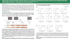 科学海报Defined Culture Conditions for Efficient Derivation Expansion and Cryopreservation of Mesenchymal Progenitor Cells from Human Pluripotent Stem Cells

 沪公网安备31010102008431号