Kamei K-i et al. (MAY 2010)
Lab on a chip 10 9 1113--9
Microfluidic image cytometry for quantitative single-cell profiling of human pluripotent stem cells in chemically defined conditions.
Microfluidic image cytometry (MIC) has been developed to study phenotypes of various hPSC lines by screening several chemically defined serum/feeder-free conditions. A chemically defined hPSC culture was established using 20 ng mL(-1) of bFGF on 20 microg mL(-1) of Matrigel to grow hPSCs over a week in an undifferentiated state. Following hPSC culture,we conducted quantitative MIC to perform a single cell profiling of simultaneously detected protein expression (OCT4 and SSEA1). Using clustering analysis,we were able to systematically compare the characteristics of various hPSC lines in different conditions.
View Publication
产品类型:
产品号#:
05850
05857
05870
05875
85850
85857
85870
85875
产品名:
mTeSR™1
mTeSR™1
Shimakura Y et al. (JAN 2000)
Stem cells (Dayton,Ohio) 18 3 183--9
Murine stromal cell line HESS-5 maintains reconstituting ability of Ex vivo-generated hematopoietic stem cells from human bone marrow and cytokine-mobilized peripheral blood.
Human bone marrow (BM) or mobilized peripheral blood (mPB) CD34(+) cells have been shown to loose their stem cell quality during culture period more easily than those from cord blood (CB). We previously reported that human umbilical CB stem cells could effectively be expanded in the presence of human recombinant cytokines and a newly established murine bone marrow stromal cell line HESS-5. In this study we assessed the efficacy of this xenogeneic coculture system using human BM and mPB CD34(+) cells as materials. We measured the generation of CD34(+)CD38(-) cells and colony-forming units,and assessed severe-combined immunodeficient mouse-repopulating cell (SRC) activity using cells five days after serum-free cytokine-containing culture in the presence or the absence of a direct contact with HESS-5 cells. As compared with the stroma-free culture,the xenogeneic coculture was significantly superior on expansion of CD34(+)CD38(-) cells and colony-forming cells and on maintenance of SRC activity. The PKH26 study demonstrated that cell division was promoted faster in cells cocultured with HESS-5 cells than in cells cultured without HESS-5 cells. These results indicate that HESS-5 supports rapid generation of primitive progenitor cells (PPC) and maintains reconstituting ability of newly generated stem cells during ex vivo culture irrespective of the source of samples. This xenogeneic coculture system will be useful for ex vivo manipulation such as gene transduction to promote cell division and the generation of PPC and to prevent loss of stem cell quality.
View Publication
产品类型:
产品号#:
04064
04034
04044
产品名:
MethoCult™ H4034 Optimum 入门试剂盒
MethoCult™ H4034 Optimum
MethoCult™ H4034 Optimum
Raffaghello L et al. (JAN 2008)
Stem cells (Dayton,Ohio) 26 1 151--62
Human mesenchymal stem cells inhibit neutrophil apoptosis: a model for neutrophil preservation in the bone marrow niche.
Mesenchymal stem cells (MSC) establish close interactions with bone marrow sinusoids in a putative perivascular niche. These vessels contain a large storage pool of mature nonproliferating neutrophils. Here,we have investigated the effects of human bone marrow MSC on neutrophil survival and effector functions. MSC from healthy donors,at very low MSC:neutrophil ratios (up to 1:500),significantly inhibited apoptosis of resting and interleukin (IL)-8-activated neutrophils and dampened N-formyl-l-methionin-l-leucyl-l-phenylalanine (f-MLP)-induced respiratory burst. The antiapoptotic activity of MSC did not require cell-to-cell contact,as shown by transwell experiments. Antibody neutralization experiments demonstrated that the key MSC-derived soluble factor responsible for neutrophil protection from apoptosis was IL-6,which signaled by activating STAT-3 transcription factor. Furthermore,IL-6 expression was detected in MSC by real-time reverse transcription-polymerase chain reaction and enzyme-linked immunosorbent assay. Finally,recombinant IL-6 was found to protect neutrophils from apoptosis in a dose-dependent manner. MSC had no effect on neutrophil phagocytosis,expression of adhesion molecules,and chemotaxis in response to IL-8,f-MLP,or C5a. These results support the following conclusions: (a) in the bone marrow niche,MSC likely protect neutrophils of the storage pool from apoptosis,preserving their effector functions and preventing the excessive or inappropriate activation of the oxidative metabolism,and (b) a novel mechanism whereby the inflammatory potential of activated neutrophils is harnessed by inhibition of apoptosis and reactive oxygen species production without impairing phagocytosis and chemotaxis has been identified.
View Publication
产品类型:
产品号#:
05401
05402
05411
产品名:
MesenCult™ MSC 基础培养基(人)
MesenCult™ MSC刺激添加物(人)
MesenCult™ 增殖试剂盒(人)
Swartz EW et al. (NOV 2016)
STEM CELLS Translational Medicine 5 11 1461--1472
A Novel Protocol for Directed Differentiation of C9orf72-Associated Human Induced Pluripotent Stem Cells Into Contractile Skeletal Myotubes
Induced pluripotent stem cells (iPSCs) offer an unlimited resource of cells to be used for the study of underlying molecular biology of disease,therapeutic drug screening,and transplant-based regenerative medicine. However,methods for the directed differentiation of skeletal muscle for these purposes remain scarce and incomplete. Here,we present a novel,small molecule-based protocol for the generation of multinucleated skeletal myotubes using eight independent iPSC lines. Through combinatorial inhibition of phosphoinositide 3-kinase (PI3K) and glycogen synthase kinase 3β (GSK3β) with addition of bone morphogenic protein 4 (BMP4) and fibroblast growth factor 2 (FGF2),we report up to 64% conversion of iPSCs into the myogenic program by day 36 as indicated by MYOG+ cell populations. These cells began to exhibit spontaneous contractions as early as 34 days in vitro in the presence of a serum-free medium formulation. We used this protocol to obtain iPSC-derived muscle cells from frontotemporal dementia (FTD) patients harboring C9orf72 hexanucleotide repeat expansions (rGGGGCC),sporadic FTD,and unaffected controls. iPSCs derived from rGGGGCC carriers contained RNA foci but did not vary in differentiation efficiency when compared to unaffected controls nor display mislocalized TDP-43 after as many as 120 days in vitro. This study presents a rapid,efficient,and transgene-free method for generating multinucleated skeletal myotubes from iPSCs and a resource for further modeling the role of skeletal muscle in amyotrophic lateral sclerosis and other motor neuron diseases. SIGNIFICANCE Protocols to produce skeletal myotubes for disease modeling or therapy are scarce and incomplete. The present study efficiently generates functional skeletal myotubes from human induced pluripotent stem cells using a small molecule-based approach. Using this strategy,terminal myogenic induction of up to 64% in 36 days and spontaneously contractile myotubes within 34 days were achieved. Myotubes derived from patients carrying the C9orf72 repeat expansion show no change in differentiation efficiency and normal TDP-43 localization after as many as 120 days in vitro when compared to unaffected controls. This study provides an efficient,novel protocol for the generation of skeletal myotubes from human induced pluripotent stem cells that may serve as a valuable tool in drug discovery and modeling of musculoskeletal and neuromuscular diseases.
View Publication
产品类型:
产品号#:
05832
72302
72304
72307
72308
78006
78006.1
78006.2
78005
78005.1
78005.2
78005.3
34811
34815
34850
34821
34825
34860
05835
05839
100-1044
产品名:
STEMdiff™ 神经花环选择试剂
Y-27632(二盐酸盐)
Y-27632(二盐酸盐)
Y-27632(二盐酸盐)
Y-27632(二盐酸盐)
重组人EGF
重组人EGF
重组人EGF
重组人BDNF
重组人BDNF
重组人BDNF
重组人BDNF
AggreWell™ 800 24孔板,1个
AggreWell™ 800 24孔板,5个
AggreWell™ 800 24孔板启动套装
AggreWell™ 800 6孔板,1个
AggreWell™ 800 6孔板,5个
AggreWell™ 800 6孔板启动套装
STEMdiff™ 神经诱导培养基
STEMdiff™ 神经诱导培养基
Y-27632(二盐酸盐)
Gadkari R et al. (JUL 2014)
Regenerative medicine 9 4 453--465
Human embryonic stem cell derived-mesenchymal stem cells: an alternative mesenchymal stem cell source for regenerative medicine therapy.
AIM To enumerate and characterize mesenchymal stem cells (MSC) derived from human embryonic stem cells (hESC) for clinical application. MATERIALS & METHODS hESC were differentiated into hESC-MSC and characterized by the expression of surface markers using flow cytometry. hESC-MSC were evaluated with respect to growth kinetics,colony-forming potential,as well as osteogenic and adipogenic differentiation capacity. Immunosuppressive effects were assessed using peripheral blood mononuclear cell (PBMC) proliferation and cytotoxicity assays. RESULTS hESC-MSC showed similar morphology,and cell surface markers as adipose (AMSC) and bone marrow-derived MSC (BMSC). hESC-MSC exhibited a higher growth rate during early in vitro expansion and equivalent adipogenic and osteogenic differentiation and colony-forming potential as AMSC and BMSC. hESC-MSC demonstrated similar immunosuppressive effects as AMSC and BMSC. CONCLUSION hESC-MSC were comparable to BMSC and AMSC and hence can be used as an alternative source of MSC for clinical applications.
View Publication
产品类型:
产品号#:
产品名:
Park H-JJ et al. (MAY 2015)
Biomaterials 50 1 127--139
Bio-inspired oligovitronectin-grafted surface for enhanced self-renewal and long-term maintenance of human pluripotent stem cells under feeder-free conditions.
Current protocols for human pluripotent stem cell (hPSC) expansion require feeder cells or matrices from animal sources that have been the major obstacle to obtain clinical grade hPSCs due to safety issues,difficulty in quality control,and high expense. Thus,feeder-free,chemically defined synthetic platforms have been developed,but are mostly confined to typical polystyrene culture plates. Here,we report a chemically defined,material-independent,bio-inspired surface coating allowing for feeder-free expansion and maintenance of self-renewal and pluripotency of hPSCs on various polymer substrates and devices. Polydopamine (pDA)-mediated immobilization of vitronectin (VN) peptides results in surface functionalization of VN-dimer/pDA conjugates. The engineered surfaces facilitate adhesion,proliferation,and colony formation of hPSCs via enhanced focal adhesion,cell-cell interaction,and biophysical signals,providing a chemically defined,xeno-free culture system for clonal expansion and long-term maintenance of hPSCs. This surface engineering enables the application of clinically-relevant hPSCs to a variety of biomedical systems such as tissue-engineering scaffolds and medical devices.
View Publication
产品类型:
产品号#:
05850
05857
05870
05875
85850
85857
85870
85875
产品名:
mTeSR™1
mTeSR™1
Bak XY et al. (NOV 2011)
Human gene therapy 22 11 1365--77
Human embryonic stem cell-derived mesenchymal stem cells as cellular delivery vehicles for prodrug gene therapy of glioblastoma.
Mesenchymal stem cells (MSCs) possess tumor-tropic properties and consequently have been used to deliver therapeutic agents for cancer treatment. Their potential in cancer therapy highlights the need for a consistent and renewable source for the production of uniform human MSCs suitable for clinical applications. In this study,we seek to investigate whether human embryonic stem cells can be used as a cell source to fulfill this goal. We generated MSC-like cells from two human embryonic stem cell lines,HuES9 and H1,and observed that MSC-like cells derived from human embryonic stem cells were able to migrate into human glioma intracranial xenografts after being injected into the cerebral hemisphere contralateral to the tumor inoculation site. We engineered these cells with baculoviral and lentiviral vectors,respectively,for transient and stable expression of the herpes simplex virus thymidine kinase gene. In tumor-bearing mice the engineered MSC-like cells were capable of inhibiting tumor growth and prolonging survival in the presence of ganciclovir after they were injected either directly into the xenografts or into the opposite hemisphere. Our findings suggest that human embryonic stem cell-derived MSCs may be a viable and attractive alternative for large-scale derivation of targeting vehicles for cancer therapy.
View Publication
产品类型:
产品号#:
05850
05857
05870
05875
85850
85857
85870
85875
产品名:
mTeSR™1
mTeSR™1
Takayama N et al. (DEC 2010)
The Journal of experimental medicine 207 13 2817--30
Transient activation of c-MYC expression is critical for efficient platelet generation from human induced pluripotent stem cells.
Human (h) induced pluripotent stem cells (iPSCs) are a potentially abundant source of blood cells,but how best to select iPSC clones suitable for this purpose from among the many clones that can be simultaneously established from an identical source is not clear. Using an in vitro culture system yielding a hematopoietic niche that concentrates hematopoietic progenitors,we show that the pattern of c-MYC reactivation after reprogramming influences platelet generation from hiPSCs. During differentiation,reduction of c-MYC expression after initial reactivation of c-MYC expression in selected hiPSC clones was associated with more efficient in vitro generation of CD41a(+)CD42b(+) platelets. This effect was recapitulated in virus integration-free hiPSCs using a doxycycline-controlled c-MYC expression vector. In vivo imaging revealed that these CD42b(+) platelets were present in thrombi after laser-induced vessel wall injury. In contrast,sustained and excessive c-MYC expression in megakaryocytes was accompanied by increased p14 (ARF) and p16 (INK4A) expression,decreased GATA1 expression,and impaired production of functional platelets. These findings suggest that the pattern of c-MYC expression,particularly its later decline,is key to producing functional platelets from selected iPSC clones.
View Publication
产品类型:
产品号#:
04434
04444
产品名:
MethoCult™ H4434 Classic
MethoCult™ H4434 Classic
Peters DT et al. (MAY 2016)
Development (Cambridge,England) 143 9 1475--81
Asialoglycoprotein receptor 1 is a specific cell-surface marker for isolating hepatocytes derived from human pluripotent stem cells.
Hepatocyte-like cells (HLCs) are derived from human pluripotent stem cells (hPSCs) in vitro,but differentiation protocols commonly give rise to a heterogeneous mixture of cells. This variability confounds the evaluation of in vitro functional assays performed using HLCs. Increased differentiation efficiency and more accurate approximation of the in vivo hepatocyte gene expression profile would improve the utility of hPSCs. Towards this goal,we demonstrate the purification of a subpopulation of functional HLCs using the hepatocyte surface marker asialoglycoprotein receptor 1 (ASGR1). We analyzed the expression profile of ASGR1-positive cells by microarray,and tested their ability to perform mature hepatocyte functions (albumin and urea secretion,cytochrome activity). By these measures,ASGR1-positive HLCs are enriched for the gene expression profile and functional characteristics of primary hepatocytes compared with unsorted HLCs. We have demonstrated that ASGR1-positive sorting isolates a functional subpopulation of HLCs from among the heterogeneous cellular population produced by directed differentiation.
View Publication
产品类型:
产品号#:
05850
05857
05870
05875
85850
85857
85870
85875
产品名:
mTeSR™1
mTeSR™1
Yan Y et al. (JUN 2016)
Acta Biomaterialia 42 114--126
Neural patterning of human induced pluripotent stem cells in 3-D cultures for studying biomolecule-directed differential cellular responses
Introduction Appropriate neural patterning of human induced pluripotent stem cells (hiPSCs) is critical to generate specific neural cells/tissues and even mini-brains that are physiologically relevant to model neurological diseases. However,the capacity of signaling factors that regulate 3-D neural tissue patterning in vitro and differential responses of the resulting neural populations to various biomolecules have not yet been fully understood. Methods By tuning neural patterning of hiPSCs with small molecules targeting sonic hedgehog (SHH) signaling,this study generated different 3-D neuronal cultures that were mainly comprised of either cortical glutamatergic neurons or motor neurons. Results Abundant glutamatergic neurons were observed following the treatment with an antagonist of SHH signaling,cyclopamine,while Islet-1 and HB9-expressing motor neurons were enriched by an SHH agonist,purmorphamine. In neurons derived with different neural patterning factors,whole-cell patch clamp recordings showed similar voltage-gated Na+/K+ currents,depolarization-evoked action potentials and spontaneous excitatory post-synaptic currents. Moreover,these different neuronal populations exhibited differential responses to three classes of biomolecules,including (1) matrix metalloproteinase inhibitors that affect extracellular matrix remodeling; (2) N-methyl-D-aspartate that induces general neurotoxicity; and (3) amyloid ?? (1???42) oligomers that cause neuronal subtype-specific neurotoxicity. Conclusions This study should advance our understanding of hiPSC self-organization and neural tissue development and provide a transformative approach to establish 3-D models for neurological disease modeling and drug discovery. Statement of Significance Appropriate neural patterning of human induced pluripotent stem cells (hiPSCs) is critical to generate specific neural cells,tissues and even mini-brains that are physiologically relevant to model neurological diseases. However,the capability of sonic hedgehog-related small molecules to tune different neuronal subtypes in 3-D differentiation from hiPSCs and the differential cellular responses of region-specific neuronal subtypes to various biomolecules have not been fully investigated. By tuning neural patterning of hiPSCs with small molecules targeting sonic hedgehog signaling,this study provides knowledge on the differential susceptibility of region-specific neuronal subtypes derived from hiPSCs to different biomolecules in extracellular matrix remodeling and neurotoxicity. The findings are significant for understanding 3-D neural patterning of hiPSCs for the applications in brain organoid formation,neurological disease modeling,and drug discovery.
View Publication
产品类型:
产品号#:
05850
05857
05870
05875
85850
85857
85870
85875
产品名:
mTeSR™1
mTeSR™1
Nath SC et al. (SEP 2016)
Bioprocess and biosystems engineering
Culture medium refinement by dialysis for the expansion of human induced pluripotent stem cells in suspension culture.
Human induced pluripotent stem cells (hiPSCs) secrete essential autocrine factors that are removed along with toxic metabolites when the growth medium is exchanged daily. In this study,after determining the minimum inhibitory level of lactic acid for hiPSCs,a medium refining system was constructed by which toxic metabolites were removed from used culture medium and autocrine factors as well as other growth factors were recycled. Specifically,about 87 % of the basic fibroblast growth factor and 80 % of transforming growth factor beta 1 were retained in the refined medium after dialysis. The refined medium efficiently potentiated the proliferation of hiPS cells in adherent culture. When the refining system was used to refresh medium in suspension culture,a final cell density of (1.1 ± 0.1) × 10(6) cells mL(-1) was obtained,with 99.5 ± 0.2 % OCT 3/4 and 78.3 ± 1.1 % TRA-1-60 expression,on day 4 of culture. These levels of expression were similar to those observed in the conventional suspension culture. With this method,culture medium refinement by dialysis was established to remove toxic metabolites,recycle autocrine factors as well as other growth factors,and reduce the use of macromolecules for the expansion of hiPSCs in suspension culture.
View Publication

 EasySep™小鼠TIL(CD45)正选试剂盒


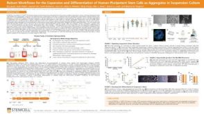 科学海报Robust Workflows for the Expansion and Differentiation of Human Pluripotent Stem Cells as Aggregates in Suspension Culture
 沪公网安备31010102008431号