
 EasySep™小鼠TIL(CD45)正选试剂盒


搜索结果: 'methocult media formulations for human hematopoietic cells serum containing'
-

产品类型:
产品号#:
03231
产品名:
MethoCult™ M3231
-
产品类型:
产品号#:
01700
01705
01701
01702
产品名:
ALDEFLUOR™ 试剂盒
ALDEFLUOR™ DEAB试剂, 1.5 mM, 1 mL
ALDEFLUOR™检测缓冲液
-
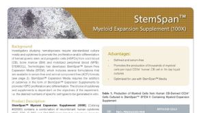 产品手册StemSpan™ Media and Supplements for the Expansion and Differentiation of Granulocytes and Monocytes
产品类型:
品牌:
StemSpan
产品号#:
02693
02694
产品名:
StemSpan™髓系扩增添加物 (100X)
StemSpan™髓系扩增添加物II (100X)
-
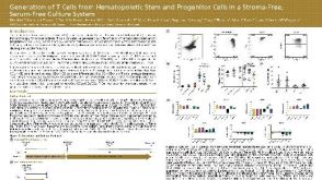 科学海报Generation of T Cells from Hematopoietic Stem and Progenitor Cells in a Stroma-Free, Serum-Free Culture System
产品类型:
产品号#:
09940
09605
09915
09925
09930
产品名:
StemSpan™ T细胞生成试剂盒
StemSpan™ SFEM II
StemSpan™淋系祖细胞扩增添加物(10X)
StemSpan™淋系祖细胞分化包板材料(100X)
StemSpan™ T细胞祖细胞成熟添加物(10X)
-
产品类型:
产品号#:
05850
05857
05870
05875
85850
85857
85870
85875
产品名:
mTeSR™1
mTeSR™1
-
产品类型:
产品号#:
05850
05857
05870
05875
85850
85857
85870
85875
产品名:
mTeSR™1
mTeSR™1
-
产品类型:
产品号#:
05850
05857
05870
05875
85850
85857
85870
85875
产品名:
mTeSR™1
mTeSR™1
-
产品类型:
产品号#:
19052
19052RF
产品名:
EasySep™人CD4+ T细胞富集试剂盒
RoboSep™ 人CD4+ T细胞富集试剂盒含滤芯吸头
-
产品类型:
产品号#:
05401
05402
05404
05411
产品名:
MesenCult™ MSC 基础培养基(人)
MesenCult™ MSC刺激添加物(人)
MesenCult™ 增殖试剂盒(人)
-
产品类型:
产品号#:
05850
05857
05870
05875
09500
85850
85857
85870
85875
产品名:
BIT 9500血清替代物
mTeSR™1
mTeSR™1
-
产品类型:
产品号#:
01700
01705
01701
01702
产品名:
ALDEFLUOR™ 试剂盒
ALDEFLUOR™ DEAB试剂, 1.5 mM, 1 mL
ALDEFLUOR™检测缓冲液
-
产品类型:
产品号#:
05850
05857
05870
05875
85850
85857
85870
85875
产品名:
mTeSR™1
mTeSR™1

 沪公网安备31010102008431号