Santoni de Sio FR et al. (JUN 2006)
Blood 107 11 4257--65
Proteasome activity restricts lentiviral gene transfer into hematopoietic stem cells and is down-regulated by cytokines that enhance transduction.
The therapeutic potential of hematopoietic stem cell (HSC) gene therapy can be fully exploited only by reaching efficient gene transfer into HSCs without compromising their biologic properties. Although HSCs can be transduced by HIV-derived lentiviral vectors (LVs) in short ex vivo culture,they display low permissivity to the vector,requiring cytokine stimulation to reach high-frequency transduction. Using stringent assays of competitive xenograft repopulation,we show that early-acting cytokines synergistically enhanced human HSC gene transfer by LVs without impairing engraftment and repopulation capacity. Using S-phase suicide assays,we show that transduction enhancement by cytokines was not dependent on cell cycle progression and that LVs can transduce quiescent HSCs. Pharmacologic inhibition of the proteasome during transduction dramatically enhanced HSC gene transfer,allowing the reach of very high levels of vector integration in their progeny in vivo. Thus,LVs are effectively restricted at a postentry step by the activity of this proteolytic complex. Unexpectedly,cytokine stimulation rapidly and substantially down-regulated proteasome activity in hematopoietic progenitors,highlighting one mechanism by which cytokines may enhance permissiveness to LV gene transfer. These findings demonstrate that antiviral responses ultimately mediated by proteasomes strongly limit the efficiency of HSC transduction by LVs and establish improved conditions for HSC-based gene therapy.
View Publication
产品类型:
产品号#:
09600
09650
产品名:
StemSpan™ SFEM
StemSpan™ SFEM
Gü et al. (DEC 2010)
Cytotherapy 12 8 1006--12
Evaluation of mobilized peripheral stem cells according to CD34 and aldehyde dehydrogenase expression and effect of SSC(lo) ALDH(br) cells on hematopoietic recovery.
BACKGROUND AIMS: We evaluated hematopoietic stem cells according to CD34 expression and aldehyde dehydrogenase (ALDH) activity in peripheral blood and apheresis product samples from patients after mobilization with granulocyte-colony-stimulating factor (G-CSF) alone or G-CSF after high-dose cyclophosphamide (4 g/m²) once daily,intravenously on day 1). We also investigated the relationship between the number of SSC(lo) CD45(dim) CD34(hi) cells,SSC(lo) ALDH(br) cells and engraftment. METHODS: Thirty patients (20 males and 10 females),who were candidates for autologous peripheral blood stem cell transplantation,were included in the study. Cyclophosphamide + G-CSF was used for 17 and G-CSF alone for 24 mobilizations. Primary diagnoses were multiple myeloma (n = 14),Hodgkin's lymphoma (n = 7),non-Hodgkin's lymphoma (n = 2),acute myloid leukemia (n = 2),chronic lymphocytic leukemia (n = 1) and germ cell testis tumor (n = 1). RESULTS: Numbers of SSC(lo) CD45(dim) CD34(hi) cells and SSC(lo) ALDH(br) cells were highly correlated in both peripheral blood and apheresis products (P textless 0.001). We could not find a relationship between the transplanted SSC(lo) CD45(dim) CD34(hi) cell dose or SSC(lo) ALDH(br) cell dose and platelet or neutrophil recovery. The optimal thresholds for SSC(lo) CD45(dim) CD34(hi) cells were 5.40 × 10�?�/kg for neutrophil recovery and 7.22 x 10�?�/kg for platelet recovery. The optimal thresholds for SSC(lo) ALDH(br) cells were 6.53 x 10�?�/kg for neutrophil recovery and 8.72 x 10�?�/kg platelet recovery. CONCLUSIONS: According to our data,numbers of SSC(lo) ALDH(br) cells are in very good agreement with numbers of SSC(lo) CD45(dim) CD34(hi) cells and can be a predictor of stem cell mobilization.
View Publication
产品类型:
产品号#:
01700
01705
01701
01702
产品名:
ALDEFLUOR™ 试剂盒
ALDEFLUOR™ DEAB试剂, 1.5 mM, 1 mL
ALDEFLUOR™检测缓冲液
Lawrence HJ et al. (DEC 2005)
Blood 106 12 3988--94
Loss of expression of the Hoxa-9 homeobox gene impairs the proliferation and repopulating ability of hematopoietic stem cells.
The homeobox gene Hoxa-9 is normally expressed in primitive bone marrow cells,and overexpression of Hoxa-9 markedly expands hematopoietic stem cells,suggesting a function in early hematopoiesis. We present evidence for major functional defects in Hoxa-9-/- hematopoietic stem cells. Hoxa-9-/- marrow cells have normal numbers of immunophenotypic stem cells (Lin(-)c-kit(+)flk-2(-)Sca-1+ [KLFS] cells). However,sublethally irradiated Hoxa-9-/- mice develop persistent pancytopenia,indicating unusual sensitivity to ionizing irradiation. In competitive transplantation assays,Hoxa-9-/- cells showed an 8-fold reduction in multilineage long-term repopulating ability,a defect not seen in marrow cells deficient for the adjacent Hoxa-10 gene. Single-cell cultures of KLFS cells showed a 4-fold reduction in large high-proliferation potential colonies. In liquid cultures,Hoxa-9-deficient Lin(-)Sca-1(+) cells showed slowed proliferation (a 5-fold reduction in cell numbers at day 8) and delayed emergence of committed progenitors (a 5-fold decrease in colony-forming cells). Slowing of proliferation was accompanied by a delay in myeloid maturation,with a decrease in Gr-1hiMac-1hi cells at the end of the culture. Retroviral transduction with a Hoxa-9 expression vector dramatically enhanced the cytokine-driven proliferation and in vivo engraftment of Hoxa-9-/- marrow cells. Hoxa-9 appears to be specifically required for normal hematopoietic stem cell function both in vitro and in vivo.
View Publication
产品类型:
产品号#:
03231
09600
09650
产品名:
MethoCult™ M3231
StemSpan™ SFEM
StemSpan™ SFEM
Wu W et al. (JUN 2015)
Analytica chimica acta 881 124--30
Strand displacement amplification for ultrasensitive detection of human pluripotent stem cells.
Human pluripotent stem cells (hPSCs),such as embryonic stem cells (ESCs) and induced pluripotent stem cells (iPSCs),provide a powerful model system for studies of cellular identity and early mammalian development,which hold great promise for regenerative medicine. It is necessary to develop a convenient method to discriminate hPSCs from other cells in clinics and basic research. Herein,a simple and reliable biosensor for stem cell detection was established. In this biosensor system,stage-specific embryonic antigen-3 (SSEA-3) and stage-specific embryonic antigen-4 (SSEA-4) were used to mark human pluripotent stem cells (hPSCs). Antibody specific for SSEA-3 was coated onto magnetic beads for hPSCs enrichment,and antibody specific for SSEA-4 was conjugated with carboxyl-modified tDNA sequence which was used as template for strand displacement amplification (SDA). The amplified single strand DNA (ssDNA) was detected with a lateral flow biosensor (LFB). This biosensor is capable of detecting a minimum of 19 human embryonic stem cells by a strip reader and 100 human embryonic stem cells by the naked eye within 80min. This approach has also shown excellent specificity to distinguish hPSCs from other types of cells,showing that it is promising for specific and handy detection of human pluripotent stem cells.
View Publication
产品类型:
产品号#:
05850
05857
05870
05875
85850
85857
85870
85875
产品名:
mTeSR™1
mTeSR™1
Tohyama S et al. (APR 2016)
Cell Metabolism 23 4 663--674
Glutamine Oxidation Is Indispensable for Survival of Human Pluripotent Stem Cells
Summary Human pluripotent stem cells (hPSCs) are uniquely dependent on aerobic glycolysis to generate ATP. However,the importance of oxidative phosphorylation (OXPHOS) has not been elucidated. Detailed amino acid profiling has revealed that glutamine is indispensable for the survival of hPSCs. Under glucose- and glutamine-depleted conditions,hPSCs quickly died due to the loss of ATP. Metabolome analyses showed that hPSCs oxidized pyruvate poorly and that glutamine was the main energy source for OXPHOS. hPSCs were unable to utilize pyruvate-derived citrate due to negligible expression of aconitase 2 (ACO2) and isocitrate dehydrogenase 2/3 (IDH2/3) and high expression of ATP-citrate lyase. Cardiomyocytes with mature mitochondria were not able to survive without glucose and glutamine,although they were able to use lactate to synthesize pyruvate and glutamate. This distinguishing feature of hPSC metabolism allows preparation of clinical-grade cell sources free of undifferentiated hPSCs,which prevents tumor formation during stem cell therapy.
View Publication
产品类型:
产品号#:
05850
05857
05870
05875
85850
85857
85870
85875
产品名:
mTeSR™1
mTeSR™1
N. Albinger et al. (apr 2022)
Blood cancer journal 12 4 61
Primary CD33-targeting CAR-NK cells for the treatment of acute myeloid leukemia.
Acute myeloid leukemia (AML) is a malignant disorder derived from neoplastic myeloid progenitor cells characterized by abnormal proliferation and differentiation. Although novel therapeutics have recently been introduced,AML remains a therapeutic challenge with insufficient cure rates. In the last years,immune-directed therapies such as chimeric antigen receptor (CAR)-T cells were introduced,which showed outstanding clinical activity against B-cell malignancies including acute lymphoblastic leukemia (ALL). However,the application of CAR-T cells appears to be challenging due to the enormous molecular heterogeneity of the disease and potential long-term suppression of hematopoiesis. Here we report on the generation of CD33-targeted CAR-modified natural killer (NK) cells by transduction of blood-derived primary NK cells using baboon envelope pseudotyped lentiviral vectors (BaEV-LVs). Transduced cells displayed stable CAR-expression,unimpeded proliferation,and increased cytotoxic activity against CD33-positive OCI-AML2 and primary AML cells in vitro. Furthermore,CD33-CAR-NK cells strongly reduced leukemic burden and prevented bone marrow engraftment of leukemic cells in OCI-AML2 xenograft mouse models without observable side effects.
View Publication
I. Gonz\'alez-Mariscal et al. (jan 2022)
Biomedicine & pharmacotherapy = Biomedecine & pharmacotherapie 145 112361
Abnormal cannabidiol ameliorates inflammation preserving pancreatic beta cells in mouse models of experimental type 1 diabetes and beta cell damage.
The atypical cannabinoid Abn-CBD improves the inflammatory status in preclinical models of several pathologies,including autoimmune diseases. However,its potential for modulating inflammation in autoimmune type 1 diabetes (T1D) is unknown. Herein we investigate whether Abn-CBD can modulate the inflammatory response during T1D onset using a mouse model of T1D (non-obese diabetic- (NOD)-mice) and of beta cell damage (streptozotocin (STZ)-injected mice). Six-week-old female NOD mice were treated with Abn-CBD (0.1-1 mg/kg) or vehicle during 12 weeks and then euthanized. Eight-to-ten-week-old male C57Bl6/J mice were pre-treated with Abn-CBD (1 mg/kg of body weight) or vehicle for 1 week,following STZ challenge,and euthanized 1 week later. Blood,pancreas,pancreatic lymph nodes (PLNs) and T cells were collected and processed for analysis. Glycemia was also monitored. In NOD mice,treatment with Abn-CBD significantly reduced the severity of insulitis and reduced the pro-inflammatory profile of CD4+ T cells compared to vehicle. Concomitantly,Abn-CBD significantly reduced islet cell apoptosis and improved glucose tolerance. In STZ-injected mice,Abn-CBD decreased circulating proinflammatory cytokines and ameliorated islet inflammation reducing intra-islet phospho-NF-$\kappa$B and TXNIP. Abn-CBD significantly reduced 2 folds intra-islet CD8+ T cells and reduced Th1/non-Th1 ratio in PLNs of STZ-injected mice. Islet cell apoptosis and intra-islet fibrosis were also significantly reduced in Abn-CBD pre-treated mice compared to vehicle. Altogether,Abn-CBD reduces circulating and intra-islet inflammation,preserving islets,thus delaying the progression of insulitis. Hence,Abn-CBD and related compounds emerge as new candidates to develop pharmacological strategies to treat the early stages of T1D.
View Publication
产品类型:
产品号#:
18000
19852
19852RF
产品名:
EasySep™磁极
EasySep™小鼠CD4+ T细胞分选试剂盒
RoboSep™ 小鼠CD4+ T细胞分选试剂盒
Ma ACH et al. (DEC 2010)
Leukemia 24 12 2090--9
A DEAB-sensitive aldehyde dehydrogenase regulates hematopoietic stem and progenitor cells development during primitive hematopoiesis in zebrafish embryos.
Although aldehyde dehydrogenase (ALDH) activity has become a surrogate of hematopoietic stem and progenitor cells (HSPCs),its function during hematopoiesis was unclear. Here,we examined its role in zebrafish hematopoiesis based on pharmacological inhibition and morpholino (MO) knockdown. Zebrafish embryos were treated with diethylaminobenzaldehyde (DEAB,1 μmol/l) between 0- and 48 hour-post-fertilization (hpf). MOs targeting aldhs were injected between 1 and 4-cell stage. The effects on hematopoiesis were evaluated at different stages. DEAB treatment between 0 and 18 hpf increased gene expression associated with HSPC (scl,lmo2),erythropoiesis (gata1,α- and β-eHb) and myelopoiesis (spi1) as well as gfp(+) cells in dissociated Tg(gata1:gfp) embryos. The effects were ameliorated by all-trans retinoic acid (1 nmol/l). Definitive hematopoiesis and the erythromyeloid precursors were unaffected. In all,14 out of 15 zebrafish aldhs were detectable by reverse transcription PCR in 18 hpf embryos,of which only aldh1a2 and aldh16a1 were expressed in sites pertinent to hematopoiesis. Molecular targeting by MOs was demonstrated for 15 aldhs,but none of them,even in combined aldh1a2 and aldh1a3 knockdown,recapitulated the hematopoietic expansion in DEAB-treated embryos. In conclusion,DEAB expands HSPC population during primitive hematopoiesis through inhibition of aldh and retinoic acid synthesis. The specific aldh isoform(s) remains to be determined.
View Publication
产品类型:
产品号#:
01700
01705
01702
产品名:
ALDEFLUOR™ 试剂盒
ALDEFLUOR™ DEAB试剂, 1.5 mM, 1 mL
ALDEFLUOR™检测缓冲液
W. Xing et al. (Jul 2025)
Stem Cell Research & Therapy 16 4
Deletion of p18 INK4c enhances both osteogenesis and hematopoietic supportive capacity of bone marrow mesenchymal stromal cells
p18 INK4 C (CDKN2C,encoded by p18 INK4c or Cdkn2c ) is an early G1-phase cyclin-dependent kinase inhibitor protein. Previous studies demonstrated enhanced self-renewal capacity of hematopoietic stem cells (HSCs) in p18 −/− mice compared to wild-type (WT) mice. Given the critical role of bone marrow niche cells-particularly mesenchymal stromal cells (MSCs)-in hematopoiesis,this study investigated the functional alterations of p18 −/− MSCs and their impact on hematopoietic support. Bone marrow derived MSCs were isolated from p18 −/− and WT mice. Their proliferation and differentiation capacities were assessed,followed by evaluation of hematopoietic support using cobblestone area-forming cell assay and long-term culture-initiating cell assay. RNA sequencing was performed to analyze the transcriptional profile of p18 −/− MSCs,with a focus on differentially expressed genes (DEGs). Key pathways associated with hematopoietic support were identified using Ingenuity Pathway Analysis. A candidate protein was quantified by ELISA,and its functional role in hematopoietic support was validated via a modified coculture system. p18 −/− MSCs displayed an increased proliferation rate,preferential differentiation toward osteogenesis over adipogenesis,and enhanced hematopoietic support. RNA sequencing analysis identified 137 DEGs,with secreted phosphoprotein 1 ( Spp1,encoding osteopontin,Opn) being significantly upregulated in p18 −/− MSCs. Elevated Opn levels were confirmed in both bone marrow and MSC-conditioned media from p18 −/− mice. Functional validation further demonstrated that Opn enhanced the hematopoietic supportive capacity of MSCs in vitro. p18 deficiency promotes osteogenic differentiation and enhances the hematopoietic supportive function of MSCs,likely mediated by Opn upregulation. These findings suggest a potential therapeutic strategy for improving bone regeneration and HSC expansion. The online version contains supplementary material available at 10.1186/s13287-025-04402-6.
View Publication
产品类型:
产品号#:
03434
03444
产品名:
MethoCult™ GF M3434
MethoCult™ GF M3434
Reya T et al. (MAY 2003)
Nature 423 6938 409--14
A role for Wnt signalling in self-renewal of haematopoietic stem cells.
Haematopoietic stem cells (HSCs) have the ability to renew themselves and to give rise to all lineages of the blood; however,the signals that regulate HSC self-renewal remain unclear. Here we show that the Wnt signalling pathway has an important role in this process. Overexpression of activated beta-catenin expands the pool of HSCs in long-term cultures by both phenotype and function. Furthermore,HSCs in their normal microenvironment activate a LEF-1/TCF reporter,which indicates that HCSs respond to Wnt signalling in vivo. To demonstrate the physiological significance of this pathway for HSC proliferation we show that the ectopic expression of axin or a frizzled ligand-binding domain,inhibitors of the Wnt signalling pathway,leads to inhibition of HSC growth in vitro and reduced reconstitution in vivo. Furthermore,activation of Wnt signalling in HSCs induces increased expression of HoxB4 and Notch1,genes previously implicated in self-renewal of HSCs. We conclude that the Wnt signalling pathway is critical for normal HSC homeostasis in vitro and in vivo,and provide insight into a potential molecular hierarchy of regulation of HSC development.
View Publication

 EasySep™小鼠TIL(CD45)正选试剂盒


 实验方案How to Prepare Conditioned Medium for the Expansion of Epithelial Cells
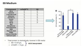

 沪公网安备31010102008431号