Maherali N et al. (SEP 2008)
Cell stem cell 3 3 340--5
A high-efficiency system for the generation and study of human induced pluripotent stem cells.
Direct reprogramming of human fibroblasts to a pluripotent state has been achieved through ectopic expression of the transcription factors OCT4,SOX2,and either cMYC and KLF4 or NANOG and LIN28. Little is known,however,about the mechanisms by which reprogramming occurs,which is in part limited by the low efficiency of conversion. To this end,we sought to create a doxycycline-inducible lentiviral system to convert primary human fibroblasts and keratinocytes into human induced pluripotent stem cells (hiPSCs). hiPSCs generated with this system were molecularly and functionally similar to human embryonic stem cells (hESCs),demonstrated by gene expression profiles,DNA methylation status,and differentiation potential. While expression of the viral transgenes was required for several weeks in fibroblasts,we found that 10 days was sufficient for the reprogramming of keratinocytes. Using our inducible system,we developed a strategy to induce hiPSC formation at high frequency. Upon addition of doxycycline to hiPSC-derived differentiated cells,we obtained secondary" hiPSCs at a frequency at least 100-fold greater than the initial conversion. The ability to reprogram cells at high efficiency provides a unique platform to dissect the underlying molecular and biochemical processes that accompany nuclear reprogramming."
View Publication
产品类型:
产品号#:
72742
产品名:
强力霉素(盐酸盐)
Ohgushi M et al. (AUG 2010)
Cell stem cell 7 2 225--39
Molecular pathway and cell state responsible for dissociation-induced apoptosis in human pluripotent stem cells.
Human embryonic stem cells (hESCs),unlike mouse ones (mESCs),are vulnerable to apoptosis upon dissociation. Here,we show that the apoptosis,which is of a nonanoikis type,is caused by ROCK-dependent hyperactivation of actomyosin and efficiently suppressed by the myosin inhibitor Blebbistatin. The actomyosin hyperactivation is triggered by the loss of E-cadherin-dependent intercellular contact and also observed in dissociated mouse epiblast-derived pluripotent cells but not in mESCs. We reveal that Abr,a unique Rho-GEF family factor containing a functional Rac-GAP domain,is an indispensable upstream regulator of the apoptosis and ROCK/myosin hyperactivation. Rho activation coupled with Rac inhibition is induced in hESCs upon dissociation,but not in Abr-depleted hESCs or mESCs. Furthermore,artificial Rho or ROCK activation with Rac inhibition restores the vulnerability of Abr-depleted hESCs to dissociation-induced apoptosis. Thus,the Abr-dependent Rho-high/Rac-low" state plays a decisive role in initiating the dissociation-induced actomyosin hyperactivation and apoptosis in hESCs."
View Publication
产品类型:
产品号#:
72402
72404
产品名:
(-)-Blebbistatin
(-)-Blebbistatin
Du S-HH et al. (AUG 2015)
Journal of bioscience and bioengineering 120 2 210--217
Human iPS cell-derived fibroblast-like cells as feeder layers for iPS cell derivation and expansion
Mouse embryonic fibroblasts (MEFs) are commonly used as feeder cells for the generation of human induced pluripotent stem cells (hiPSCs). However,medical applications of cell derivatives of hiPSCs generated with a MEF feeder system run the risk of having xeno-factor contamination due to long-term cell culturing under an animal factor-containing environment. We developed a new method for the derivation of human fibroblast-like cells (FLCs) from a previously established hiPSC line in an FLC differentiation medium. The method was based on direct differentiation of hiPSCs seeded on Matrigel followed by expansion of differentiating cells on gelatin. Using inactivated FLCs as feeder layers,primary human foreskin fibroblasts were successfully reprogrammed into a state of pluripotency by Oct4,Sox2 Klf4,and c-Myc (OSKM) transcription factor genes,with a reprogramming efficiency under an optimized condition superior to that obtained on MEF feeder layers. Furthermore,the FLCs were more effective in supporting the growth of human pluripotent stem cells. The pluripotency and differentiation capability of the cells cultured on FLC feeder layers were well retained. Our results suggest that FLCs are a safe alternative to MEFs for hiPSC generation and expansion,especially in the clinical settings wherein hiPSC derivatives will be used for medical treatment.
View Publication
产品类型:
产品号#:
05850
05857
05870
05875
07923
85850
85857
85870
85875
产品名:
Dispase (1 U/mL)
mTeSR™1
mTeSR™1
Liu Y et al. (MAY 2011)
Nature protocols 6 5 640--55
OLIG gene targeting in human pluripotent stem cells for motor neuron and oligodendrocyte differentiation.
Pluripotent stem cells can be genetically labeled to facilitate differentiation studies. In this paper,we describe a gene-targeting protocol to knock in a GFP cassette into key gene loci in human pluripotent stem cells (hPSCs),and then use the genetically tagged hPSCs to guide in vitro differentiation,immunocytochemical and electrophysiological profiling and in vivo characterization after cell transplantation. The Olig transcription factors have key roles in the transcription regulatory pathways for the genesis of motor neurons (MNs) and oligodendrocytes (OLs). We have generated OLIG2-GFP hPSC reporter lines that reliably mark MNs and OLs for monitoring their sequential differentiation from hPSCs. The expression of the GFP reporter recapitulates the endogenous expression of OLIG genes. The in vitro characterization of fluorescence-activated cell sorting-purified cells is consistent with cells of the MN or OL lineages,depending on the stages at which they are collected. This protocol is efficient and reliable and usually takes 5-7 months to complete. The genetic tagging-differentiation methodology used herein provides a general framework for similar work for differentiation of hPSCs into other lineages.
View Publication
产品类型:
产品号#:
05850
05857
05870
05875
85850
85857
85870
85875
产品名:
mTeSR™1
mTeSR™1
Wang Z et al. (APR 2012)
Cell stem cell 10 4 440--454
Distinct lineage specification roles for NANOG, OCT4, and SOX2 in human embryonic stem cells.
Nanog,Oct4,and Sox2 are the core regulators of mouse (m)ESC pluripotency. Although their basic importance in human (h)ESCs has been demonstrated,the mechanistic functions are not well defined. Here,we identify general and cell-line-specific requirements for NANOG,OCT4,and SOX2 in hESCs. We show that OCT4 regulates,and interacts with,the BMP4 pathway to specify four developmental fates. High levels of OCT4 enable self-renewal in the absence of BMP4 but specify mesendoderm in the presence of BMP4. Low levels of OCT4 induce embryonic ectoderm differentiation in the absence of BMP4 but specify extraembryonic lineages in the presence of BMP4. NANOG represses embryonic ectoderm differentiation but has little effect on other lineages,whereas SOX2 and SOX3 are redundant and repress mesendoderm differentiation. Thus,instead of being panrepressors of differentiation,each factor controls specific cell fates. Our study revises the view of how self-renewal is orchestrated in hESCs.
View Publication
产品类型:
产品号#:
05850
05857
05870
05875
85850
85857
85870
85875
产品名:
mTeSR™1
mTeSR™1
(Apr 2025)
International Journal of Molecular Sciences 26 7
Ex Vivo Plasma Application on Human Brain Microvascular Endothelial-like Cells for Blood–Brain Barrier Modeling
hiPSC-derived blood–brain barrier (BBB) models are valuable for pharmacological and physiological studies,yet their translational potential is limited due to insufficient cell phenotypes and the neglection of the complex environment of the BBB. This study evaluates the plasma compatibility with hiPSC-derived microvascular endothelial-like cells to enhance the translational potential of in vitro BBB models. Therefore,plasma samples (sodium/lithium heparin,citrate,EDTA) and serum from healthy donors were tested on hiPSC-derived microvascular endothelial-like cells at concentrations of 100%,75%,and 50%. After 24 h,cell viability parameters were assessed. The impact of heparin-anticoagulated plasmas was further evaluated regarding barrier function and endothelial phenotype of differentiated endothelial-like cells. Finally,sodium-heparin plasma was tested in an isogenic triple-culture BBB model with continuous TEER measurements for 72 h. Only the application of heparin-anticoagulated plasmas did not significantly alter viability parameters compared to medium. Furthermore,heparin plasmas improved barrier function without increasing cell density and induced a von Willebrand factor signal. Finally,continuous TEER measurements of the triple-culture model confirmed the positive impact of sodium-heparin plasma on barrier function. Consequently,heparin-anticoagulated plasmas were proven to be compatible with hiPSC-derived microvascular endothelial-like cells. Thereby,the translational potential of BBB models can be substantially improved in the future.
View Publication
产品类型:
产品号#:
100-0276
100-1130
产品名:
mTeSR™ Plus
mTeSR™ Plus
F. Tang et al. (Oct 2024)
Stem Cell Research & Therapy 15
Genetically engineered human induced pluripotent stem cells for the production of brain-targeting extracellular vesicles
Extracellular vesicles (EVs) are cell-secreted membrane vesicles that have become a promising,natural nanoparticle system for delivering either naturally carried or exogenously loaded therapeutic molecules. Among reported cell sources for EV manufacture,human induced pluripotent stem cells (hiPSCs) offer numerous advantages. However,hiPSC-EVs only have a moderate ability for brain delivery. Herein,we sought to develop a stable hiPSC line for producing EVs with substantially enhanced brain targeting by genetic engineering to overexpress rabies viral glycoprotein (RVG) peptide fused to the N terminus of lysosomal associated membrane protein 2B (RVG-Lamp2B) which has been shown capable of boosting the brain delivery of EVs via the nicotinic acetylcholine receptor. An RVG-Lamp2B-HA expression cassette was knocked into the AAVS1 safe harbor locus of a control hiPSC line using the CRISPR/Cas9-assisted homologous recombination. Western blot was used to detect the expression of RVG-Lamp2B-HA in RVG-edited hiPSCs as well as EVs derived from RVG-edited hiPSCs. Uptake of EVs by SH-SY5Y cells in the presence of various endocytic inhibitors was analyzed using flow cytometry. Biodistribution and brain delivery of intravenously injected control and RVG-modified EVs in wild-type mice were examined using ex vivo fluorescent imaging. Here we report that an RVG-Lamp2B-HA expression cassette was knocked into the AAVS1 safe harbor locus of a control hiPSC line using the CRISPR/Cas9-assisted homologous recombination. The RVG-edited iPSCs have normal karyotype,express pluripotency markers,and have differentiation potential. Expression of RVG-Lamp2B-HA was detected in total cell extracts as well as EVs derived from RVG-edited (vs. control) hiPSCs. The RVG-modified EVs enter neuronal cells via distinct endocytic pathways,compared with control EVs. The biodistribution study confirmed that EVs derived from RVG-edited hiPSCs possess higher brain delivery efficiency. Taken together,we have established stable,genetically engineered hiPSCs for producing EVs with RVG expression,offering the improved ability for brain-targeted drug delivery. The online version contains supplementary material available at 10.1186/s13287-024-03955-2.
View Publication
产品类型:
产品号#:
05230
产品名:
STEMdiff™ 三胚层分化试剂盒
Pearce DJ and Bonnet D (SEP 2007)
Experimental hematology 35 9 1437--46
The combined use of Hoechst efflux ability and aldehyde dehydrogenase activity to identify murine and human hematopoietic stem cells.
OBJECTIVE: In murine hematopoietic tissue,direct repopulation experiments have demonstrated that the side population (SP) represents a remarkable enrichment of hematopoietic stem cells. Human SP has been phenotyped as negative for lineage antigens as well as CD34. However,in the 9 years since the original publication,no long-term hematopoietic reconstitution has been reported for the adult human SP/CD34(-) subset. Elevated levels of aldehyde dehydrogenase (ALDH) have been demonstrated in murine and human progenitor cells when compared to other hematopoietic cells. METHODS: Here,we report the phenotype of human cord blood SP cells. We established the technique of simultaneous phenotyping,Hoechst exclusion,and ALDH labeling on murine tissues. We then performed the simultaneous analysis of phenotype,SP,and ALDH activity on human cord blood and bone marrow cells. Finally,we analyzed the phenotype and functional potential of human cord blood ALDH(+) cells to determine whether Lin(-)/CD34(-) cells are identified via this technique. RESULTS: We demonstrate that human Lin(-)/CD34(-)/ALDH(+) cells are capable of long-term repopulation. Although the SP technique identifies cells that overlap with the ALDH(+) cell population,this is restricted to the CD34(+) cell subset. CONCLUSION: Hoechst exclusion ability does not seem to be the method of choice for the isolation of human hematopoietic stem cells.
View Publication
ErbB4 Activated p38$$ MAPK Isoform Mediates Early Cardiogenesis Through NKx2.5 in Human Pluripotent Stem Cells
Activation of ErbB4 receptor signaling is instrumental in heart development,lack of which results in embryonic lethality. However,mechanism governing its intracellular signaling remains elusive. Using human pluripotent stem cells,we show that ErbB4 is critical for cardiogenesis whereby its genetic knockdown results in loss of cardiomyocytes. Phospho-proteome profiling and Western blot studies attribute this loss to inactivation of p38$\$ isoform which physically interacts with NKx2.5 and GATA4 transcription factors. Post-cardiomyocyte formation p38$\$/NKx2.5 downregulation is followed by p38$\$/MEF2c upregulation suggesting stage-specific developmental roles of p38 MAPK isoforms. Knockdown of p38$\$ similarly disrupts cardiomyocyte formation in spite of the presence of NKx2.5. Cell fractionation and NKx2.5 phosphorylation studies suggest inhibition of ErbB4-p38$\$ hinders NKx2.5 nuclear translocation during early cardiogenesis. This study reveals a novel pathway that directly links ErbB4 and p38$\$ the transcriptional machinery of NKx2.5-GATA4 complex which is critical for cardiomyocyte formation during mammalian heart development.
View Publication
Chen G et al. (AUG 2010)
Cell stem cell 7 2 240--8
Actin-myosin contractility is responsible for the reduced viability of dissociated human embryonic stem cells.
Human ESCs are the pluripotent precursor of the three embryonic germ layers. Human ESCs exhibit basal-apical polarity,junctional complexes,integrin-dependent matrix adhesion,and E-cadherin-dependent cell-cell adhesion,all characteristics shared by the epiblast epithelium of the intact mammalian embryo. After disruption of epithelial structures,programmed cell death is commonly observed. If individualized human ESCs are prevented from reattaching and forming colonies,their viability is significantly reduced. Here,we show that actin-myosin contraction is a critical effector of the cell death response to human ESC dissociation. Inhibition of myosin heavy chain ATPase,downregulation of myosin heavy chain,and downregulation of myosin light chain all increase survival and cloning efficiency of individualized human ESCs. ROCK inhibition decreases phosphorylation of myosin light chain,suggesting that inhibition of actin-myosin contraction is also the mechanism through which ROCK inhibitors increase cloning efficiency of human ESCs.
View Publication
产品类型:
产品号#:
05850
05857
05870
05875
72402
72404
85850
85857
85870
85875
产品名:
(-)-Blebbistatin
(-)-Blebbistatin
mTeSR™1
mTeSR™1
Carpentier A et al. (MAR 2016)
Stem Cell Research 16 3 640--650
Hepatic differentiation of human pluripotent stem cells in miniaturized format suitable for high-throughput screen
The establishment of protocols to differentiate human pluripotent stem cells (hPSCs) including embryonic (ESC) and induced pluripotent (iPSC) stem cells into functional hepatocyte-like cells (HLCs) creates new opportunities to study liver metabolism,genetic diseases and infection of hepatotropic viruses (hepatitis B and C viruses) in the context of specific genetic background. While supporting efficient differentiation to HLCs,the published protocols are limited in terms of differentiation into fully mature hepatocytes and in a smaller-well format. This limitation handicaps the application of these cells to high-throughput assays. Here we describe a protocol allowing efficient and consistent hepatic differentiation of hPSCs in 384-well plates into functional hepatocyte-like cells,which remain differentiated for more than 3 weeks. This protocol affords the unique opportunity to miniaturize the hPSC-based differentiation technology and facilitates screening for molecules in modulating liver differentiation,metabolism,genetic network,and response to infection or other external stimuli.
View Publication

 EasySep™小鼠TIL(CD45)正选试剂盒


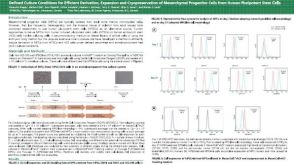 科学海报Defined Culture Conditions for Efficient Derivation Expansion and Cryopreservation of Mesenchymal Progenitor Cells from Human Pluripotent Stem Cells
 沪公网安备31010102008431号