Siatskas C et al. (OCT 2005)
FASEB journal : official publication of the Federation of American Societies for Experimental Biology 19 12 1752--4
Specific pharmacological dimerization of KDR in lentivirally transduced human hematopoietic cells activates anti-apoptotic and proliferative mechanisms.
Selective and regulatable expansion of transduced cells could augment gene therapy for many disorders. The activation of modified growth factor receptors via synthetic chemical inducers of dimerization allows for the coordinated growth of transduced cells. This system can also provide information on specific receptor-mediated signaling without interference from other family members. Although several receptor subunits have been investigated in this context,little is known about the precise molecular events associated with dimerizer-initiated signaling. We have constructed and expressed an AP20187-regulated KDR chimeric receptor in human TF1 cells and analyzed activation of this gene switch using functional,biochemical,and microarray analyses. When deprived of natural ligands,GM-CSF,interleukin-3,or erythropoietin,AP20187 prevented apoptosis of transduced TF1 cells,induced dose-dependent proliferation,and supported long-term growth. In addition,AP20187 stimulation activated the signaling molecules associated with mitogen-activated protein kinase and phosphatidyl-inositol 3-kinase/Akt pathways. Microarray analysis determined that a number of transcripts involved in a variety of cellular processes were differentially expressed. Notably,mRNAs affiliated with heat stress,including Hsp70 and Hsp105,were up-regulated. Functional assays showed that Hsp70 and Hsp105 protected transduced TF1 cells from apoptosis and premature senescence,in part through regulation of Akt. These observations delineate specific roles for kinase insert domain-containing receptor,or KDR,signaling and suggest strategies to endow genetically modified cells with a survival advantage enabling the generation of adequate cell numbers for therapeutic outcomes.
View Publication
Expansion of hematopoietic progenitor cell populations in stirred suspension bioreactors of normal human bone marrow cells.
We have investigated the potential of stirred suspension cultures to support hematopoiesis from starting innocula of normal human bone marrow cells. Initial studies showed that the short-term maintenance of both colony-forming cell (CFC) numbers and their precursors,detected as long-term culture-initiating cells (LTC-IC),could be achieved as well in stirred suspension cultures as in static cultures. Neither of these progenitor cell populations was affected in either type of culture when porous microcarriers were added to provide an increased surface for adherent cell attachment. Supplementation of the medium with 10 ng/ml of Steel factor (SF) and 2 ng/ml of interleukin-3 (IL-3) resulted in a significant expansion of LTC-IC,CFC and total cell numbers in stirred cultures. Both the duration and ultimate magnitude of these expansions were correlated with the initial cell density and after 4 weeks the number of LTC-IC and CFC present in stirred cultures initiated with the highest starting cell concentration tested reflected average increases of 7- and 22-fold,respectively,above input values. Stirred suspension cultures offer the combined advantages of homogeneity and lack of dependence on the formation and maintenance of an adherent cell layer. Our results suggest their applicability to the development of scaled-up bioreactor systems for clinical procedures requiring the production of primitive hematopoietic cell populations. In addition,stirred suspension cultures may offer a new tool for the analysis of hematopoietic regulatory mechanisms.
View Publication
产品类型:
产品号#:
05150
05350
产品名:
MyeloCult™ H5100
Hess DA et al. (MAR 2006)
Blood 107 5 2162--9
Selection based on CD133 and high aldehyde dehydrogenase activity isolates long-term reconstituting human hematopoietic stem cells.
The development of novel cell-based therapies requires understanding of distinct human hematopoietic stem and progenitor cell populations. We recently isolated reconstituting hematopoietic stem cells (HSCs) by lineage depletion and purification based on high aldehyde dehydrogenase activity (ALDH(hi)Lin- cells). Here,we further dissected the ALDH(hi)-Lin- population by selection for CD133,a surface molecule expressed on progenitors from hematopoietic,endothelial,and neural lineages. ALDH(hi)CD133+Lin- cells were primarily CD34+,but also included CD34-CD38-CD133+ cells,a phenotype previously associated with repopulating function. Both ALDH(hi)CD133-Lin- and ALDH(hi)CD133+Lin- cells demonstrated distinct clonogenic progenitor function in vitro,whereas only the ALDH(hi)CD133+Lin- population seeded the murine bone marrow 48 hours after transplantation. Significant human cell repopulation was observed only in NOD/SCID and NOD/SCID beta2M-null mice that received transplants of ALDH(hi)CD133+Lin- cells. Limiting dilution analysis demonstrated a 10-fold increase in the frequency of NOD/SCID repopulating cells compared with CD133+Lin- cells,suggesting that high ALDH activity further purified cells with repopulating function. Transplanted ALDH(hi)CD133+Lin- cells also maintained primitive hematopoietic phenotypes (CD34+CD38-) and demonstrated enhanced repopulating function in recipients of serial,secondary transplants. Cell selection based on ALDH activity and CD133 expression provides a novel purification of HSCs with long-term repopulating function and may be considered an alternative to CD34 cell selection for stem cell therapies.
View Publication
产品类型:
产品号#:
01700
01705
01701
01702
产品名:
ALDEFLUOR™ 试剂盒
ALDEFLUOR™ DEAB试剂, 1.5 mM, 1 mL
ALDEFLUOR™检测缓冲液
Su YR et al. (AUG 2008)
Arteriosclerosis,thrombosis,and vascular biology 28 8 1439--46
Lentiviral transduction of apoAI into hematopoietic progenitor cells and macrophages: applications to cell therapy of atherosclerosis.
OBJECTIVE: We used genetically engineered mouse hematopoietic progenitor cells (HPCs) to investigate the therapeutic effects of human apoAI on atherosclerosis in apoE(-/-) mice. METHODS AND RESULTS: Lentiviral constructs expressing either human apoAI (LV-apoAI) or green fluorescent protein (LV-GFP) cDNA under a macrophage specific promoter (CD68) were generated and used for ex vivo transduction of mouse HPCs and macrophages. The transduction efficiency was textgreater25% for HPCs and textgreater70% for macrophages. ApoAI was found in the macrophage culture media,mostly associated with the HDL fraction. Interestingly,a significant increase in mRNA and protein levels for ATP binding cassette A1 (ABCA1) and ABCG1 were found in apoAI-expressing macrophages after acLDL loading. Expression of apoAI significantly increased cholesterol efflux in wild-type and apoE(-/-) macrophages. HPCs transduced with LV-apoAI ex vivo and then transplanted into apoE(-/-) mice caused a 50% reduction in atherosclerotic lesion area compared to GFP controls,without influencing plasma HDL-C levels. CONCLUSIONS: Lentiviral transduction of apoAI into HPCs reduces atherosclerosis in apoE(-/-) mice. Expression of apoAI in macrophages improves cholesterol trafficking in wild-type apoE-producing macrophages and causes upregulation of ABCA1 and ABCG1. These novel observations set the stage for a cell therapy approach to atherosclerosis regression,exploiting the cooperation between apoE and apoAI to maximize cholesterol exit from the plaque.
View Publication
产品类型:
产品号#:
09600
09650
18756
18756RF
18757
18757RF
产品名:
StemSpan™ SFEM
StemSpan™ SFEM
EasySep™小鼠SCA1正选试剂盒
RoboSep™ 小鼠SCA1正选试剂盒含滤芯吸头
EasySep™小鼠CD117(cKIT)正选试剂盒
RoboSep™ 小鼠CD117(cKIT)正选试剂盒含滤芯吸头
M. Lora et al. (Apr 2025)
Clinical and Translational Science 18 5
Low Dose Methotrexate Has Divergent Effects on Cycling and Resting Human Hematopoietic Stem and Progenitor Cells
Low dose methotrexate (LD‐MTX) remains the gold standard in rheumatoid arthritis (RA) therapy. Multiple mechanisms on a variety of immune cells contribute to the anti‐inflammatory effects of LD‐MTX. Inflammatory signaling is deeply implicated in hematopoiesis by regulating hematopoietic stem and progenitor cell (HSPC) fate decisions; raising the question of whether HSPC are also modulated by LD‐MTX. This is the first study to characterize the effects of LD‐MTX on HSPC. CD34 + HSPC were isolated from healthy donors' non‐mobilized peripheral blood. Resting and/or cycling HSPCs were treated with LD‐MTX [dose equivalent to that used in RA patients]. Flow cytometry was performed to assess HSPC viability,cell cycle,surface abundance of reduced folate carrier 1 (RFC1),proliferation,reactive oxygen species (ROS) levels,DNA double‐strand breaks,p38 activation,and CD34 + subpopulations. HSPC clonogenicity was tested in colony‐forming cell assays. Our results indicate that in cycling HSPC,membrane RFC1 is upregulated and,following LD‐MTX treatment,they accumulate more intracellular MTX than resting HSPC. In cycling HSPC,LD‐MTX inhibits HSPC expansion by promoting S‐phase cell‐cycle arrest,increases intracellular HSPC ROS levels and DNA damage,and reduces HSPC viability. Those effects involve the activation of the p38 MAPK pathway and are rescued by folinic acid. The effects of LD‐MTX are more evident in CD34 + CD38High progenitors. In non‐cycling HSPC,LD‐MTX also reduces the proliferative response while preserving their clonogenicity. In summary,HSPC uptake LD‐MTX differentially according to their cycling state. In turn,LD‐MTX results in reduced proliferation and the preservation of HSPC clonogenicity.
View Publication
产品类型:
产品号#:
04034
04044
产品名:
MethoCult™ H4034 Optimum
MethoCult™ H4034 Optimum
Kuroki MM et al. ( 2005)
Anticancer Research 25 6A 3733--9
Preparation of human IgG and IgM monoclonal antibodies for MK-1/Ep-CAM by using human immunoglobulin gene-transferred mouse and gene cloning of their variable regions.
For antibody-based therapy of cancer,monoclonal antibodies (mAbs) of human origin are superior to mouse,mouse/human chimeric or humanized mAbs,because of their minimum immunogenicity to humans and their efficient collaboration with human effector cells. In the present study,human mAbs were prepared against a pancarcinoma antigen,MK-1 (Ep-CAM),using a genetically-engineered mouse (KM mouse) that contains the human immunoglobulin genes. Spleen cells from KM mice,immunized with recombinant MK-1,were fused with P3-U1 mouse myeloma cells. Of 44 anti-MK-1 clones analyzed,two were of IgG4 and the others of IgM clones. Although the two IgG4 clones were suggested to recognize the same antigenic determinant or two closely located determinants,their VK regions were encoded by different light-chain genes while their VH sequences were identical. The two IgG4 and one of the IgM clones tested revealed antibody-dependent cell-mediated cytotoxicity and complement-dependent cytotoxicity,respectively,against MK-1-expressing cells in vitro,suggesting that these fully human mAbs produced against MK-1 and their V-region genes,which are applicable for the preparation of engineered antibody fragments that may be useful for antibody-based therapy of cancer.
View Publication
产品类型:
产品号#:
03800
03801
03802
03803
03804
03805
03806
产品名:
ClonaCell™-HY杂交瘤试剂盒
ClonaCell™-HY培养基A
ClonaCell™-HY 培养基 B
ClonaCell™-HY 培养基 C
ClonaCell™-HY 培养基 D
ClonaCell™-HY 培养基 E
ClonaCell™-HY PEG
Bartulos O et al. (JUL 2016)
JCI insight 1 10
ISL1 cardiovascular progenitor cells for cardiac repair after myocardial infarction.
Cardiovascular progenitor cells (CPCs) expressing the ISL1-LIM-homeodomain transcription factor contribute developmentally to cardiomyocytes in all 4 chambers of the heart. Here,we show that ISL1-CPCs can be applied to myocardial regeneration following injury. We used a rapid 3D methylcellulose approach to form murine and human ISL1-CPC spheroids that engrafted after myocardial infarction in murine hearts,where they differentiated into cardiomyocytes and endothelial cells,integrating into the myocardium and forming new blood vessels. ISL1-CPC spheroid-treated mice exhibited reduced infarct area and increased blood vessel formation compared with control animals. Moreover,left ventricular (LV) contractile function was significantly better in mice transplanted with ISL1-CPCs 4 weeks after injury than that in control animals. These results provide proof-of-concept of a cardiac repair strategy employing ISL1-CPCs that,based on our previous lineage-tracing studies,are committed to forming heart tissue,in combination with a robust methylcellulose spheroid-based delivery approach.
View Publication
产品类型:
产品号#:
05850
05857
05870
05875
85850
85857
85870
85875
产品名:
mTeSR™1
mTeSR™1
Kameoka S et al. (JAN 2014)
Toxicological Sciences 137 1 76--90
A High-Throughput Screen for Teratogens Using Human Pluripotent Stem Cells
There is need in the pharmaceutical and chemical industries for high-throughput human cell-based assays for identifying hazardous chemicals,thereby reducing the overall reliance on animal studies for predicting the risk of toxic responses in humans. Despite instances of human-specific teratogens such as thalidomide,the use of human cell-teratogenicity assays has just started to be explored. Herein,a human pluripotent stem cell test (hPST) for identifying teratogens is described,benchmarking the in vitro findings to traditional preclinical toxicology teratogenicity studies and when available to teratogenic outcomes in humans. The hPST method employs a 3-day monolayer directed differentiation of human embryonic stem cells. The teratogenic risk of a compound is gauged by measuring the reduction in nuclear translocation of the transcription factor SOX17 in mesendodermal cells. Decreased nuclear SOX17 in the hPST model was strongly correlated with in vivo teratogenicity. Specifically,71 drug-like compounds with known in vivo effects,including thalidomide,were examined in the hPST. A threshold of 5μM demonstrated 94% accuracy (97% sensitivity and 92% specificity). Furthermore,15 environmental toxicants with physicochemical properties distinct from small molecule pharmaceutical agents were examined and a similarly strong concordance with teratogenicity outcomes from in vivo studies was observed. Finally,to assess the suitability of the hPST for high-throughput screens,a small library of 300 kinase inhibitors was tested,demonstrating the hPST platform's utility for interrogating teratogenic mechanisms and drug safety prediction. Thus,the hPST assay is a robust predictor of teratogenicity and appears to be an improvement over existing in vitro models.
View Publication
产品类型:
产品号#:
05850
05857
05870
05875
07923
07920
85850
85857
85870
85875
07922
产品名:
Dispase (1 U/mL)
ACCUTASE™
mTeSR™1
mTeSR™1
ACCUTASE™
A. J. Cole et al. (May 2025)
Nature Communications 16
A chimeric viral platform for directed evolution in mammalian cells
Directed evolution is a process of mutation and artificial selection to breed biomolecules with new or improved activity. Directed evolution platforms are primarily prokaryotic or yeast-based,and stable mammalian systems have been challenging to establish and apply. To this end,we develop PROTein Evolution Using Selection (PROTEUS),a platform that uses chimeric virus-like vesicles to enable extended mammalian directed evolution campaigns without loss of system integrity. This platform is stable and can generate sufficient diversity for directed evolution in mammalian systems. Using PROTEUS,we alter the doxycycline responsiveness of tetracycline-controlled transactivators,generating a more sensitive TetON-4G tool for gene regulation with mammalian-specific adaptations. PROTEUS is also compatible with intracellular nanobody evolution,and we use it to evolve a DNA damage-responsive anti-p53 nanobody. Overall,PROTEUS is an efficient and stable platform to direct evolution of biomolecules within mammalian cells. Subject terms: Synthetic biology,Synthetic biology,Molecular evolution,Next-generation sequencing
View Publication
产品类型:
产品号#:
100-0483
100-0484
产品名:
Hausser Scientificᵀᴹ 明线血球计数板
ReLeSR™
Singbrant S et al. (JUN 2010)
Blood 115 23 4689--98
Canonical BMP signaling is dispensable for hematopoietic stem cell function in both adult and fetal liver hematopoiesis, but essential to preserve colon architecture.
Numerous publications have described the importance of bone morphogenetic protein (BMP) signaling in the specification of hematopoietic tissue in developing embryos. Here we investigate the full role of canonical BMP signaling in both adult and fetal liver hematopoiesis using conditional knockout strategies because conventional disruption of components of the BMP signaling pathway result in early death of the embryo. By targeting both Smad1 and Smad5,we have generated a double-knockout mouse with complete disruption of canonical BMP signaling. Interestingly,concurrent deletion of Smad1 and Smad5 results in death because of extrahematopoietic pathologic changes in the colon. However,Smad1/Smad5-deficient bone marrow cells can compete normally with wild-type cells and display unaffected self-renewal and differentiation capacity when transplanted into lethally irradiated recipients. Moreover,although BMP receptor expression is increased in fetal liver,fetal liver cells deficient in both Smad1 and Smad5 remain competent to long-term reconstitute lethally irradiated recipients in a multilineage manner. In conclusion,canonical BMP signaling is not required to maintain either adult or fetal liver hematopoiesis,despite its crucial role in the initial patterning of hematopoiesis in early embryonic development.
View Publication

 EasySep™小鼠TIL(CD45)正选试剂盒


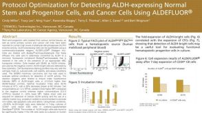 科学海报Protocol Optimization for Detecting ALDH-Expressing Normal Stem and Progenitor Cells and Cancer Cells Using ALDEFLUOR™
 技术公告StemSpan™ Medium and Supplements for the Generation of T Cells from Cord Blood-Derived CD34+ Cells
 沪公网安备31010102008431号