Hockemeyer D et al. (SEP 2008)
Cell stem cell 3 3 346--53
A drug-inducible system for direct reprogramming of human somatic cells to pluripotency.
Current approaches to reprogram human somatic cells to pluripotent iPSCs utilize viral transduction of different combinations of transcription factors. These protocols are highly inefficient because only a small fraction of cells carry the appropriate number and stoichiometry of proviral insertions to initiate the reprogramming process. Here we have generated genetically homogeneous secondary" somatic cells�
View Publication
产品类型:
产品号#:
72742
产品名:
强力霉素(盐酸盐)
Horikiri T et al. ( 2017)
PloS one 12 1 e0170342
SOX10-Nano-Lantern Reporter Human iPS Cells; A Versatile Tool for Neural Crest Research.
The neural crest is a source to produce multipotent neural crest stem cells that have a potential to differentiate into diverse cell types. The transcription factor SOX10 is expressed through early neural crest progenitors and stem cells in vertebrates. Here we report the generation of SOX10-Nano-lantern (NL) reporter human induced pluripotent stem cells (hiPS) by using CRISPR/Cas9 systems,that are beneficial to investigate the generation and maintenance of neural crest progenitor cells. SOX10-NL positive cells are produced transiently from hiPS cells by treatment with TGFβ inhibitor SB431542 and GSK3 inhibitor CHIR99021. We found that all SOX10-NL-positive cells expressed an early neural crest marker NGFR,however SOX10-NL-positive cells purified from differentiated hiPS cells progressively attenuate their NL-expression under proliferation. We therefore attempted to maintain SOX10-NL-positive cells with additional signaling on the plane and sphere culture conditions. These SOX10-NL cells provide us to investigate mass culture with neural crest cells for stem cell research.
View Publication
Expansion of hematopoietic progenitor cell populations in stirred suspension bioreactors of normal human bone marrow cells.
We have investigated the potential of stirred suspension cultures to support hematopoiesis from starting innocula of normal human bone marrow cells. Initial studies showed that the short-term maintenance of both colony-forming cell (CFC) numbers and their precursors,detected as long-term culture-initiating cells (LTC-IC),could be achieved as well in stirred suspension cultures as in static cultures. Neither of these progenitor cell populations was affected in either type of culture when porous microcarriers were added to provide an increased surface for adherent cell attachment. Supplementation of the medium with 10 ng/ml of Steel factor (SF) and 2 ng/ml of interleukin-3 (IL-3) resulted in a significant expansion of LTC-IC,CFC and total cell numbers in stirred cultures. Both the duration and ultimate magnitude of these expansions were correlated with the initial cell density and after 4 weeks the number of LTC-IC and CFC present in stirred cultures initiated with the highest starting cell concentration tested reflected average increases of 7- and 22-fold,respectively,above input values. Stirred suspension cultures offer the combined advantages of homogeneity and lack of dependence on the formation and maintenance of an adherent cell layer. Our results suggest their applicability to the development of scaled-up bioreactor systems for clinical procedures requiring the production of primitive hematopoietic cell populations. In addition,stirred suspension cultures may offer a new tool for the analysis of hematopoietic regulatory mechanisms.
View Publication
产品类型:
产品号#:
05150
05350
产品名:
MyeloCult™ H5100
Hess DA et al. (MAR 2006)
Blood 107 5 2162--9
Selection based on CD133 and high aldehyde dehydrogenase activity isolates long-term reconstituting human hematopoietic stem cells.
The development of novel cell-based therapies requires understanding of distinct human hematopoietic stem and progenitor cell populations. We recently isolated reconstituting hematopoietic stem cells (HSCs) by lineage depletion and purification based on high aldehyde dehydrogenase activity (ALDH(hi)Lin- cells). Here,we further dissected the ALDH(hi)-Lin- population by selection for CD133,a surface molecule expressed on progenitors from hematopoietic,endothelial,and neural lineages. ALDH(hi)CD133+Lin- cells were primarily CD34+,but also included CD34-CD38-CD133+ cells,a phenotype previously associated with repopulating function. Both ALDH(hi)CD133-Lin- and ALDH(hi)CD133+Lin- cells demonstrated distinct clonogenic progenitor function in vitro,whereas only the ALDH(hi)CD133+Lin- population seeded the murine bone marrow 48 hours after transplantation. Significant human cell repopulation was observed only in NOD/SCID and NOD/SCID beta2M-null mice that received transplants of ALDH(hi)CD133+Lin- cells. Limiting dilution analysis demonstrated a 10-fold increase in the frequency of NOD/SCID repopulating cells compared with CD133+Lin- cells,suggesting that high ALDH activity further purified cells with repopulating function. Transplanted ALDH(hi)CD133+Lin- cells also maintained primitive hematopoietic phenotypes (CD34+CD38-) and demonstrated enhanced repopulating function in recipients of serial,secondary transplants. Cell selection based on ALDH activity and CD133 expression provides a novel purification of HSCs with long-term repopulating function and may be considered an alternative to CD34 cell selection for stem cell therapies.
View Publication
产品类型:
产品号#:
01700
01705
01701
01702
产品名:
ALDEFLUOR™ 试剂盒
ALDEFLUOR™ DEAB试剂, 1.5 mM, 1 mL
ALDEFLUOR™检测缓冲液
M. Lora et al. (Apr 2025)
Clinical and Translational Science 18 5
Low Dose Methotrexate Has Divergent Effects on Cycling and Resting Human Hematopoietic Stem and Progenitor Cells
Low dose methotrexate (LD‐MTX) remains the gold standard in rheumatoid arthritis (RA) therapy. Multiple mechanisms on a variety of immune cells contribute to the anti‐inflammatory effects of LD‐MTX. Inflammatory signaling is deeply implicated in hematopoiesis by regulating hematopoietic stem and progenitor cell (HSPC) fate decisions; raising the question of whether HSPC are also modulated by LD‐MTX. This is the first study to characterize the effects of LD‐MTX on HSPC. CD34 + HSPC were isolated from healthy donors' non‐mobilized peripheral blood. Resting and/or cycling HSPCs were treated with LD‐MTX [dose equivalent to that used in RA patients]. Flow cytometry was performed to assess HSPC viability,cell cycle,surface abundance of reduced folate carrier 1 (RFC1),proliferation,reactive oxygen species (ROS) levels,DNA double‐strand breaks,p38 activation,and CD34 + subpopulations. HSPC clonogenicity was tested in colony‐forming cell assays. Our results indicate that in cycling HSPC,membrane RFC1 is upregulated and,following LD‐MTX treatment,they accumulate more intracellular MTX than resting HSPC. In cycling HSPC,LD‐MTX inhibits HSPC expansion by promoting S‐phase cell‐cycle arrest,increases intracellular HSPC ROS levels and DNA damage,and reduces HSPC viability. Those effects involve the activation of the p38 MAPK pathway and are rescued by folinic acid. The effects of LD‐MTX are more evident in CD34 + CD38High progenitors. In non‐cycling HSPC,LD‐MTX also reduces the proliferative response while preserving their clonogenicity. In summary,HSPC uptake LD‐MTX differentially according to their cycling state. In turn,LD‐MTX results in reduced proliferation and the preservation of HSPC clonogenicity.
View Publication
产品类型:
产品号#:
04034
04044
产品名:
MethoCult™ H4034 Optimum
MethoCult™ H4034 Optimum
Liu J et al. (NOV 2015)
Nature Protocols 10 11 1842--59
Efficient delivery of nuclease proteins for genome editing in human stem cells and primary cells.
Targeted nucleases,including zinc-finger nucleases (ZFNs),transcription activator-like (TAL) effector nucleases (TALENs) and clustered regularly interspaced short palindromic repeat (CRISPR)/CRISPR-associated protein 9 (Cas9),have provided researchers with the ability to manipulate nearly any genomic sequence in human cells and model organisms. However,realizing the full potential of these genome-modifying technologies requires their safe and efficient delivery into relevant cell types. Unlike methods that rely on expression from nucleic acids,the direct delivery of nuclease proteins to cells provides rapid action and fast turnover,leading to fewer off-target effects while maintaining high rates of targeted modification. These features make nuclease protein delivery particularly well suited for precision genome engineering. Here we describe procedures for implementing protein-based genome editing in human embryonic stem cells and primary cells. Protocols for the expression,purification and delivery of ZFN proteins,which are intrinsically cell-permeable; TALEN proteins,which can be internalized via conjugation with cell-penetrating peptide moieties; and Cas9 ribonucleoprotein,whose nucleofection into cells facilitates rapid induction of multiplexed modifications,are described,along with procedures for evaluating nuclease protein activity. Once they are constructed,nuclease proteins can be expressed and purified within 6 d,and they can be used to induce genomic modifications in human cells within 2 d.
View Publication
产品类型:
产品号#:
05850
05857
05870
05875
07920
17952
17952RF
19052
19052RF
18000
15470
15450
15420
15460
15425
15465
15430
15415
85850
85857
85870
85875
100-0696
07922
产品名:
ACCUTASE™
EasySep™人CD4+ T细胞分选试剂盒
RoboSep™ 人CD4+ T细胞分选试剂盒
EasySep™人CD4+ T细胞富集试剂盒
RoboSep™ 人CD4+ T细胞富集试剂盒含滤芯吸头
EasySep™磁极
mTeSR™1
mTeSR™1
EasySep™人CD4+ T细胞分离试剂盒
ACCUTASE™
D. T. Claiborne et al. (Jan 2025)
Nature Communications 16
High frequency CCR5 editing in human hematopoietic stem progenitor cells protects xenograft mice from HIV infection
The only cure of HIV has been achieved in a small number of people who received a hematopoietic stem cell transplant (HSCT) comprising allogeneic cells carrying a rare,naturally occurring,homozygous deletion in the CCR5 gene. The rarity of the mutation and the significant morbidity and mortality of such allogeneic transplants precludes widespread adoption of this HIV cure. Here,we show the application of CRISPR/Cas9 to achieve >90% CCR5 editing in human,mobilized hematopoietic stem progenitor cells (HSPC),resulting in a transplant that undergoes normal hematopoiesis,produces CCR5 null T cells,and renders xenograft mice refractory to HIV infection. Titration studies transplanting decreasing frequencies of CCR5 edited HSPCs demonstrate that <90% CCR5 editing confers decreasing protective benefit that becomes negligible between 54% and 26%. Our study demonstrates the feasibility of using CRISPR/Cas9/RNP to produce an HSPC transplant with high frequency CCR5 editing that is refractory to HIV replication. These results raise the potential of using CRISPR/Cas9 to produce a curative autologous HSCT and bring us closer to the development of a cure for HIV infection. Subject terms: HIV infections,CRISPR-Cas9 genome editing,Retrovirus,Translational research
View Publication
产品类型:
产品号#:
04034
04044
22001
22005
22006
22007
22008
22009
22011
22012
产品名:
MethoCult™ H4034 Optimum
MethoCult™ H4034 Optimum
STEMvision™ 人脐带血7-天CFU分析包
STEMvision™ 彩色人脐带血14-天CFU分析包
STEMvision™ 彩色人骨髓14-天CFU分析包
STEMvision™ 彩色人动员外周血14-天CFU分析包
STEMvision™ 小鼠总CFU分析包
STEMvision™ 小鼠髓系CFU分析包
STEMvision™ 小鼠红系CFU分析包
STEMvision™ 小鼠CFU分析包(髓系和红系)
Mateizel I et al. (OCT 2009)
Human reproduction (Oxford,England) 24 10 2477--89
Characterization of CD30 expression in human embryonic stem cell lines cultured in serum-free media and passaged mechanically
BACKGROUND: The presence of chromosomal abnormalities could have a negative impact for human embryonic stem cell (hESC) applications both in regenerative medicine and in research. A biomarker that allows the identification of chromosomal abnormalities induced in hESC in culture before they take over the culture would represent an important tool for defining optimal culture conditions for hESC. Here we investigate the expression of CD30,reported to be a biomarker of hESCs with abnormal karyotype,in undifferentiated and spontaneously differentiated hESC.backslashnbackslashnMETHODS AND RESULTS: hESC were derived and cultured on mouse fibroblasts in KO-SR containing medium (serum free media) and passaged mechanically. Our results based on analysis at mRNA (RT-PCR) and protein (fluorescence-activated cell sorting and immunocytochemistry) level show that CD30 is expressed in undifferentiated hESC,even at very early passages,without any correlation with the presence of chromosomal anomalies. We also show that the expression of CD30 is rapidly lost during early spontaneous differentiation of hESC.backslashnbackslashnCONCLUSION: We conclude that CD30 expression in hESC cultures is probably a consequence of culture conditions,and that KO-SR may play a role. In addition,the expression of so-called 'stemness' markers does not change in undifferentiated hESC during long-term culture or when cells acquire chromosomal abnormalities.
View Publication
产品类型:
产品号#:
05850
05857
05870
05875
85850
85857
85870
85875
产品名:
mTeSR™1
mTeSR™1
Srour EF et al. (APR 2005)
Blood 105 8 3109--16
Modulation of in vitro proliferation kinetics and primitive hematopoietic potential of individual human CD34+CD38-/lo cells in G0.
Whether cytokines can modulate the fate of primitive hematopoietic progenitor cells (HPCs) through successive in vitro cell divisions has not been established. Single human marrow CD34+CD38-/lo cells in the G0 phase of cell cycle were cultured under 7 different cytokine combinations,monitored for proliferation on days 3,5,and 7,then assayed for long-term culture-initiating cell (LTC-IC) function on day 7. LTC-IC function was then retrospectively correlated with prior number of in vitro cell divisions to determine whether maintenance of LTC-IC function after in vitro cell division is dependent on cytokine exposure. In the presence of proliferation progression signals,initial cell division was independent of cytokine stimulation,suggesting that entry of primitive HPCs into the cell cycle is a stochastic property. However,kinetics of proliferation beyond day 3 and maintenance of LTC-IC function were sensitive to cytokine stimulation,such that LTC-IC underwent an initial long cell cycle,followed by more synchronized shorter cycles varying in length depending on the cytokine combination. Nonobese diabetic/severe combined immunodeficiency (NOD/SCID) transplantation studies revealed analogous results to those obtained with LTC-ICs. These data suggest that although exit from quiescence and commitment to proliferation might be stochastic,kinetics of proliferation,and possibly fate of primitive HPCs,might be modulated by extrinsic factors.
View Publication
产品类型:
产品号#:
05150
产品名:
MyeloCult™ H5100
(Dec 2024)
Stem Cell Research & Therapy 15 16
Enhanced fetal hemoglobin production via dual-beneficial mutation editing of the HBG promoter in hematopoietic stem and progenitor cells for β-hemoglobinopathies
BackgroundSickle cell disease (SCD) and β-thalassemia patients with elevated gamma globin (HBG1/G2) levels exhibit mild or no symptoms. To recapitulate this natural phenomenon,the most coveted gene therapy approach is to edit the regulatory sequences of HBG1/G2 to reactivate them. By editing more than one regulatory sequence in the HBG promoter,the production of fetal hemoglobin (HbF) can be significantly increased. However,achieving this goal requires precise nucleotide conversions in hematopoietic stem and progenitor cells (HSPCs) at therapeutic efficiency,which remains a challenge.MethodsWe employed Cas9 RNP-ssODN-mediated homology-directed repair (HDR) gene editing to mimic two naturally occurring HBG promoter point mutations; -175T > C,associated with high HbF levels,and −158 C > T,a common polymorphism in the Indian population that induces HbF under erythropoietic stress,in HSPCs.ResultsAsymmetric,nontarget ssODN induced high rates of complete HDR conversions,with at least 15% of HSPCs exhibiting both the −175T > C and −158 C > T mutations. Optimized conditions and treatment with the small molecule AZD-7648 increased this rate,with up to 57% of long-term engrafting human HSPCs in NBSGW mice containing at least one beneficial mutation. Functionally,in vivo erythroblasts exhibited high levels of HbF,which was sufficient to reverse the cellular phenotype of β-thalassemia. Further support through bone marrow MSC co-culture boosted complete HDR conversion rates to exceed 80%,with minimal InDels,improved cell viability,and induced fetal hemoglobin levels similar to those of Cas9 RNP-mediated indels at BCL11A enhancer and HBG promoter.ConclusionsCas9 RNP-ssODN-based nucleotide conversion at the HBG promoter offers a promising gene therapy approach to ameliorate the phenotypes of β-thalassemia and SCD. The developed approach can simplify and broaden applications that require the cointroduction of multiple nucleotide modifications in HSPCs.Supplementary InformationThe online version contains supplementary material available at 10.1186/s13287-024-04117-0.
View Publication

 EasySep™小鼠TIL(CD45)正选试剂盒


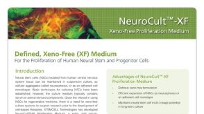 产品手册NeuroCult™-XF: Xeno-Free Culture Medium for the Proliferation of Human Neural Stem Cells

 沪公网安备31010102008431号