Borowiak M et al. (APR 2009)
Cell stem cell 4 4 348--58
Small molecules efficiently direct endodermal differentiation of mouse and human embryonic stem cells.
An essential step for therapeutic and research applications of stem cells is the ability to differentiate them into specific cell types. Endodermal cell derivatives,including lung,liver,and pancreas,are of interest for regenerative medicine,but efforts to produce these cells have been met with only modest success. In a screen of 4000 compounds,two cell-permeable small molecules were indentified that direct differentiation of ESCs into the endodermal lineage. These compounds induce nearly 80% of ESCs to form definitive endoderm,a higher efficiency than that achieved by Activin A or Nodal,commonly used protein inducers of endoderm. The chemically induced endoderm expresses multiple endodermal markers,can participate in normal development when injected into developing embryos,and can form pancreatic progenitors. The application of small molecules to differentiate mouse and human ESCs into endoderm represents a step toward achieving a reproducible and efficient production of desired ESC derivatives.
View Publication
产品类型:
产品号#:
72312
72314
72512
72514
产品名:
(-) -Indolactam V(吲哚内酰胺 V)
IDE1
IDE1
T. W. Braun et al. (mar 2022)
STAR protocols 3 1 101070
FACS and immunomagnetic isolation of early erythroid progenitor cells from mouse fetal liver.
Early erythroid progenitors are transit-amplifying cells with high proliferative capacity committed to undergoing red cell differentiation. CD71/CD24low progenitors are less mature and have greater proliferative capacity than CD71/CD24high. We present protocols for isolation of CD71/CD24low progenitors from mouse fetal liver using both fluorescence-activated cell sorting (FACS) and immunomagnetic enrichment. CD71/CD24low progenitors isolated with both approaches show similar transcriptomes at single-cell resolution and exhibit characteristic proliferative responses to glucocorticoids. For complete details on the use and execution of this protocol,please refer to Li et al. (2019).
View Publication
Feeder-independent culture systems for human pluripotent stem cells.
The continued success of pluripotent stem cell research is ultimately dependent on access to reliable and defined reagents for the consistent culture and cryopreservation of undifferentiated,pluripotent cells. The development of defined and feeder-independent culture media has provided a platform for greater reproducibility and standardization in this field. Here we provide detailed protocols for the use of mTeSR™1 and TeSR™2 with various cell culture matrices as well as defined cryopreservation protocols for human embryonic and human induced pluripotent stem cells.
View Publication
产品类型:
产品号#:
05850
05857
05870
05875
85850
85857
85870
85875
产品名:
mTeSR™1
mTeSR™1
Marchetti S et al. (MAY 2002)
Journal of cell science 115 Pt 10 2075--85
Endothelial cells genetically selected from differentiating mouse embryonic stem cells incorporate at sites of neovascularization in vivo.
Large scale purification of endothelial cells is of great interest as it could improve tissue transplantation,reperfusion of ischemic tissues and treatment of pathologies in which an endothelial cell dysfunction exists. In this study,we describe a novel genetic approach that selects for endothelial cells from differentiating embryonic stem (ES) cells. Our strategy is based on the establishment of ES-cell clones that carry an integrated puromycin resistance gene under the control of a vascular endothelium-specific promoter,tie-1. Using EGFP as a reporter gene,we first confirmed the endothelial specificity of the tie-1 promoter in the embryoid body model and in cells differentiated in 2D cultures. Subsequently,tie-1-EGFP ES cells were used as recipients for the tie-1-driven puror transgene. The resulting stable clones were expanded and differentiated for seven days in the presence of VEGF before puromycin selection. As expected,puromycin-resistant cells were positive for EGFP and also expressed several endothelial markers,including CD31,CD34,VEGFR-1,VEGFR-2,Tie-1,VE-cadherin and ICAM-2. Release from the puromycin selection resulted in the appearance of alpha-smooth muscle actin-positive cells. Such cells became more numerous when the population was cultured on laminin-1 or in the presence of TGF-beta1,two known inducers of smooth muscle cell differentiation. The hypothesis that endothelial cells or their progenitors may differentiate towards a smooth muscle cell phenotype was further supported by the presence of cells expressing both CD31 and alpha-smooth muscle actin markers. Finally,we show that purified endothelial cells can incorporate into the neovasculature of transplanted tumors in nude mice. Taken together,these results suggest that application of endothelial lineage selection to differentiating ES cells may become a useful approach for future pro-angiogenic and endothelial cell replacement therapies.
View Publication
产品类型:
产品号#:
06902
06952
00321
00322
00323
00324
00325
产品名:
Chen C et al. (NOV 2016)
JCI insight 1 19 e88632
Humanized neuronal chimeric mouse brain generated by neonatally engrafted human iPSC-derived primitive neural progenitor cells.
The creation of a humanized chimeric mouse nervous system permits the study of human neural development and disease pathogenesis using human cells in vivo. Humanized glial chimeric mice with the brain and spinal cord being colonized by human glial cells have been successfully generated. However,generation of humanized chimeric mouse brains repopulated by human neurons to possess a high degree of chimerism have not been well studied. Here we created humanized neuronal chimeric mouse brains by neonatally engrafting the distinct and highly neurogenic human induced pluripotent stem cell (hiPSC)-derived rosette-type primitive neural progenitors. These neural progenitors predominantly differentiate to neurons,which disperse widely throughout the mouse brain with infiltration of the cerebral cortex and hippocampus at 6 and 13 months after transplantation. Building upon the hiPSC technology,we propose that this potentially unique humanized neuronal chimeric mouse model will provide profound opportunities to define the structure,function,and plasticity of neural networks containing human neurons derived from a broad variety of neurological disorders.
View Publication
Nardosinone Improves the Proliferation, Migration and Selective Differentiation of Mouse Embryonic Neural Stem Cells
In this study,we investigated the impact of Nardosinone,a bioactive component in Nardostachys root,on the proliferation and differentiation of neural stem cells. The neural stem cells were isolated from cerebrums of embryonic day 14 CD1 mice. The proliferation of cells was monitored using the cell counting kit-8 assay,bromodeoxyuridine incorporation and cell cycle analysis. Cell migration and differentiation were investigated with the neurosphere assay and cell specific markers,respectively. The results showed that Nardosinone promotes cells proliferation and increases cells migration distance in a dose-dependent manner. Nardosinone also induces the selective differentiation of neural stem cells to neurons and oligodendrocytes,as indicated by the expression of microtubule-associated protein-2 and myelin basic protein,respectively. Nardosinone also increases the expression of phospho-extracellular signal-regulated kinase and phospho-cAMP response element binding protein during proliferation and differentiation. In conclusion,this study reveals the regulatory effects of Nardosinone on neural stem cells,which may have significant implications for the treatment of brain injury and neurodegenerative diseases.
View Publication
产品类型:
产品号#:
05700
05702
05704
产品名:
NeuroCult™ 基础培养基(小鼠和大鼠)
NeuroCult™扩增试剂盒(小鼠和大鼠)
NeuroCult™ 分化试剂盒(小鼠和大鼠)
M. G. Bracha et al. (Jul 2025)
Frontiers in Immunology 16 8
Mouse B cells engineered to express an anti-HPV antibody elicit anti-tumor T cell responses
Transplantation of engineered B cells has demonstrated efficacy in HIV disease models. B cell engineering may also be utilized for the treatment of cancer. Recent studies have highlighted that B cell activity is associated with favorable clinical outcomes in oncology. In mice,polyclonal B cells have been shown to elicit anti-cancer responses. As a potential novel cell therapy,we demonstrate that engineering B cells to target a tumor-associated antigen enhances polyclonal anti-tumor responses. We observe that engineered B cells expressing an anti-HPV B cell receptor internalize the antigen,enabling subsequent activation of oncoantigen-specific T cells. Secreted antibodies from engineered B cells form immune complexes,which are taken up by antigen-presenting cells to further promote T cell activation. Engineered B cells hold promise as novel,multi-modal cell therapies and open new avenues in solid tumor targeting.
View Publication
产品类型:
产品号#:
100-1003
产品名:
ImmunoCult™ 小鼠B细胞扩增试剂盒
Stier S et al. (AUG 2003)
Blood 102 4 1260--6
Ex vivo targeting of p21Cip1/Waf1 permits relative expansion of human hematopoietic stem cells.
Relative quiescence is a defining characteristic of hematopoietic stem cells. Reasoning that inhibitory tone dominates control of stem cell cycling,we previously showed that mice engineered to be deficient in the cyclin-dependent kinase inhibitor,p21Cip1/Waf1 (p21),have an increased stem cell pool under homeostatic conditions. Since p21 was necessary to maintain stem cell quiescence and its absence sufficient to permit increased murine stem cell cycling,we tested whether reduction of p21 alone in human adult-derived stem cells could affect stem cell proliferation. We demonstrate here that interrupting p21 expression ex vivo resulted in expanded stem cell number and in vivo stem cell function compared with control,manipulated cells. Further,we demonstrate full multilineage reconstitution capability in cells where p21 expression was knocked down. Therefore,lifting the brake on cell proliferation by altering cell cycle checkpoints provides an alternative paradigm for increasing hematopoietic stem cell numbers. This approach may be useful for relative ex vivo human stem cell expansion.
View Publication
产品类型:
产品号#:
05150
04435
04445
产品名:
MyeloCult™ H5100
MethoCult™ H4435 Enriched
MethoCult™ H4435 Enriched
Boussaad I et al. (AUG 2011)
Journal of virology 85 15 7710--8
Wild-type measles virus interferes with short-term engraftment of human CD34+ hematopoietic progenitor cells.
Transient lymphopenia is a hallmark of measles virus (MV)-induced immunosuppression. To address to what extent replenishment of the peripheral lymphocyte compartment from bone marrow (BM) progenitor/stem cells might be affected,we analyzed the interaction of wild-type MV with hematopoietic stem and progenitor cells (HS/PCs) and stroma cells in vitro. Infection of human CD34(+) HS/PCs or stroma cells with wild-type MV is highly inefficient yet noncytolytic. It occurs independently of CD150 in stroma cells but also in HS/PCs,where infection is established in CD34(+) CD150(-) and CD34(+) CD150(+) (in humans representing HS/PC oligopotent precursors) subsets. Stroma cells and HS/PCs can mutually transmit MV and may thereby create a possible niche for continuous viral exchange in the BM. Infected lymphocytes homing to this compartment may serve as sources for HS/PC or stroma cell infection,as reflected by highly efficient transmission of MV from both populations in cocultures with MV-infected B or T cells. Though MV exposure does not detectably affect the viability,expansion,and colony-forming activity of either CD150(+) or CD150(-) HS/PCs in vitro,it efficiently interferes with short- but not long-term hematopoietic reconstitution in NOD/SCID mice. Altogether,these findings support the hypothesis that MV accession of the BM compartment by infected lymphocytes may contribute to peripheral blood mononuclear cell lymphopenia at the level of BM suppression.
View Publication
产品类型:
产品号#:
04434
04444
产品名:
MethoCult™ H4434 Classic
MethoCult™ H4434 Classic
Tzeng Y-S et al. (JAN 2011)
Blood 117 2 429--39
Loss of Cxcl12/Sdf-1 in adult mice decreases the quiescent state of hematopoietic stem/progenitor cells and alters the pattern of hematopoietic regeneration after myelosuppression.
The C-X-C-type chemokine Cxcl12,also known as stromal cell-derived factor-1,plays a critical role in hematopoiesis during fetal development. However,the functional requirement of Cxcl12 in the adult hematopoietic stem/progenitor cell (HSPC) regulation was still unclear. In this report,we developed a murine Cxcl12 conditional deletion model in which the target gene can be deleted at the adult stage. We found that loss of stroma-secreted Cxcl12 in the adult led to expansion of the HSPC population as well as a reduction in long-term quiescent stem cells. In Cxcl12-deficient bone marrow,HSPCs were absent along the endosteal surface,and blood cell regeneration occurred predominantly in the perisinusoidal space after 5-fluorouracil myelosuppression challenge. Our results indicate that Cxcl12 is required for HSPC homeostasis regulation and is an important factor for osteoblastic niche organization in adult stage bone marrow.
View Publication

 EasySep™小鼠TIL(CD45)正选试剂盒


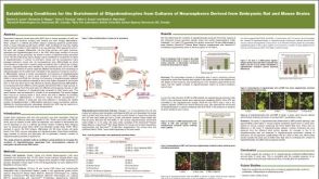 科学海报Establishing Conditions for the Enrichment of Oligodendrocytes from Cultures of Neurospheres Derived from Embryonic Rat and Mouse Brains
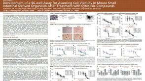 科学海报Development of a 96-well Assay for Assessing Cell Viability in Mouse Small Intestinal-Derived Organoids after Treatment with Cytotoxic Compounds
 沪公网安备31010102008431号