Modular tissue-in-a-CUBE platform to model blood-brain barrier (BBB) and brain interaction
With the advent of increasingly sophisticated organoids,there is growing demand for technology to replicate the interactions between multiple tissues or organs. This is challenging to achieve,however,due to the varying culture conditions of the different cell types that make up each tissue. Current methods often require complicated microfluidic setups,but fragile tissue samples tend not to fare well with rough handling. Furthermore,the more complicated the human system to be replicated,the more difficult the model becomes to operate. Here,we present the development of a multi-tissue chip platform that takes advantage of the modularity and convenient handling ability of a CUBE device. We first developed a blood-brain barrier-in-a-CUBE by layering astrocytes,pericytes,and brain microvascular endothelial cells in the CUBE,and confirmed the expression and function of important tight junction and transporter proteins in the blood-brain barrier model. Then,we demonstrated the application of integrating Tissue-in-a-CUBE with a chip in simulating the in vitro testing of the permeability of a drug through the blood-brain barrier to the brain and its effect on treating the glioblastoma brain cancer model. We anticipate that this platform can be adapted for use with organoids to build complex human systems in vitro by the combination of multiple simple CUBE units. Development of platform to integrate multiple Tissue-in-a-CUBEs in a chip for tissue-tissue interaction,demonstrated by simulating the testing of the permeability and effect of a cancer drug in a BBB-Brain cancer model.
View Publication
产品类型:
产品号#:
100-0483
100-0484
100-0276
100-1130
产品名:
Hausser Scientificᵀᴹ 明线血球计数板
ReLeSR™
mTeSR™ Plus
mTeSR™ Plus
(Jul 2025)
Molecular Metabolism 99 10
Complete loss of PAX4 causes transient neonatal diabetes in humans
ObjectiveGene discovery studies in individuals with diabetes diagnosed within 6 months of life (neonatal diabetes,NDM) can provide unique insights into the development and function of human pancreatic beta-cells.MethodsWe performed genome sequencing in a cohort of 43 consanguineous individuals with NDM in whom all the known genetic causes had previously been excluded. We used quantitative PCR and RNA-sequencing in CRISPR-edited human induced pluripotent stem cells (iPSCs),and CUT&RUN-sequencing in EndoC-?H1 cells to investigate the effect of PAX4 loss on human pancreatic development.ResultsWe describe the identification of homozygous PAX4 loss-of-function variants in 2 individuals with transient NDM: a p.(Arg126?) stop-gain variant and a c.-352_104del deletion affecting the first 4 PAX4 exons. We confirmed the p.(Arg126?) variant causes nonsense mediated decay in CRISPR-edited iPSC-derived pancreatic endoderm cells. Integrated analysis of CUT&RUN-sequencing in EndoC-?H1 cells and RNA-sequencing in PAX4-depleted islet stem cell models identified genes directly regulated by PAX4 involved in both pancreatic islet development and glucose-stimulated insulin secretion.ConclusionWe report the first human cases of complete loss of PAX4,establishing it as a novel cause of NDM and highlighting its role in human beta cell development. Both probands had transient NDM which remitted in early infancy but relapsed at the ages of 2.4 and 6.7 years,demonstrating that in contrast to mouse models,PAX4 is not essential for the development of human pancreatic beta-cells. Highlights•Homozygous loss-of-function variants in PAX4 are a novel genetic cause of transient neonatal diabetes.•PAX4 directly regulates genes involved in pancreatic beta cell development and glucose-sensitive insulin secretion.•The role of PAX4 in humans differs to that observed in mouse and is not essential for beta cell development.
View Publication
产品类型:
产品号#:
85850
85857
产品名:
mTeSR™1
mTeSR™1
(Jun 2025)
Cellular and Molecular Life Sciences: CMLS 82 1
The ADCY1-mediated cAMP signaling pathway mediates functional effects of montelukast treatment in brain organoids
Montelukast (MTK) is a drug widely used for treating allergic rhinitis and asthma. However,severe neuropsychiatric adverse events related to MTK have been reported,with limited understanding of the underlying mechanisms. Here we leveraged human forebrain organoids (hFOs) and showed that MTK exposure in hFOs downregulated the expression of genes associated with multiple neuronal functions and neuropsychiatric disorders. The following integrative analysis highlighted adenylate cyclase 1 (ADCY1),a main regulator of the cAMP signaling pathway,as a hub gene mediating the functional effects of MTK exposure. We also showed that MTK exposure resulted in a reduction of cAMP and neuroactivities,and caused neural maturation defects. These cellular phenotypes could be recapitulated by treating hFOs with ST034307,a selective ADCY1 inhibitor,or partially rescued by ADCY1 overexpression in hFOs. Together,this study underscored that MTK exposure caused neuropsychiatric effects through inhibiting the ADCY1-mediated cAMP signaling pathway.Supplementary InformationThe online version contains supplementary material available at 10.1007/s00018-025-05764-z.
View Publication
Laminin-associated integrins mediate Diffuse Intrinsic Pontine Glioma infiltration and therapy response within a neural assembloid model
Diffuse Intrinsic Pontine Glioma (DIPG) is a highly aggressive and fatal pediatric brain cancer. One pre-requisite for tumor cells to infiltrate is adhesion to extracellular matrix (ECM) components. However,it remains largely unknown which ECM proteins are critical in enabling DIPG adhesion and migration and which integrin receptors mediate these processes. Here,we identify laminin as a key ECM protein that supports robust DIPG cell adhesion and migration. To study DIPG infiltration,we developed a DIPG-neural assembloid model,which is composed of a DIPG spheroid fused to a human induced pluripotent stem cell-derived neural organoid. Using this assembloid model,we demonstrate that knockdown of laminin-associated integrins significantly impedes DIPG infiltration. Moreover,laminin-associated integrin knockdown improves DIPG response to radiation and HDAC inhibitor treatment within the DIPG-neural assembloids. These findings reveal the critical role of laminin-associated integrins in mediating DIPG progression and drug response. The results also provide evidence that disrupting integrin receptors may offer a novel therapeutic strategy to enhance DIPG treatment outcomes. Finally,these results establish DIPG-neural assembloid models as a powerful tool to study DIPG disease progression and enable drug discovery.Supplementary InformationThe online version contains supplementary material available at 10.1186/s40478-024-01765-4.
View Publication
产品类型:
产品号#:
34811
34815
34821
34825
34850
34860
100-0276
100-1130
产品名:
AggreWell™ 800 24孔板,1个
AggreWell™ 800 24孔板,5个
AggreWell™ 800 6孔板,1个
AggreWell™ 800 6孔板,5个
AggreWell™ 800 24孔板启动套装
AggreWell™ 800 6孔板启动套装
mTeSR™ Plus
mTeSR™ Plus
(Mar 2025)
Journal of Neuroinflammation 22
Bystander neuronal progenitors in forebrain organoids promote protective antiviral responses
Neurotropic viruses are the most common cause of infectious encephalitis and highly target neurons for infection. Our understanding of the intrinsic capacity of neuronal innate immune responses to mediate protective antiviral responses remains incomplete. Here,we evaluated the role of intercellular crosstalk in mediating intrinsic neuronal immunity and its contribution to limiting viral infection. We found that in the absence of viral antagonism,neurons transcriptionally induce robust interferon signaling and can effectively signal to uninfected bystander neurons. Yet,in two-dimensional cultures,this dynamic response did not restrict viral spread. Interestingly,this differed in the context of viral infection in three-dimensional forebrain organoids with complex neuronal subtypes and cellular organization,where we observed protective capacity. We showed antiviral crosstalk between infected neurons and bystander neural progenitors is mediated by type I interferon signaling. Using spatial transcriptomics,we then uncovered regions containing bystander neural progenitors that expressed distinct antiviral genes,revealing critical underpinnings of protective antiviral responses among neuronal subtypes. These findings underscore the importance of interneuronal communication in protective antiviral immunity in the brain and implicate key contributions to protective antiviral signaling.Supplementary InformationThe online version contains supplementary material available at 10.1186/s12974-025-03381-y.
View Publication
Alzheimer’s disease protective allele of Clusterin modulates neuronal excitability through lipid-droplet-mediated neuron-glia communication
BackgroundGenome-wide association studies (GWAS) of Alzheimer’s disease (AD) have identified a plethora of risk loci. However,the disease variants/genes and the underlying mechanisms have not been extensively studied.MethodsBulk ATAC-seq was performed in induced pluripotent stem cells (iPSCs) differentiated various brain cell types to identify allele-specific open chromatin (ASoC) SNPs. CRISPR-Cas9 editing generated isogenic pairs,which were then differentiated into glutamatergic neurons (iGlut). Transcriptomic analysis and functional studies of iGlut co-cultured with mouse astrocytes assessed neuronal excitability and lipid droplet formation.ResultsWe identified a putative causal SNP of CLU that impacted neuronal chromatin accessibility to transcription-factor(s),with the AD protective allele upregulating neuronal CLU and promoting neuron excitability. And,neuronal CLU facilitated neuron-to-glia lipid transfer and astrocytic lipid droplet formation coupled with reactive oxygen species (ROS) accumulation. These changes caused astrocytes to uptake less glutamate thereby altering neuron excitability.ConclusionsFor a strong AD-associated locus near Clusterin (CLU),we connected an AD protective allele to a role of neuronal CLU in promoting neuron excitability through lipid-mediated neuron-glia communication. Our study provides insights into how CLU confers resilience to AD through neuron-glia interactions.Supplementary InformationThe online version contains supplementary material available at 10.1186/s13024-025-00840-1.
View Publication
产品类型:
产品号#:
100-0483
100-0484
100-0276
100-1130
产品名:
Hausser Scientificᵀᴹ 明线血球计数板
ReLeSR™
mTeSR™ Plus
mTeSR™ Plus
(Jul 2024)
Cell reports 43 7
Mechanomemory of nucleoplasm and RNA polymerase II after chromatin stretching by a microinjected magnetic nanoparticle force
SUMMARY Increasing evidence suggests that the mechanics of chromatin and nucleoplasm regulate gene transcription and nuclear function. However,how the chromatin and nucleoplasm sense and respond to forces remains elusive. Here,we employed a strategy of applying forces directly to the chromatin of a cell via a microinjected 200-nm anti-H2B-antibody-coated ferromagnetic nanoparticle (FMNP) and an anti-immunoglobulin G (IgG)-antibody-coated or an uncoated FMNP. The chromatin behaved as a viscoelastic gel-like structure and the nucleoplasm was a softer viscoelastic structure at loading frequencies of 0.1–5 Hz. Protein diffusivity of the chromatin,nucleoplasm,and RNA polymerase II (RNA Pol II) and RNA Pol II activity were upregulated in a chromatin-stretching-dependent manner and stayed upregulated for tens of minutes after force cessation. Chromatin stiffness increased,but the mechanomemory duration of chromatin diffusivity decreased,with substrate stiffness. These findings may provide a mechanomemory mechanism of transcription upregulation and have implications on cell and nuclear functions. Graphical abstract In brief Rashid et al. show that chromatin and nucleoplasm in cells behave as viscoelastic materials. Chromatin stretching mediates the mechanomemory of chromatin and nucleoplasm diffusivity as well as of RNA polymerase II activity. The mechanomemory of RNA polymerase II activity provides a mechanism for sustained transcription upregulation tens of minutes after force cessation.
View Publication
产品类型:
产品号#:
85850
85857
产品名:
mTeSR™1
mTeSR™1
S. C. Chow et al. (may 1995)
FEBS letters 364 2 134--8
Involvement of multiple proteases during Fas-mediated apoptosis in T lymphocytes.
The mechanism of Fas antigen-mediated apoptosis is at present unclear. We show here that the 100,000 x g supernatant from cell lysates prepared from anti-Fas-stimulated JUR-KAT T cells,induces chromatin fragmentation in isolated nuclei with concomitant morphological changes typically seen in apoptosis. The formation of this apoptotic nuclei promoting activity (ANPA) in JURKAT T cells after Fas antigen ligation was blocked by the serine protease inhibitors,TPCK and DCI,and by the interleukin 1-beta-converting enzyme inhibitor,VAD-FMK. In addition,chromatin degradation and morphological changes mediated by the ANPA in isolated nuclei were inhibited by TPCK,but not by DCI or VAD-FMK. These results suggest that Fas-mediated apoptosis in T cells involves the activation of a cascade of proteases.
View Publication
产品类型:
产品号#:
100-0534
100-0535
产品名:
Z-VAD-FMK
Z-VAD-FMK
Z. Diaz et al. (feb 2005)
Blood 105 3 1237--45
Trolox selectively enhances arsenic-mediated oxidative stress and apoptosis in APL and other malignant cell lines.
Although arsenic trioxide (As(2)O(3)) is an effective therapy in acute promyelocytic leukemia (APL),its use in other malignancies is limited by the toxicity of concentrations required to induce apoptosis in non-APL tumor cells. We looked for agents that would synergize with As(2)O(3) to induce apoptosis in malignant cells,but not in normal cells. We found that trolox (6-hydroxy-2,5,7,8-tetramethylchroman-2-carboxylic acid),a widely known antioxidant,enhances As(2)O(3)-mediated apoptosis in APL,myeloma,and breast cancer cells. Treatment with As(2)O(3) and trolox increased intracellular oxidative stress,as evidenced by heme oxygenase-1 (HO-1) protein levels,c-Jun terminal kinase (JNK) activation,and protein and lipid oxidation. The synergistic effects of trolox may be specific to As(2)O(3),as trolox does not add to toxicity induced by other chemotherapeutic drugs. We explored the mechanism of this synergy using electron paramagnetic resonance and observed the formation of trolox radicals when trolox was combined with As(2)O(3),but not with doxorubicin. Importantly,trolox protected nonmalignant cells from As(2)O(3)-mediated cytotoxicity. Our data provide the first evidence that trolox may extend the therapeutic spectrum of As(2)O(3). Furthermore,the combination of As(2)O(3) and trolox shows potential specificity for tumor cells,suggesting it may not increase the toxicity associated with As(2)O(3) monotherapy in vivo.
View Publication
产品类型:
产品号#:
100-0572
100-0573
产品名:
Trolox
Trolox
J. S. Knight et al. (dec 2015)
Annals of the rheumatic diseases 74 12 2199--206
Peptidylarginine deiminase inhibition disrupts NET formation and protects against kidney, skin and vascular disease in lupus-prone MRL/lpr mice.
OBJECTIVES An imbalance between neutrophil extracellular trap (NET) formation and degradation has been described in systemic lupus erythematosus (SLE),potentially contributing to autoantigen externalisation,type I interferon synthesis and endothelial damage. We have demonstrated that peptidylarginine deiminase (PAD) inhibition reduces NET formation and protects against lupus-related vascular damage in the New Zealand Mixed model of lupus. However,another strategy for inhibiting NETs--knockout of NOX2--accelerates lupus in a different murine model,MRL/lpr. Here,we test the effects of PAD inhibition on MRL/lpr mice in order to clarify whether some NET inhibitory pathways may be consistently therapeutic across models of SLE. METHODS NET formation and autoantibodies to NETs were characterised in lupus-prone MRL/lpr mice. MRL/lpr mice were also treated with two different PAD inhibitors,Cl-amidine and the newly described BB-Cl-amidine. NET formation,endothelial function,interferon signature,nephritis and skin disease were examined in treated mice. RESULTS Neutrophils from MRL/lpr mice demonstrate accelerated NET formation compared with controls. MRL/lpr mice also form autoantibodies to NETs and have evidence of endothelial dysfunction. PAD inhibition markedly improves endothelial function,while downregulating the expression of type I interferon-regulated genes. PAD inhibition also reduces proteinuria and immune complex deposition in the kidneys,while protecting against skin disease. CONCLUSIONS PAD inhibition reduces NET formation,while protecting against lupus-related damage to the vasculature,kidneys and skin in various lupus models. The strategy by which NETs are inhibited will have to be carefully considered if human studies are to be undertaken.
View Publication

 EasySep™小鼠TIL(CD45)正选试剂盒


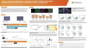 科学海报A Novel mRNA-LNP Platform to Rapidly Generate Functional Forebrain Neurons from hPSCs Using NGN2

 沪公网安备31010102008431号