Y. Kim et al. (May 2020)
FASEB Journal 34 6965-6983
Microtubule-associated protein 2 mediates induction of long-term potentiation in hippocampal neurons
Microtubule-associated protein (MAP) 2 has been perceived as a static cytoskeletal protein enriched in neuronal dendritic shafts. Emerging evidence indicates dynamic functions for various MAPs in activity-dependent synaptic plasticity. However,it is unclear how MAP2 is associated with synaptic plasticity mechanisms. Here,we demonstrate that specific silencing of high-molecular-weight MAP2 in vivo abolished induction of long-term potentiation (LTP) in the Schaffer collateral pathway of CA1 pyramidal neurons and in vitro blocked LTP-induced surface delivery of AMPA receptors and spine enlargement. In mature hippocampal neurons,we observed rapid translocation of a subpopulation of MAP2,present in dendritic shafts,to spines following LTP stimulation. Time-lapse confocal imaging showed that spine translocation of MAP2 was coupled with LTP-induced spine enlargement. Consistently,immunogold electron microscopy revealed that LTP stimulation of the Schaffer collateral pathway promoted MAP2 labeling in spine heads of CA1 neurons. This translocation depended on NMDA receptor activation and Ras-MAPK signaling. Furthermore,LTP stimulation led to an increase in surface-expressed AMPA receptors specifically in the neurons with MAP2 spine translocation. Altogether,this study indicates a novel role for MAP2 in LTP mechanisms and suggests that MAP2 participates in activity-dependent synaptic plasticity in mature hippocampal networks.
View Publication
Y. Chang et al. (jan 2022)
Allergy,asthma & immunology research 14 1 99--116
OASL1-Mediated Inhibition of Type I IFN Reduces Influenza A Infection-Induced Airway Inflammation by Regulating ILC2s.
PURPOSE Three observations drove this study. First,2'-5'-oligoadenylate synthetase-like protein (OASL) is a negative regulator of type I interferon (IFN). Second,type I IFN plays a central role during virus infections and the pathogenesis of various diseases,including asthma. Third,influenza A virus (IAV) causes non-eosinophilic asthma. To evaluate the potential relationships between OASL,type I IFN,and pulmonary innate immune cells in IAV-induced acute airway inflammation by using Oasl1-/- mice. METHODS Asthma was induced in wild-type (WT) and Oasl1-/- mice with IAV or ovalbumin (OVA). Airway hyperreactivity (AHR) and immune cell infiltration in the bronchoalveolar lavage (BAL) fluids were measured. The immune cells in the lungs were analyzed by flow cytometry. To investigate the ability of type I IFN to shape the response of lung type 2 innate lymphoid cells (ILC2s),IFN-$\alpha$ was treated intratracheally. Plasmacytoid dendritic cells (pDCs) sorted from bone marrow and ILC2s sorted from lungs of naive mice were co-cultured with/without interferon-alpha receptor subunit 1 (IFNAR-1)-blocking antibodies. RESULTS In the IAV-induced asthma model,Oasl1-/- mice developed greater AHR and immune cell infiltration in the BAL fluids than WT mice. This was not observed in OVA-induced asthma,a standard model of allergen-induced asthma. The lungs of infected Oasl1-/- mice also had elevated DC numbers and Ifna expression and depressed IAV-induced ILC2 responses,namely,proliferation and type 2 cytokine and amphiregulin production. Intratracheal administration of type I IFN in na{\{i}}ve mice suppressed lung ILC2 production of type 2 cytokines and amphiregulin. Co-culture of ILC2s with pDCs showed that pDCs inhibit the function of ILC2s by secreting type I IFN. CONCLUSIONS OASL1 may impede the IAV-induced acute airway inflammation that drives AHR by inhibiting IAV-induced type I IFN production from lung DCs thereby preserving the functions of lung ILC2s including their amphiregulin production."
View Publication
产品类型:
产品号#:
19764
19875
19764RF
产品名:
EasySep™小鼠浆细胞样DC分选试剂盒
EasySep™小鼠Pan-ILC富集试剂盒
RoboSep™ 小鼠浆细胞样DC分选试剂盒
S. Biswas et al. ( 2022)
Frontiers in immunology 13 875320
Pharmacological Inhibition of MALT1 Ameliorates Autoimmune Pathogenesis and Can Be Uncoupled From Effects on Regulatory T-Cells.
MALT1 forms part of a central signaling node downstream of immunoreceptor tyrosine-based activation motif (ITAM)-containing receptors,across a broad range of immune cell subsets,and regulates NF-$\kappa$B driven transcriptional responses via dual scaffolding-protease activity. Allosteric inhibition of MALT1 activity has demonstrated benefit in animal models of inflammation. However,development of MALT1 inhibitors to treat autoimmune and inflammatory diseases (A&ID) has been hindered by reports linking MALT1 inhibition and genetic loss-of-function to reductions in regulatory T-cell (Treg) numbers and development of auto-inflammatory syndromes. Using an allosteric MALT1 inhibitor,we investigated the consequence of pharmacological inhibition of MALT1 on proinflammatory cells compared to regulatory T-cells. Consistent with its known role in ITAM-driven responses,MALT1 inhibition suppressed proinflammatory cytokine production from activated human T-cells and monocyte-derived macrophages,and attenuated B-cell proliferation. Oral administration of a MALT1 inhibitor reduced disease severity and synovial cytokine production in a rat collagen-induced arthritis model. Interestingly,reduction in splenic Treg numbers was less pronounced in the context of inflammation compared with na{\{i}}ve animals. Additionally in the context of the disease model we observed an uncoupling of anti-inflammatory effects of MALT1 inhibition from Treg reduction with lower systemic concentrations of inhibitor needed to reduce disease severity compared to that required to reduce Treg numbers. MALT1 inhibition did not affect suppressive function of human Tregs in vitro. These data indicate that anti-inflammatory efficacy can be achieved with MALT1 inhibition without impacting the number or function of Tregs further supporting the potential of MALT1 inhibition in the treatment of autoimmune disease."
View Publication
产品类型:
产品号#:
100-0785
19654
19654RF
产品名:
ImmunoCult™ 人CD3/CD28/CD2 T细胞激活剂
EasySep™ Direct 人 PBMC 分选试剂盒
RoboSep™ Direct 人 PBMC 分选试剂盒
J. Wu et al. ( 2022)
Pathology oncology research : POR 28 1610555
Enhancing Natural Killer Cell-Mediated Cancer Immunotherapy by the Biological Macromolecule Nocardia rubra Cell-Wall Skeleton.
The biological macromolecule Nocardia rubra cell-wall skeleton (Nr-CWS) has well-established immune-stimulating and anti-tumor activities. However,the role of Nr-CWS on natural killer (NK) cells remains unclear. Here,we explore the function and related mechanisms of Nr-CWS on NK cells. Using a tumor-bearing model,we show that Nr-CWS has slightly effect on solid tumor. In addition,using a tumor metastasis model,we show that Nr-CWS suppresses the lung metastasis induced by B16F10 melanoma cells in mice,which indicates that Nr-CWS may up-regulate the function of NK cells. Further investigation demonstrated that Nr-CWS can increase the expression of TRAIL and FasL on spleen NK cells from Nr-CWS treated B16F10 tumor metastasis mice. The spleen index and serum levels of TNF-$\alpha$,IFN-$\gamma$,and IL-2 in B16F10 tumor metastasis mice treated with Nr-CWS were significantly increased. In vitro,the studies using purified or sorted NK cells revealed that Nr-CWS increases the expression of CD69,TRAIL,and FasL,decreases the expression of CD27,and enhances NK cell cytotoxicity. The intracellular expression of IFN-$\gamma$,TNF-$\alpha$,perforin (prf),granzyme-B (GrzB),and secreted TNF-$\alpha$,IFN-$\gamma$,IL-6 of the cultured NK cells were significantly increased after treatment with Nr-CWS. Overall,the findings indicate that Nr-CWS could suppress the lung metastasis induced by B16F10 melanoma cells,which may be exerted through its effect on NK cells by promoting NK cell terminal differentiation (CD27lowCD11bhigh),and up-regulating the production of cytokines and cytotoxic molecules.
View Publication
产品类型:
产品号#:
19855
19855RF
产品名:
EasySep™小鼠NK细胞分选试剂盒
RoboSep™ 小鼠NK细胞分选试剂盒
M. Soutto et al. ( 2019)
Nature communications 10 1 3039
Activation of STAT3 signaling is mediated by TFF1 silencing in gastric neoplasia.
TFF1,a secreted protein,plays an essential role in keeping the integrity of gastric mucosa and its barrier function. Loss of TFF1 expression in the TFF1-knockout (KO) mouse leads to a pro-inflammatory phenotype with a cascade of gastric lesions that include low-grade dysplasia,high-grade dysplasia,and adenocarcinomas. In this study,we demonstrate nuclear localization of p-STATY705,with significant overexpression of several STAT3 target genes in gastric glands from the TFF1-KO mice. We also show frequent loss of TFF1 with nuclear localization of STAT3 in human gastric cancers. The reconstitution of TFF1 protein in human gastric cancer cells and 3D gastric glands organoids from TFF1-KO mice abrogates IL6-induced nuclear p-STAT3Y705 expression. Reconstitution of TFF1 inhibits IL6-induced STAT3 transcription activity,suppressing expression of its target genes. TFF1 blocks IL6R$\alpha$-GP130 complex formation through interfering with binding of IL6 to its receptor IL6R$\alpha$. These findings demonstrate a functional role of TFF1 in suppressing gastric tumorigenesis by impeding the IL6-STAT3 pro-inflammatory signaling axis.
View Publication
Modular tissue-in-a-CUBE platform to model blood-brain barrier (BBB) and brain interaction
With the advent of increasingly sophisticated organoids,there is growing demand for technology to replicate the interactions between multiple tissues or organs. This is challenging to achieve,however,due to the varying culture conditions of the different cell types that make up each tissue. Current methods often require complicated microfluidic setups,but fragile tissue samples tend not to fare well with rough handling. Furthermore,the more complicated the human system to be replicated,the more difficult the model becomes to operate. Here,we present the development of a multi-tissue chip platform that takes advantage of the modularity and convenient handling ability of a CUBE device. We first developed a blood-brain barrier-in-a-CUBE by layering astrocytes,pericytes,and brain microvascular endothelial cells in the CUBE,and confirmed the expression and function of important tight junction and transporter proteins in the blood-brain barrier model. Then,we demonstrated the application of integrating Tissue-in-a-CUBE with a chip in simulating the in vitro testing of the permeability of a drug through the blood-brain barrier to the brain and its effect on treating the glioblastoma brain cancer model. We anticipate that this platform can be adapted for use with organoids to build complex human systems in vitro by the combination of multiple simple CUBE units. Development of platform to integrate multiple Tissue-in-a-CUBEs in a chip for tissue-tissue interaction,demonstrated by simulating the testing of the permeability and effect of a cancer drug in a BBB-Brain cancer model.
View Publication
产品类型:
产品号#:
100-0483
100-0484
100-0276
100-1130
产品名:
Hausser Scientificᵀᴹ 明线血球计数板
ReLeSR™
mTeSR™ Plus
mTeSR™ Plus
(Jul 2025)
Molecular Metabolism 99 10
Complete loss of PAX4 causes transient neonatal diabetes in humans
ObjectiveGene discovery studies in individuals with diabetes diagnosed within 6 months of life (neonatal diabetes,NDM) can provide unique insights into the development and function of human pancreatic beta-cells.MethodsWe performed genome sequencing in a cohort of 43 consanguineous individuals with NDM in whom all the known genetic causes had previously been excluded. We used quantitative PCR and RNA-sequencing in CRISPR-edited human induced pluripotent stem cells (iPSCs),and CUT&RUN-sequencing in EndoC-?H1 cells to investigate the effect of PAX4 loss on human pancreatic development.ResultsWe describe the identification of homozygous PAX4 loss-of-function variants in 2 individuals with transient NDM: a p.(Arg126?) stop-gain variant and a c.-352_104del deletion affecting the first 4 PAX4 exons. We confirmed the p.(Arg126?) variant causes nonsense mediated decay in CRISPR-edited iPSC-derived pancreatic endoderm cells. Integrated analysis of CUT&RUN-sequencing in EndoC-?H1 cells and RNA-sequencing in PAX4-depleted islet stem cell models identified genes directly regulated by PAX4 involved in both pancreatic islet development and glucose-stimulated insulin secretion.ConclusionWe report the first human cases of complete loss of PAX4,establishing it as a novel cause of NDM and highlighting its role in human beta cell development. Both probands had transient NDM which remitted in early infancy but relapsed at the ages of 2.4 and 6.7 years,demonstrating that in contrast to mouse models,PAX4 is not essential for the development of human pancreatic beta-cells. Highlights•Homozygous loss-of-function variants in PAX4 are a novel genetic cause of transient neonatal diabetes.•PAX4 directly regulates genes involved in pancreatic beta cell development and glucose-sensitive insulin secretion.•The role of PAX4 in humans differs to that observed in mouse and is not essential for beta cell development.
View Publication
产品类型:
产品号#:
85850
85857
产品名:
mTeSR™1
mTeSR™1
(Jun 2025)
Cellular and Molecular Life Sciences: CMLS 82 1
The ADCY1-mediated cAMP signaling pathway mediates functional effects of montelukast treatment in brain organoids
Montelukast (MTK) is a drug widely used for treating allergic rhinitis and asthma. However,severe neuropsychiatric adverse events related to MTK have been reported,with limited understanding of the underlying mechanisms. Here we leveraged human forebrain organoids (hFOs) and showed that MTK exposure in hFOs downregulated the expression of genes associated with multiple neuronal functions and neuropsychiatric disorders. The following integrative analysis highlighted adenylate cyclase 1 (ADCY1),a main regulator of the cAMP signaling pathway,as a hub gene mediating the functional effects of MTK exposure. We also showed that MTK exposure resulted in a reduction of cAMP and neuroactivities,and caused neural maturation defects. These cellular phenotypes could be recapitulated by treating hFOs with ST034307,a selective ADCY1 inhibitor,or partially rescued by ADCY1 overexpression in hFOs. Together,this study underscored that MTK exposure caused neuropsychiatric effects through inhibiting the ADCY1-mediated cAMP signaling pathway.Supplementary InformationThe online version contains supplementary material available at 10.1007/s00018-025-05764-z.
View Publication

 EasySep™小鼠TIL(CD45)正选试剂盒


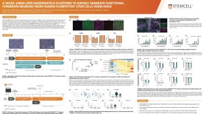 科学海报A Novel mRNA-LNP Platform to Rapidly Generate Functional Forebrain Neurons from hPSCs Using NGN2

 沪公网安备31010102008431号