
 EasySep™小鼠TIL(CD45)正选试剂盒


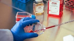
搜索结果: 'methocult media formulations for human hematopoietic cells serum containing'
-
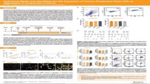 科学海报Optimized Media and Workflow for the Expansion of Human Pluripotent Stem Cells as Aggregates in Suspension Cultures
产品类型:
Conference:
ISSCR 2019
产品号#:
产品名:
发布日期: 07/05/2019 -

产品类型:
产品号#:
09600
09650
产品名:
StemSpan™ SFEM
StemSpan™ SFEM
-
产品类型:
产品号#:
22001
22005
22006
22007
22008
22009
22011
22012
22013
产品名:
STEMvision™ 人脐带血7-天CFU分析包
STEMvision™ 彩色人脐带血14-天CFU分析包
STEMvision™ 彩色人骨髓14-天CFU分析包
STEMvision™ 彩色人动员外周血14-天CFU分析包
STEMvision™ 小鼠总CFU分析包
STEMvision™ 小鼠髓系CFU分析包
STEMvision™ 小鼠红系CFU分析包
STEMvision™ 小鼠CFU分析包(髓系和红系)
-
 实验方案Transitioning from Feeder-Free Media to mTeSR™ Plus for Human Pluripotent Stem Cell Culture
产品类型:
研究方向:
干细胞生物学
产品号#:
产品名:
发布日期: 02/10/2020 -
产品类型:
产品号#:
09600
09650
产品名:
StemSpan™ SFEM
StemSpan™ SFEM
-
产品类型:
产品号#:
产品名:
-
产品类型:
产品号#:
05850
05857
05870
05875
85850
85857
85870
85875
产品名:
mTeSR™1
mTeSR™1
-
技术手册Human Colony-Forming Unit (CFU) Assays Using MethoCult™
产品类型:
产品号#:
00217
00217UK
00215
00215GER
00215UK
00602
00603
00606
00607
00608
00609
04436
04064
04100
04230
04236
04431
04434
04444
04464
04531
04535
04545
04536
04564
09300
04035
04330
04034
04044
04435
04445
04534
04544
04437
04447
00215US.4
00215CA.3
00215GER.3
0
产品名:
MethoCult™ SF H4436
MethoCult™ H4034 Optimum 入门试剂盒
MethoCult™ H4100
MethoCult™ H4230
MethoCult™ SF H4236
MethoCult™ H4431
MethoCult™ H4434 Classic
MethoCult™ H4434 Classic
MethoCult™ H4434 Classic 套装
MethoCult™ H4531
MethoCult™ H4535 Enriched,不含EPO
MethoCult™ H4535 Enriched,不含EPO
MethoCult™ SF H4536
MethoCult™ H4534 Classic 无 EPO 入门试剂盒
含有10% 牛血清白蛋白(BSA)的 Iscove's MDM
MethoCult™ 不含EPO的H4035 Optimum
MethoCult™ H4330
MethoCult™ H4034 Optimum
MethoCult™ H4034 Optimum
MethoCult™ H4435 Enriched
MethoCult™ H4435 Enriched
MethoCult™ H4534 Classic(不含 EPO)
MethoCult™ H4534 Classic(不含 EPO)
MethoCult™ Express
MethoCult™ Express
造血祖细胞检测标准化培训课程
-
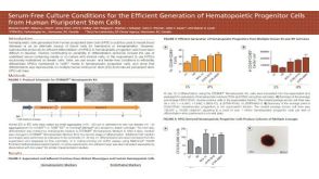 科学海报Efficient Differentiation of Human Pluripotent Stem Cells to Hematopoietic Progenitor Cells in Serum-Free Culture Conditions
产品类型:
Conference:
ISEH 2016
产品号#:
05310
产品名:
STEMdiff™ 造血试剂盒
-
产品类型:
产品号#:
05230
产品名:
STEMdiff™ 三胚层分化试剂盒
-
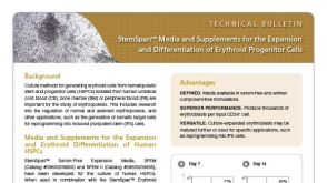 技术公告StemSpan™ Media and Supplements for the Expansion and Differentiation of Erythroid Progenitor Cells
产品类型:
产品号#:
02692
09600
09650
09605
09655
09955
产品名:
StemSpan™红系扩增添加物 (100X)
StemSpan™ SFEM
StemSpan™ SFEM
StemSpan™ SFEM II
StemSpan™ SFEM II
发布日期: 02/01/2016

 沪公网安备31010102008431号