Hoxa3 promotes the differentiation of hematopoietic progenitor cells into proangiogenic Gr-1+CD11b+ myeloid cells.
Injury induces the recruitment of bone marrow-derived cells (BMDCs) that contribute to the repair and regeneration process. The behavior of BMDCs in injured tissue has a profound effect on repair,but the regulation of BMDC behavior is poorly understood. Aberrant recruitment/retention of these cells in wounds of diabetic patients and animal models is associated with chronic inflammation and impaired healing. BMD Gr-1(+)CD11b(+) cells function as immune suppressor cells and contribute significantly to tumor-induced neovascularization. Here we report that Gr-1(+)CD11b(+) cells also contribute to injury-induced neovascularization,but show altered recruitment/retention kinetics in the diabetic environment. Moreover,diabetic-derived Gr-1(+)CD11b(+) cells fail to stimulate neovascularization in vivo and have aberrant proliferative,chemotaxis,adhesion,and differentiation potential. Previously we demonstrated that gene transfer of HOXA3 to wounds of diabetic mice is taken up by and expressed by recruited BMDCs. This is associated with a suppressed inflammatory response,enhanced neovascularization,and accelerated wound healing. Here we show that sustained expression of Hoxa3 in diabetic-derived BMD Gr-1(+)CD11b(+) cells reverses their diabetic phenotype. These findings demonstrate that manipulation of adult stem/progenitor cells ex vivo could be used as a potential therapy in patients with impaired wound healing.
View Publication
产品类型:
产品号#:
03434
03444
产品名:
MethoCult™ GF M3434
MethoCult™ GF M3434
T. J. Lynch et al. (MAY 2018)
Cell stem cell 22 5 653--667.e5
Submucosal Gland Myoepithelial Cells Are Reserve Stem Cells That Can Regenerate Mouse Tracheal Epithelium.
The mouse trachea is thought to contain two distinct stem cell compartments that contribute to airway repair-basal cells in the surface airway epithelium (SAE) and an unknown submucosal gland (SMG) cell type. Whether a lineage relationship exists between these two stem cell compartments remains unclear. Using lineage tracing of glandular myoepithelial cells (MECs),we demonstrate that MECs can give rise to seven cell types of the SAE and SMGs following severe airway injury. MECs progressively adopted a basal cell phenotype on the SAE and established lasting progenitors capable of further regeneration following reinjury. MECs activate Wnt-regulated transcription factors (Lef-1/TCF7) following injury and Lef-1 induction in cultured MECs promoted transition to a basal cell phenotype. Surprisingly,dose-dependent MEC conditional activation of Lef-1 in vivo promoted self-limited airway regeneration in the absence of injury. Thus,modulating the Lef-1 transcriptional program in MEC-derived progenitors may have regenerative medicine applications for lung diseases.
View Publication
产品类型:
产品号#:
05001
05021
05022
产品名:
PneumaCult™-ALI 培养基
PneumaCult™-ALI 培养基含12 mm Transwell®插件
PneumaCult™-ALI 培养基含6.5 mm Transwell®插件
Fukuta M et al. (DEC 2014)
PLoS ONE 9 12 e112291
Derivation of mesenchymal stromal cells from pluripotent stem cells through a neural crest lineage using small molecule compounds with defined media
Neural crest cells (NCCs) are an embryonic migratory cell population with the ability to differentiate into a wide variety of cell types that contribute to the craniofacial skeleton,cornea,peripheral nervous system,and skin pigmentation. This ability suggests the promising role of NCCs as a source for cell-based therapy. Although several methods have been used to induce human NCCs (hNCCs) from human pluripotent stem cells (hPSCs),such as embryonic stem cells (ESCs) and induced pluripotent stem cells (iPSCs),further modifications are required to improve the robustness,efficacy,and simplicity of these methods. Chemically defined medium (CDM) was used as the basal medium in the induction and maintenance steps. By optimizing the culture conditions,the combination of the GSK3β inhibitor and TGFβ inhibitor with a minimum growth factor (insulin) very efficiently induced hNCCs (70-80%) from hPSCs. The induced hNCCs expressed cranial NCC-related genes and stably proliferated in CDM supplemented with EGF and FGF2 up to at least 10 passages without changes being observed in the major gene expression profiles. Differentiation properties were confirmed for peripheral neurons,glia,melanocytes,and corneal endothelial cells. In addition,cells with differentiation characteristics similar to multipotent mesenchymal stromal cells (MSCs) were induced from hNCCs using CDM specific for human MSCs. Our simple and robust induction protocol using small molecule compounds with defined media enabled the generation of hNCCs as an intermediate material producing terminally differentiated cells for cell-based innovative medicine.
View Publication
产品类型:
产品号#:
05850
05857
05870
05875
85850
85857
85870
85875
产品名:
mTeSR™1
mTeSR™1
K. Maneechai et al. (Sep 2024)
Heliyon 10 19
Generation of ex vivo autologous hematopoietic stem cell-derived T lymphocytes for cancer immunotherapy
CD19CAR-T cell therapy demonstrated promising outcomes in relapsed/refractory B-cell malignancies. Nonetheless,the limited T-cell function and ineffective T-cell apheresis for therapeutic purposes are still concern in heavily pretreated patients. We investigated the feasibility of generating hematopoietic stem cell-derived T lymphocytes (HSC-T) for cancer immunotherapy. The patients’ autologous peripheral blood HSCs were enriched for CD34 + and CD3 + cells. The CD34 + cells were then cultured following three steps of lymphoid progenitor differentiation,T-cell differentiation,and T-cell maturation processes. HSC-T cells were successfully generated with robust fold expansion of 3735 times. After lymphoid progenitor differentiation,CD5 + and CD7 + cells remarkably increased (65–84 %) while CD34 + cells consequentially declined. The mature CD3 + cells were detected up to 40 % and 90 % on days 42 and 52,respectively. The majority of HSC-T population was naïve phenotype compared to CD3-T cells (73 % vs 34 %) and CD8:CD4 ratio was 2:1. The higher level of cytokine and cytotoxic granule secretion in HSC-T was observed after activation. HSC-T cells were assessed for clinical application and found that CD19CAR-transduced HSC-T cells demonstrated higher cytokine secretion and a trend of superior cytotoxicity against CD19 + target cells compared to control CAR-T cells. A chronic antigen stimulation assay revealed similar T-cell proliferation,stemness,and exhaustion phenotypes among CAR-T cell types. In conclusions,autologous HSC-T was feasible to generate with preserved T-cell efficacy. The HSC-T cells are potentially utilized as an alternative option for cellular immunotherapy.
View Publication
产品类型:
产品号#:
09600
09605
09650
09655
产品名:
StemSpan™ SFEM
StemSpan™ SFEM II
StemSpan™ SFEM
StemSpan™ SFEM II
Zhang L et al. (APR 2016)
Human Reproduction 31 4 832--843
Protein kinase A inhibitor, H89, enhances survival and clonogenicity of dissociated human embryonic stem cells through Rho-associated coiled-coil containing protein kinase (ROCK) inhibition
H89 inhibits the dissociation-induced phosphorylation of PKA and two substrates of Rho-associated coiled-coil containing protein kinase (ROCK),myosin light chain (MLC2) and myosin phosphatase target subunit 1 (MYPT1),significantly increases cell survival and colony formation,and strongly depresses dissociation-induced cell death and cell blebbing without affecting the pluripotency of hESCs and their differentiation in vitro.
View Publication
产品类型:
产品号#:
05835
05839
产品名:
STEMdiff™ 神经诱导培养基
STEMdiff™ 神经诱导培养基
Olmsted-Davis EA et al. (DEC 2003)
Proceedings of the National Academy of Sciences of the United States of America 100 26 15877--82
Primitive adult hematopoietic stem cells can function as osteoblast precursors.
Osteoblasts are continually recruited from stem cell pools to maintain bone. Although their immediate precursor is a plastic-adherent mesenchymal stem cell able to generate tissues other than bone,increasing evidence suggests the existence of a more primitive cell that can differentiate to both hematopoietic and mesenchymal cells. We show here that the side population" (SP) of marrow stem cells�
View Publication
产品类型:
产品号#:
05501
05502
产品名:
Heringer-Walther S et al. (JUN 2009)
Haematologica 94 6 857--60
Angiotensin-(1-7) stimulates hematopoietic progenitor cells in vitro and in vivo.
Effects of angiotensin (Ang)-(1-7),an AngII metabolite,on bone marrow-derived hematopoietic cells were studied. We identified Ang-(1-7) to stimulate proliferation of human CD34(+) and mononuclear cells in vitro. Under in vivo conditions,we monitored proliferation and differentiation of human cord blood mononuclear cells in NOD/SCID mice. Ang-(1-7) stimulated differentially human cells in bone marrow and accumulated them in the spleen. The number of HLA-I(+) and CD34(+) cells in the bone marrow was increased 42-fold and 600-fold,respectively. These results indicate a decisive impact of Ang-(1-7) on hematopoiesis and its promising therapeutic potential in diseases requiring progenitor stimulation.
View Publication
Functional analysis of leukemia-associated PTPN11 mutations in primary hematopoietic cells.
PTPN11 encodes the protein tyrosine phosphatase SHP-2,which relays signals from growth factor receptors to Ras and other effectors. Germline PTPN11 mutations underlie about 50% of Noonan syndrome (NS),a developmental disorder that is associated with an elevated risk of juvenile myelomonocytic leukemia (JMML). Somatic PTPN11 mutations were recently identified in about 35% of patients with JMML; these mutations introduce amino acid substitutions that are largely distinct from those found in NS. We assessed the functional consequences of leukemia-associated PTPN11 mutations in murine hematopoietic cells. Expressing an E76K SHP-2 protein induced a hypersensitive pattern of granulocyte-macrophage colony-forming unit (CFU-GM) colony growth in response to granulocyte-macrophage colony-stimulating factor (GM-CSF) and interleukin 3 (IL-3) that was dependent on SHP-2 catalytic activity. E76K SHP-2 expression also enhanced the growth of immature progenitor cells with high replating potential,perturbed erythroid growth,and impaired normal differentiation in liquid cultures. In addition,leukemia-associated SHP-2 mutations conferred a stronger phenotype than a germline mutation found in patients with NS. Mutant SHP-2 proteins induce aberrant growth in multiple hematopoietic compartments,which supports a primary role of hyperactive Ras in the pathogenesis of JMML.
View Publication
产品类型:
产品号#:
03231
03334
03434
03444
09600
09650
产品名:
MethoCult™ M3231
MethoCult™ M3334
MethoCult™ GF M3434
MethoCult™ GF M3434
StemSpan™ SFEM
StemSpan™ SFEM
Goldman FD et al. (MAY 2008)
Blood 111 9 4523--31
Characterization of primitive hematopoietic cells from patients with dyskeratosis congenita.
Dyskeratosis congenita (DC) is an inherited bone marrow (BM) failure syndrome associated with mutations in telomerase genes and the acquisition of shortened telomeres in blood cells. To investigate the basis of the compromised hematopoiesis seen in DC,we analyzed cells from granulocyte colony-stimulating factor mobilized peripheral blood (mPB) collections from 5 members of a family with autosomal dominant DC with a hTERC mutation. Premobilization BM samples were hypocellular,and percentages of CD34(+) cells in marrow and mPB collections were significantly below values for age-matched controls in 4 DC subjects. Directly clonogenic cells,although present at normal frequencies within the CD34(+) subset,were therefore absolutely decreased. In contrast,even the frequency of long-term culture-initiating cells within the CD34(+) DC mPB cells was decreased,and the telomere lengths of these cells were also markedly reduced. Nevertheless,the different lineages of mature cells were produced in normal numbers in vitro. These results suggest that marrow failure in DC is caused by a reduction in the ability of hematopoietic stem cells to sustain their numbers due to telomere impairment rather than a qualitative defect in their commitment to specific lineages or in the ability of their lineage-restricted progeny to execute normal differentiation programs.
View Publication
Liu W et al. (DEC 2014)
Cell death and differentiation 4 12 1950--1960
BRD4 regulates Nanog expression in mouse embryonic stem cells and preimplantation embryos.
Bromodomain-containing protein 4 (BRD4) is an important epigenetic reader implicated in the pathogenesis of a number of different cancers and other diseases. Brd4-null mouse embryos die shortly after implantation and are compromised in their ability to maintain the inner cell mass,which gives rise to embryonic stem cells (ESCs). Here we report that BRD4 regulates expression of the pluripotency factor Nanog in mouse ESCs and preimplantation embryos,as well as in human ESCs and embryonic cancer stem cells. Inhibition of BRD4 function using a chemical inhibitor,small interfering RNAs,or a dominant-negative approach suppresses Nanog expression,and abolishes the self-renewal ability of ESCs. We also find that BRD4 associates with BRG1 (brahma-related gene 1,aka Smarca4 (SWI/SNF-related,matrix-associated,actin-dependent regulator of chromatin,subfamily a,member 4)),a key regulator of ESC self-renewal and pluripotency,in the Nanog regulatory regions to regulate Nanog expression. Our study identifies Nanog as a novel BRD4 target gene,providing new insights for the biological function of BRD4 in stem cells and mouse embryos. Knowledge gained from these non-cancerous systems will facilitate future investigations of how Brd4 dysfunction leads to cancers.Cell Death and Differentiation advance online publication,22 August 2014; doi:10.1038/cdd.2014.124.
View Publication

 EasySep™小鼠TIL(CD45)正选试剂盒


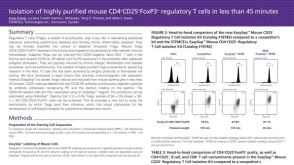 科学海报Isolation of Highly Purified Mouse CD4+CD25+Foxp3+ Regulatory T Cells in Less

 沪公网安备31010102008431号