
 EasySep™小鼠TIL(CD45)正选试剂盒


搜索结果: 'methocult media formulations for human hematopoietic cells serum containing'
-

产品类型:
产品号#:
05854
05855
100-0483
100-0484
100-0276
100-1130
产品名:
mFreSR™
mFreSR™
Hausser Scientificᵀᴹ 明线血球计数板
ReLeSR™
mTeSR™ Plus
mTeSR™ Plus
-
产品类型:
产品号#:
15271HLA
产品名:
RosetteSep™ HLA 淋系细胞富集试剂盒
-
产品类型:
产品号#:
04437
04447
产品名:
MethoCult™ Express
MethoCult™ Express
-
产品类型:
产品号#:
01700
01705
01701
01702
产品名:
ALDEFLUOR™ 试剂盒
ALDEFLUOR™ DEAB试剂, 1.5 mM, 1 mL
ALDEFLUOR™检测缓冲液
-
产品类型:
产品号#:
R1061
R1034
R1116
产品名:
-
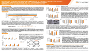
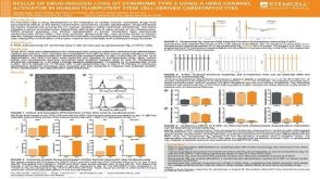 科学海报Rescue of Drug-Induced Long QT Syndrome Type 2 Using a hERG Channel Activator in Human Pluripotent Stem Cell-Derived Cardiomyocytes
产品类型:
Conference:
AHA 2018
产品号#:
05010
05020
产品名:
STEMdiff™ 心室肌细胞分化试剂盒
STEMdiff™ 心肌细胞维持培养试剂盒
-
产品类型:
产品号#:
15021
15061
产品名:
RosetteSep™人T细胞富集抗体混合物
RosetteSep™人T细胞富集抗体混合物
-
产品类型:
产品号#:
05850
05857
05870
05875
60070
60070.1
60070AD
60070AD.1
60070AZ
60070AZ.1
60070BT
60070BT.1
60070FI
60070FI.1
60070PE
60070PE.1
60070PS
60093
60093.1
60093AD
60093AD.1
60093PE
60093PE.1
85850
85857
85870
85875
产品名:
小鼠 IgG1,kappa 同型对照抗体(克隆 MOPC-21)
小鼠 IgG1,kappa 同型对照抗体(克隆 MOPC-21)
小鼠IgG1,kappa同型对照抗体(clone MOPC-21),Alexa Fluor® 488
小鼠 IgG1,kappa 同型对照抗体(克隆 MOPC-21),Alexa Fluor® 488
小鼠 IgG1,kappa 同型对照抗体(克隆 MOPC-21),APC
小鼠 IgG1,kappa 同型对照抗体(克隆 MOPC-21),APC
小鼠 IgG1,kappa 同型对照抗体(克隆 MOPC-21),Biotin
小鼠 IgG1,kappa 同型对照抗体(克隆 MOPC-21),FITC
小鼠 IgG1,kappa 同型对照抗体(克隆 MOPC-21),FITC
小鼠 IgG1,kappa 同型对照抗体(克隆 MOPC-21),PE
小鼠 IgG1,kappa 同型对照抗体(克隆 MOPC-21),PE
小鼠 IgG1,kappa 同型对照抗体(克隆 MOPC-21),PerCP-Cy5.5
抗人OCT4(OCT3)抗体,克隆3A2A20
抗人OCT4(OCT3)抗体,clone 3A2A20
抗人OCT4(OCT3)抗体,克隆3A2A20,Alexa Fluor® 488
抗人OCT4(OCT3)抗体,克隆3A2A20,Alexa Fluor® 488
抗人OCT4(OCT3)抗体,克隆3A2A20,PE
抗人OCT4(OCT3)抗体,克隆3A2A20,PE
mTeSR™1
mTeSR™1
-
产品类型:
产品号#:
05850
05857
05870
05875
85850
85857
85870
85875
产品名:
mTeSR™1
mTeSR™1
-
产品类型:
产品号#:
85850
85857
产品名:
mTeSR™1
mTeSR™1
-
产品类型:
产品号#:
100-0483
100-0484
产品名:
Hausser Scientificᵀᴹ 明线血球计数板
ReLeSR™

 沪公网安备31010102008431号