Palmer DJ et al. (JUN 2016)
Molecular Therapy — Methods & Clinical Development 3 April 16039
Helper virus-mediated downregulation of transgene expression permits production of recalcitrant helper-dependent adenoviral vector
Helper-dependent adenoviral vectors (HDAd) that express certain transgene products are impossible to produce because the transgene product is toxic to the producer cells,especially when made in large amounts during vector production. Downregulating transgene expression from the HDAd during vector production is a way to solve this problem. In this report,we show that this can be accomplished by inserting the target sequence for the adenoviral VA RNAI into the 3' untranslated region of the expression cassette in the HDAd. Thus during vector production,when the producer cells are coinfected with both the helper virus (HV) and the HDAd,the VA RNAI produced by the HV will target the transgene mRNA from the HDAd via the endogenous cellular RNAi pathway. Once the HDAd is produced and purified,transduction of the target cells results in unimpeded transgene expression because of the absence of HV. This simple and universal strategy permits for the robust production of otherwise recalcitrant HDAds.
View Publication
产品类型:
产品号#:
05850
05857
05870
05875
85850
85857
85870
85875
产品名:
mTeSR™1
mTeSR™1
Grö et al. (JUL 2016)
Blood
LFA-1 integrin antibodies inhibit leukocyte α4β1-mediated adhesion by intracellular signaling.
Binding of ICAM-1 (intercellular adhesion molecule-1) to the β2-integrin LFA-1 (leukocyte function associated antigen-1) is known to induce crosstalk to the α4β1 integrin. Using different LFA-1 monoclonal antibodies we have been able to study the requirement and mechanism of action for the crosstalk in considerable detail. LFA-1 activating antibodies and those inhibitory antibodies that signal to α4β1 induce phosphorylation of Thr-758 on the β2-chain,which is followed by binding of 14-3-3 proteins and signaling through the G protein exchange factor Tiam1. This results in dephosphorylation of Thr-788/789 on the β1-chain of α4β1 and loss of binding to its ligand VCAM-1 (vascular cell adhesion molecule-1). The results show that with LFA-1 antibodies,we can either 1) activate LFA-1 and inhibit α4β1,2) inhibit both LFA-1 and α4β1,3) inhibit LFA-1 but not α4β1 or 4) not affect LFA-1 or α4β1 These findings are important for the understanding of integrin regulation and for the interpretation of the effect of integrin antibodies and their use in clinical applications.
View Publication
产品类型:
产品号#:
10970
10990
产品名:
ImmunoCult™ 人CD3/CD28/CD2 T细胞激活剂
ImmunoCult™ 人CD3/CD28/CD2 T细胞激活剂
Brohawn DG et al. (AUG 2016)
PloS one 11 8 e0160520
RNAseq Analyses Identify Tumor Necrosis Factor-Mediated Inflammation as a Major Abnormality in ALS Spinal Cord.
ALS is a rapidly progressive,devastating neurodegenerative illness of adults that produces disabling weakness and spasticity arising from death of lower and upper motor neurons. No meaningful therapies exist to slow ALS progression,and molecular insights into pathogenesis and progression are sorely needed. In that context,we used high-depth,next generation RNA sequencing (RNAseq,Illumina) to define gene network abnormalities in RNA samples depleted of rRNA and isolated from cervical spinal cord sections of 7 ALS and 8 CTL samples. We aligned textgreater50 million 2X150 bp paired-end sequences/sample to the hg19 human genome and applied three different algorithms (Cuffdiff2,DEseq2,EdgeR) for identification of differentially expressed genes (DEG's). Ingenuity Pathways Analysis (IPA) and Weighted Gene Co-expression Network Analysis (WGCNA) identified inflammatory processes as significantly elevated in our ALS samples,with tumor necrosis factor (TNF) found to be a major pathway regulator (IPA) and TNF$$-induced protein 2 (TNFAIP2) as a major network hub" gene (WGCNA). Using the oPOSSUM algorithm�
View Publication
产品类型:
产品号#:
05850
05857
05870
05875
85850
85857
85870
85875
产品名:
mTeSR™1
mTeSR™1
Wang Q et al. (OCT 2016)
Biomaterials 105 52--65
Functional engineered human cardiac patches prepared from nature's platform improve heart function after acute myocardial infarction.
With the advent of induced pluripotent stem cells and directed differentiation techniques,it is now feasible to derive individual-specific cardiac cells for human heart tissue engineering. Here we report the generation of functional engineered human cardiac patches using human induced pluripotent stem cells-derived cardiac cells and decellularized natural heart ECM as scaffolds. The engineered human cardiac patches can be tailored to any desired size and shape and exhibited normal contractile and electrical physiology in vitro. Further,when patching on the infarct area,these patches improved heart function of rats with acute myocardial infarction in vivo. These engineered human cardiac patches can be of great value for normal and disease-specific heart tissue engineering,drug screening,and meet the demands for individual-specific heart tissues for personalized regenerative therapy of myocardial damages in the future.
View Publication
产品类型:
产品号#:
05850
05857
05870
05875
85850
85857
85870
85875
产品名:
mTeSR™1
mTeSR™1
Silva MC et al. (SEP 2016)
Stem cell reports 7 3 325--340
Human iPSC-Derived Neuronal Model of Tau-A152T Frontotemporal Dementia Reveals Tau-Mediated Mechanisms of Neuronal Vulnerability.
Frontotemporal dementia (FTD) and other tauopathies characterized by focal brain neurodegeneration and pathological accumulation of proteins are commonly associated with tau mutations. However,the mechanism of neuronal loss is not fully understood. To identify molecular events associated with tauopathy,we studied induced pluripotent stem cell (iPSC)-derived neurons from individuals carrying the tau-A152T variant. We highlight the potential of in-depth phenotyping of human neuronal cell models for pre-clinical studies and identification of modulators of endogenous tau toxicity. Through a panel of biochemical and cellular assays,A152T neurons showed accumulation,redistribution,and decreased solubility of tau. Upregulation of tau was coupled to enhanced stress-inducible markers and cell vulnerability to proteotoxic,excitotoxic,and mitochondrial stressors,which was rescued upon CRISPR/Cas9-mediated targeting of tau or by pharmacological activation of autophagy. Our findings unmask tau-mediated perturbations of specific pathways associated with neuronal vulnerability,revealing potential early disease biomarkers and therapeutic targets for FTD and other tauopathies.
View Publication
产品类型:
产品号#:
05850
05857
05870
05875
85850
85857
85870
85875
产品名:
mTeSR™1
mTeSR™1
Gao L et al. ( 2016)
PloS one 11 9 e0162149
31P NMR 2D Mapping of Creatine Kinase Forward Flux Rate in Hearts with Postinfarction Left Ventricular Remodeling in Response to Cell Therapy.
Utilizing a fast 31P magnetic resonance spectroscopy (MRS) 2-dimensional chemical shift imaging (2D-CSI) method,this study examined the heterogeneity of creatine kinase (CK) forward flux rate of hearts with postinfarction left ventricular (LV) remodeling. Immunosuppressed Yorkshire pigs were assigned to 4 groups: 1) A sham-operated normal group (SHAM,n = 6); 2) A 60 minutes distal left anterior descending coronary artery ligation and reperfusion (MI,n = 6); 3) Open patch group; ligation injury plus open fibrin patch over the site of injury (Patch,n = 6); and 4) Cell group,hiPSCs-cardiomyocytes,-endothelial cells,and -smooth muscle cells (2 million,each) were injected into the injured myocardium pass through a fibrin patch (Cell+Patch,n = 5). At 4 weeks,the creatine phosphate (PCr)/ATP ratio,CK forward flux rate (Flux PCr→ATP),and k constant of CK forward flux rate (kPCr→ATP) were severely decreased at border zone myocardium (BZ) adjacent to MI. Cell treatment results in significantly increase of PCr/ATP ratio and improve the value of kPCr→ATP and Flux PCr→ATP in BZ myocardium. Moreover,the BZ myocardial CK total activity and protein expression of CK mitochondria isozyme and CK myocardial isozyme were significantly reduced,but recovered in response to cell treatment. Thus,cell therapy results in improvement of BZ bioenergetic abnormality in hearts with postinfarction LV remodeling,which is accompanied by significantly improvements in BZ CK activity and CK isozyme expression. The fast 2D 31P MR CSI mapping can reliably measure the heterogeneity of bioenergetics in hearts with post infarction LV remodeling.
View Publication
产品类型:
产品号#:
05850
05857
05870
05875
85850
85857
85870
85875
产品名:
mTeSR™1
mTeSR™1
La Spada A et al. (DEC 2016)
The journal of histochemistry and cytochemistry : official journal of the Histochemistry Society 64 12 739--751
Cell Line Macroarray: An Alternative High-Throughput Platform to Analyze hiPSC Lines.
In the past decade,tissue microarray (TMA) technology has evolved as an innovative tool for high-throughput proteomics analysis and mainly for biomarker validation. Similarly,enormous amount of data can be obtained from the cell line macroarray (CLMA) technology,which developed from the TMA using formalin-fixed,paraffin-embedded cell pellets. Here,we applied CLMA technology in stem cell research and in particular to identify bona fide neogenerated human induced pluripotent stem cell (hiPSC) clones suitable for down the line differentiation. All hiPSC protocols generate tens of clones,which need to be tested to determine genetically stable cell lines suitable for differentiation. Screening methods generally rely on fluorescence-activated cell sorting isolation and coverslip cell growth followed by immunofluorescence; these techniques could be cumbersome. Here,we show the application of CLMA to identify neogenerated pluripotent cell colonies and neuronal differentiated cell products. We also propose the use of the automated image analyzer,TissueQuest,as a reliable tool to quickly select the best clones,based upon the level of expression of multiple pluripotent biomarkers.
View Publication
产品类型:
产品号#:
05850
05857
05870
05875
85850
85857
85870
85875
产品名:
mTeSR™1
mTeSR™1
Guo D et al. (JAN 2017)
Stem cell research 18 67--69
Creating a patient carried Men1 gene point mutation on wild type iPSCs locus mediated by CRISPR/Cas9 and ssODN.
A patient specific point mutation (c.1288GtextgreaterT) of Men1 gene was introduced into wide type iPSC line with CRISPR/Cas9 and single-stranded donor oligonucleotides carrying the mutation. The mutated iPSC line has a heterozygous c.1288GtextgreaterT mutation on exon-9 of Men1 that was confirmed by sequencing analysis. The karyotype of this line was normal and the pluripotency was demonstrated by its ability to differentiate into three germ layers. These artificially created Men1 mutation in wild type iPSC line will help to dissect out the molecular basis of two patients carried the same mutation from one family who were differentially represented hypoglycemia.
View Publication

 EasySep™小鼠TIL(CD45)正选试剂盒


 实验方案How to Generate AssemBloids™ from hPSC-Derived Dorsal and Ventral Forebrain Organoid Co-Cultures
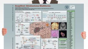 挂图SnapShot: Glioblastoma Multiforme Overview of the key concepts and mechanisms in glioblastoma multiforme biology


 沪公网安备31010102008431号