Conversion of placental hemogenic endothelial cells to hematopoietic stem and progenitor cells
Hematopoietic stem and progenitor cells (HSPCs) are critical for the treatment of blood diseases in clinic. However,the limited source of HSPCs severely hinders their clinical application. In the embryo,hematopoietic stem cells (HSCs) arise from hemogenic endothelial (HE) cells lining the major arteries in vivo. In this work,by engineering vascular niche endothelial cells (VN-ECs),we generated functional HSPCs in vitro from ECs at various sites,including the aorta-gonad-mesonephros (AGM) region and the placenta. Firstly,we converted mouse embryonic HE cells from the AGM region (aHE) into induced HSPCs (iHSPCs),which have the abilities for multilineage differentiation and self-renewal. Mechanistically,we found that VN-ECs can promote the generation of iHSPCs via secretion of CX3CL1 and IL1A. Next,through VN-EC co-culture,we showed that placental HE (pHE) cells,a type of extra-embryonic HE cells,were successfully converted into iHSPCs (pHE-iHSPCs),which have multilineage differentiation capacity,but exhibit limited self-renewal ability. Furthermore,comparative transcriptome analysis of aHE-iHSPCs and pHE-iHSPCs showed that aHE-iHSPCs highly expressed HSC-specific and self-renewal-related genes. Moreover,experimental validation showed that retinoic acid (RA) treatment promoted the transformation of pHE cells into iHSPCs that have self-renewal ability. Collectively,our results suggested that pHE cells possess the potential to transform into self-renewing iHSPCs through RA treatment,which will facilitate the clinical application of placental endothelial cells in hematopoietic cell generation. Subject terms: Haematopoietic stem cells,Haematopoietic stem cells
View Publication
产品类型:
产品号#:
03434
03444
09600
09650
产品名:
MethoCult™ GF M3434
MethoCult™ GF M3434
StemSpan™ SFEM
StemSpan™ SFEM
Walker TL et al. (JAN 2012)
PloS one 7 9 e44371
Prolactin stimulates precursor cells in the adult mouse hippocampus.
In the search for ways to combat degenerative neurological disorders,neurogenesis-stimulating factors are proving to be a promising area of research. In this study,we show that the hormonal factor prolactin (PRL) can activate a pool of latent precursor cells in the adult mouse hippocampus. Using an in vitro neurosphere assay,we found that the addition of exogenous PRL to primary adult hippocampal cells resulted in an approximate 50% increase in neurosphere number. In addition,direct infusion of PRL into the adult dentate gyrus also resulted in a significant increase in neurosphere number. Together these data indicate that exogenous PRL can increase hippocampal precursor numbers both in vitro and in vivo. Conversely,PRL null mice showed a significant reduction (approximately 80%) in the number of hippocampal-derived neurospheres. Interestingly,no deficit in precursor proliferation was observed in vivo,indicating that in this situation other niche factors can compensate for a loss in PRL. The PRL loss resulted in learning and memory deficits in the PRL null mice,as indicated by significant deficits in the standard behavioral tests requiring input from the hippocampus. This behavioral deficit was rescued by direct infusion of recombinant PRL into the hippocampus,indicating that a lack of PRL in the adult mouse hippocampus can be correlated with impaired learning and memory.
View Publication
产品类型:
产品号#:
05700
05701
05702
产品名:
NeuroCult™ 基础培养基(小鼠和大鼠)
NeuroCult™ 扩增添加物(小鼠和大鼠)
NeuroCult™扩增试剂盒(小鼠和大鼠)
Samper E et al. (APR 2002)
Blood 99 8 2767--75
Long-term repopulating ability of telomerase-deficient murine hematopoietic stem cells.
Telomere length must be tightly regulated in highly proliferative tissues,such as the lymphohematopoietic system. Under steady-state conditions,the levels and functionality of hematopoietic-committed or multipotent progenitors were not affected in late-generation telomerase-deficient mice (mTerc(-/-)) with critically short telomeres. Evaluation of self-renewal potential of mTerc(-/-) day-12 spleen colony-forming units demonstrated no alteration as compared with wildtype progenitors. However,the replating ability of mTerc(-/-) granulocyte-macrophage CFUs (CFU-GMs) was greatly reduced as compared with wildtype CFU-GMs,indicating a diminished capacity of late-generation mTerc(-/-) committed progenitors when forced to proliferate. Long-term bone marrow cultures of mTerc(-/-) bone marrow (BM) cells show a reduction in proliferative capacity; this defect can be mainly attributed to the hematopoietic,not to the stromal,mTerc(-/-) cells. In serial and competitive transplantations,mTerc(-/-) BM stem cells show reduced long-term repopulating capacity,concomitant with an increase in genetic instability compared with wildtype cells. Nevertheless,in competitive transplantations late-generation mTerc(-/-) precursors can occasionally overcome this proliferative impairment and reconstitute irradiated recipients. In summary,our results demonstrate that late-generation mTerc(-/-) BM cells with short telomeres,although exhibiting reduced proliferation ability and reduced long-term repopulating capacity,can still reconstitute myeloablated animals maintaining stem cell function.
View Publication
产品类型:
产品号#:
05350
产品名:
Zhou F-W et al. ( 2015)
PloS one 10 3 e0120281
Functional integration of human neural precursor cells in mouse cortex.
This study investigates the electrophysiological properties and functional integration of different phenotypes of transplanted human neural precursor cells (hNPCs) in immunodeficient NSG mice. Postnatal day 2 mice received unilateral injections of 100,000 GFP+ hNPCs into the right parietal cortex. Eight weeks after transplantation,1.21% of transplanted hNPCs survived. In these hNPCs,parvalbumin (PV)-,calretinin (CR)-,somatostatin (SS)-positive inhibitory interneurons and excitatory pyramidal neurons were confirmed electrophysiologically and histologically. All GFP+ hNPCs were immunoreactive with anti-human specific nuclear protein. The proportions of PV-,CR-,and SS-positive cells among GFP+ cells were 35.5%,15.7%,and 17.1%,respectively; around 15% of GFP+ cells were identified as pyramidal neurons. Those electrophysiologically and histological identified GFP+ hNPCs were shown to fire action potentials with the appropriate firing patterns for different classes of neurons and to display spontaneous excitatory and inhibitory postsynaptic currents (sEPSCs and sIPSCs). The amplitude,frequency and kinetic properties of sEPSCs and sIPSCs in different types of hNPCs were comparable to host cells of the same type. In conclusion,GFP+ hNPCs produce neurons that are competent to integrate functionally into host neocortical neuronal networks. This provides promising data on the potential for hNPCs to serve as therapeutic agents in neurological diseases with abnormal neuronal circuitry such as epilepsy.
View Publication
产品类型:
产品号#:
05750
05751
产品名:
NeuroCult™ NS-A 基础培养基(人)
NeuroCult™ NS-A 扩增试剂盒(人)
Wang F et al. (DEC 2017)
Stem Cell Research & Therapy 8 1 26
CCL11 promotes migration and proliferation of mouse neural progenitor cells
BACKGROUND Neonatal hypoxia-ischemia induces massive brain damage during the perinatal period,resulting in long-term consequences to central nervous system structural and functional maturation. Although neural progenitor cells (NPCs) migrate through the parenchyma and home in to injury sites in the rodent brain,the molecular mechanisms are unknown. We examined the role of chemokines in mediating NPC migration after neonatal hypoxic-ischemic brain injury. METHODS Nine-day-old mice were exposed to a 120-minute hypoxia following unilateral carotid occlusion. Chemokine levels were quantified in mouse brain extract. Migration and proliferation assays were performed using embryonic and infant mouse NPCs. RESULTS The neonatal hypoxic-ischemic brain injury resulted in an ipsilateral lesion,which was extended to the cortical and striatal areas. NPCs migrated toward an injured area,where a marked increase of CC chemokines was detected. In vitro studies showed that incubation of NPCs with recombinant mouse CCL11 promoted migration and proliferation. These effects were partly inhibited by a CCR3 antagonist,SB297006. CONCLUSIONS Our data implicate an important effect of CCL11 for mouse NPCs. The effective activation of NPCs may offer a promising strategy for neuroregeneration in neonatal hypoxic-ischemic brain injury.
View Publication
(Jun 2024)
Frontiers in Cell and Developmental Biology 12
Optimizing Nodal, Wnt and BMP signaling pathways for robust and efficient differentiation of human induced pluripotent stem cells to intermediate mesoderm cells
Several differentiation protocols have enabled the generation of intermediate mesoderm (IM)-derived cells from human pluripotent stem cells (hPSC). However,the substantial variability between existing protocols for generating IM cells compromises their efficiency,reproducibility,and overall success,potentially hindering the utility of urogenital system organoids. Here,we examined the role of high levels of Nodal signaling and BMP activity,as well as WNT signaling in the specification of IM cells derived from a UCSD167i-99-1 human induced pluripotent stem cells (hiPSC) line. We demonstrate that precise modulation of WNT and BMP signaling significantly enhances IM differentiation efficiency. Treatment of hPSC with 3 ?M CHIR99021 induced TBXT+/MIXL1+ mesoderm progenitor (MP) cells after 48 h of differentiation. Further treatment with a combination of 3 ?M CHIR99021 and 4 ng/mL BMP4 resulted in the generation of OSR1+/GATA3+/PAX2+ IM cells within a subsequent 48 h period. Molecular characterization of differentiated cells was confirmed through immunofluorescence staining and RT-qPCR. Hence,this study establishes a consistent and reproducible protocol for differentiating hiPSC into IM cells that faithfully recapitulates the molecular signatures of IM development. This protocol holds promise for improving the success of protocols designed to generate urogenital system organoids in vitro,with potential applications in regenerative medicine,drug discovery,and disease modeling.
View Publication
产品类型:
产品号#:
100-0276
100-1130
85850
85857
产品名:
mTeSR™ Plus
mTeSR™ Plus
mTeSR™1
mTeSR™1
Ciraci E et al. (AUG 2011)
Blood 118 8 2105--15
Adult human circulating CD34 cells can differentiate into hematopoietic and endothelial cells.
A precise identification of adult human hemangioblast is still lacking. To identify circulating precursors having the developmental potential of the hemangioblast,we established a new ex vivo long-term culture model supporting the differentiation of both hematopoietic and endothelial cell lineages. We identified from peripheral blood a population lacking the expression of CD34,lineage markers,CD45 and CD133 (CD34⁻Lin⁻CD45⁻CD133⁻ cells),endowed with the ability to differentiate after a 6-week culture into both hematopoietic and endothelial lineages. The bilineage potential of CD34⁻Lin⁻CD45⁻CD133⁻ cells was determined at the single-cell level in vitro and was confirmed by transplantation into NOD/SCID mice. In vivo,CD34⁻Lin⁻CD45⁻CD133⁻ cells showed the ability to reconstitute hematopoietic tissue and to generate functional endothelial cells that contribute to new vessel formation during tumor angiogenesis. Molecular characterization of CD34⁻Lin⁻D45⁻CD133⁻ cells unveiled a stem cell profile compatible with both hematopoietic and endothelial potentials,characterized by the expression of c-Kit and CXCR4 as well as EphB4,EphB2,and ephrinB2. Further molecular and functional characterization of CD34⁻Lin⁻CD45⁻CD133⁻ cells will help dissect their physiologic role in blood and blood vessel maintenance and repair in adult life.
View Publication
A novel role for ??-secretase in the formation of primitive streak-like intermediates from ES cells in culture
gamma-Secretase is a membrane-associated protease with multiple intracellular targets,a number of which have been shown to influence embryonic development and embryonic stem (ES) cell differentiation. This paper describes the use of the gamma-secretase inhibitor N-[N-(3,5-difluorophenacetyl)-L-alanyl]-S-phenylglycine t-butyl ester (DAPT) to evaluate the role of gamma-secretase in the differentiation of pluripotent stem cells to the germ lineages. The addition of DAPT did not prevent the formation of primitive ectoderm-like cells from ES cells in culture. In contrast,the addition of DAPT during primitive ectoderm-like cell differentiation interfered with the ability of both serum and BMP4 to induce a primitive streak-like intermediate and resulted in the preferential formation of neurectoderm. Similarly,DAPT reduced the formation of primitive streak-like intermediates from differentiating human ES cells; the culture conditions used resulted in a population enriched in human surface ectoderm. These data suggest that gamma-secretase may form part of the general pathway by which mesoderm is specified within the primitive streak. The addition of an E-cadherin neutralizing antibody was able to partially reverse the effect of DAPT,suggesting that DAPT may be preventing the formation of primitive streak-like intermediates and promoting neurectoderm differentiation by stabilizing E-cadherin and preventing its proteolysis.
View Publication
产品类型:
产品号#:
05850
05857
05870
05875
85850
85857
85870
85875
产品名:
mTeSR™1
mTeSR™1
Sun S and Wang Z (JUN 2010)
Biochemical and biophysical research communications 396 4 843--8
ALDH high adenoid cystic carcinoma cells display cancer stem cell properties and are responsible for mediating metastasis.
The cancer stem cell (CSC) theory has been proposed to explain the tumor heterogeneity and carcinogenesis process. Recent studies indicate that aldehyde dehydrogenase (ALDH) activity represents a promising CSC marker. Here,we aimed to determine whether human adenoid cystic carcinoma (AdCC) also follows CSC model by exploring the CSC properties of AdCC cells expressing high level of ALDH activity. Utilizing in-vivo series transplantation assays,we found ALDH(high) AdCC cells were capable of self-renewal and of generating tumors that recapitulate the heterogeneity of the parental tumor. Utilizing in-vitro assay,we found only ALDH(high) AdCC cells have tumorsphere-forming ability in anchorage-independent cultures. Finally,we showed ALDH(high) AdCC cells possess highly invasive capability and are responsible for mediating metastasis. These findings suggest the existence of a developmental hierarchy within human AdCC and further elucidation of the unique survival mechanism of AdCC derived CSC population may provide novel therapeutic strategies to treat AdCC.
View Publication

 EasySep™小鼠TIL(CD45)正选试剂盒


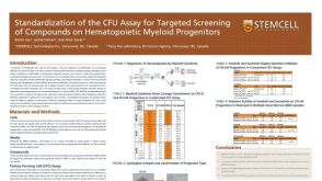 科学海报Standardization of the CFU Assay for Targeted Screening of Compounds on Hematopoietic Myeloid Progenitors
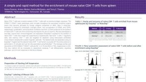 科学海报Cell Enrichment of Mouse Naïve CD4+ T Cells from Spleen
 沪公网安备31010102008431号