M. Koning et al. (Jul 2025)
NPJ Regenerative Medicine 10
Single cell transcriptomics of human kidney organoid endothelium reveals vessel growth processes and arterial maturation upon transplantation
Kidney organoids derived from human induced pluripotent stem cells lack a proper vasculature,hampering their applicability. Transplantation prevents the loss of organoid endothelial cells (ECs) observed in vitro,and promotes vascularization. In this study,we transplanted kidney organoids in chicken embryos and deployed single-cell RNA sequencing of ~12,000 organoid ECs to delineate their molecular landscape and identify key changes associated with transplantation. Transplantation significantly altered EC phenotypic composition. Consistent with angiogenesis,proliferating EC populations expanded 8 days after transplantation. Importantly,ECs underwent a major vein-to-arterial phenotypic shift. One of the transplantation-specific arterial EC populations,characterized by laminar shear stress response and Notch signalling,showed a similar transcriptome as human fetal kidney arterial/afferent arteriolar ECs. Consistently,transplantation-induced transcriptional changes involved proangiogenic and arteriogenic SOX7 transcription factor upregulation and regulon enrichment. These findings point to blood flow and candidate transcription factors such as SOX7 as possible targets to enhance kidney organoid vascularization. Subject terms: Nephrons,Transcriptomics,Angiogenesis,Angiogenesis,Stem cells,Stem-cell differentiation
View Publication
产品类型:
产品号#:
05270
05275
产品名:
STEMdiff™ APEL™2 培养基
STEMdiff™ APEL™2 培养基
Jaiswal RK et al. (MAR 2000)
The Journal of biological chemistry 275 13 9645--52
Adult human mesenchymal stem cell differentiation to the osteogenic or adipogenic lineage is regulated by mitogen-activated protein kinase.
Adult human mesenchymal stem cells are primary,multipotent cells capable of differentiating to osteocytic,chondrocytic,and adipocytic lineages when stimulated under appropriate conditions. To characterize the molecular mechanisms that regulate osteogenic differentiation,we examined the contribution of mitogen-activated protein kinase family members,ERK,JNK,and p38. Treatment of these stem cells with osteogenic supplements resulted in a sustained phase of ERK activation from day 7 to day 11 that coincided with differentiation,before decreasing to basal levels. Activation of JNK occurred much later (day 13 to day 17) in the osteogenic differentiation process. This JNK activation was associated with extracellular matrix synthesis and increased calcium deposition,the two hallmarks of bone formation. Inhibition of ERK activation by PD98059,a specific inhibitor of the ERK signaling pathway,blocked the osteogenic differentiation in a dose-dependent manner,as did transfection with a dominant negative form of MAP kinase kinase (MEK-1). Significantly,the blockage of osteogenic differentiation resulted in the adipogenic differentiation of the stem cells and the expression of adipose-specific mRNAs peroxisome proliferator-activated receptor gamma2,aP2,and lipoprotein lipase. These observations provide a potential mechanism involving MAP kinase activation in osteogenic differentiation of adult stem cells and suggest that commitment of hMSCs into osteogenic or adipogenic lineages is governed by activation or inhibition of ERK,respectively.
View Publication
产品类型:
产品号#:
72172
72174
产品名:
PD98059
PD98059
Awe JP et al. (NOV 2014)
Journal of visualized experiments : JoVE 93 e52158
Derivation and characterization of a transgene-free human induced pluripotent stem cell line and conversion into defined clinical-grade conditions.
Human induced pluripotent stem cells (hiPSCs) can be generated with lentiviral-based reprogramming methodologies. However,traces of potentially oncogenic genes remaining in actively transcribed regions of the genome,limit their potential for use in human therapeutic applications. Additionally,non-human antigens derived from stem cell reprogramming or differentiation into therapeutically relevant derivatives preclude these hiPSCs from being used in a human clinical context. In this video,we present a procedure for reprogramming and analyzing factor-free hiPSCs free of exogenous transgenes. These hiPSCs then can be analyzed for gene expression abnormalities in the specific intron containing the lentivirus. This analysis may be conducted using sensitive quantitative polymerase chain reaction (PCR),which has an advantage over less sensitive techniques previously used to detect gene expression differences. Full conversion into clinical-grade good manufacturing practice (GMP) conditions,allows human clinical relevance. Our protocol offers another methodology--provided that current safe-harbor criteria will expand and include factor-free characterized hiPSC-based derivatives for human therapeutic applications--for deriving GMP-grade hiPSCs,which should eliminate any immunogenicity risk due to non-human antigens. This protocol is broadly applicable to lentiviral reprogrammed cells of any type and provides a reproducible method for converting reprogrammed cells into GMP-grade conditions.
View Publication
产品类型:
产品号#:
05860
05880
05850
05857
05870
05875
85850
85857
85870
85875
产品名:
mTeSR™1
mTeSR™1
Kolle G et al. (OCT 2009)
Stem Cells 27 10 2446--56
Identification of human embryonic stem cell surface markers by combined membrane-polysome translation state array analysis and immunotranscriptional profiling.
Surface marker expression forms the basis for characterization and isolation of human embryonic stem cells (hESCs). Currently,there are few well-defined protein epitopes that definitively mark hESCs. Here we combine immunotranscriptional profiling of hESC lines with membrane-polysome translation state array analysis (TSAA) to determine the full set of genes encoding potential hESC surface marker proteins. Three independently isolated hESC lines (HES2,H9,and MEL1) grown under feeder and feeder-free conditions were sorted into subpopulations by fluorescence-activated cell sorting based on coimmunoreactivity to the hESC surface markers GCTM-2 and CD9. Colony-forming assays confirmed that cells displaying high coimmunoreactivity to GCTM-2 and CD9 constitute an enriched subpopulation displaying multiple stem cell properties. Following microarray profiling,820 genes were identified that were common to the GCTM-2(high)/CD9(high) stem cell-like subpopulation. Membrane-polysome TSAA analysis of hESCs identified 1,492 mRNAs encoding actively translated plasma membrane and secreted proteins. Combining these data sets,88 genes encode proteins that mark the pluripotent subpopulation,of which only four had been previously reported. Cell surface immunoreactivity was confirmed for two of these markers: TACSTD1/EPCAM and CDH3/P-Cadherin,with antibodies for EPCAM able to enrich for pluripotent hESCs. This comprehensive listing of both hESCs and spontaneous differentiation-associated transcripts and survey of translated membrane-bound and secreted proteins provides a valuable resource for future study into the role of the extracellular environment in both the maintenance of pluripotency and directed differentiation.
View Publication
产品类型:
产品号#:
05850
05857
05870
05875
85850
85857
85870
85875
产品名:
mTeSR™1
mTeSR™1
Ehnman M et al. (APR 2013)
Cancer Research 73 7 2139--2149
Distinct Effects of Ligand-Induced PDGFR and PDGFR Signaling in the Human Rhabdomyosarcoma Tumor Cell and Stroma Cell Compartments
Platelet-derived growth factor receptors (PDGFR) α and β have been suggested as potential targets for treatment of rhabdomyosarcoma,the most common soft tissue sarcoma in children. This study identifies biologic activities linked to PDGF signaling in rhabdomyosarcoma models and human sample collections. Analysis of gene expression profiles of 101 primary human rhabdomyosarcomas revealed elevated PDGF-C and -D expression in all subtypes,with PDGF-D as the solely overexpressed PDGFRβ ligand. By immunohistochemistry,PDGF-CC,PDGF-DD,and PDGFRα were found in tumor cells,whereas PDGFRβ was primarily detected in vascular stroma. These results are concordant with the biologic processes and pathways identified by data mining. While PDGF-CC/PDGFRα signaling associated with genes involved in the reactivation of developmental programs,PDGF-DD/PDGFRβ signaling related to wound healing and leukocyte differentiation. Clinicopathologic correlations further identified associations between PDGFRβ in vascular stroma and the alveolar subtype and with presence of metastases. Functional validation of our findings was carried out in molecularly distinct model systems,where therapeutic targeting reduced tumor burden in a PDGFR-dependent manner with effects on cell proliferation,vessel density,and macrophage infiltration. The PDGFR-selective inhibitor CP-673,451 regulated cell proliferation through mechanisms involving reduced phosphorylation of GSK-3α and GSK-3β. Additional tissue culture studies showed a PDGFR-dependent regulation of rhabdosphere formation/cancer cell stemness,differentiation,senescence,and apoptosis. In summary,the study shows a clinically relevant distinction in PDGF signaling in human rhabdomyosarcoma and also suggests continued exploration of the influence of stromal PDGFRs on sarcoma progression.
View Publication
产品类型:
产品号#:
05750
05751
产品名:
NeuroCult™ NS-A 基础培养基(人)
NeuroCult™ NS-A 扩增试剂盒(人)
Garcia-Bates TM et al. (MAR 2016)
Journal of immunology (Baltimore,Md. : 1950) 196 6 2870--8
Enhanced Cytotoxic CD8 T Cell Priming Using Dendritic Cell-Expressing Human Papillomavirus-16 E6/E7-p16INK4 Fusion Protein with Sequenced Anti-Programmed Death-1.
The incidence of human papillomavirus (HPV)-related head and neck squamous cell carcinoma has increased in recent decades,though HPV prevention vaccines may reduce this rise in the future. HPV-related cancers express the viral oncoproteins E6 and E7. The latter inactivates the tumor suppressor protein retinoblastoma (Rb),which leads to the overexpression of p16(INK4) protein,providing unique Ags for therapeutic HPV-specific cancer vaccination. We developed potential adenoviral vaccines that express a fusion protein of HPV-16 E6 and E7 (Ad.E6E7) alone or fused with p16 (Ad.E6E7p16) and also encoding an anti-programmed death (PD)-1 Ab. Human monocyte-derived dendritic cells (DC) transduced with Ad.E6E7 or Ad.E6E7p16 with or without Ad.αPD1 were used to activate autologous CD8 CTL in vitro. CTL responses were tested against naturally HPV-infected head and neck squamous cell carcinoma cells using IFN-γ ELISPOT and [(51)Cr]release assay. Surprisingly,stimulation and antitumor activity of CTL were increased after incubation with Ad.E6E7p16-transduced DC (DC.E6E7p16) compared with Ad.E6E7 (DC.E6E7),a result that may be due to an effect of p16 on cyclin-dependent kinase 4 levels and IL-12 secretion by DC. Moreover,the beneficial effect was most prominent when anti-PD-1 was introduced during the second round of stimulation (after initial priming). These data suggest that careful sequencing of Ad.E6E7.p16 with Ad.αPD1 could improve antitumor immunity against HPV-related tumors and that p16 may enhance the immunogenicity of DC,through cyclin-dependent pathways,Th1 cytokine secretion,and by adding a nonviral Ag highly overexpressed in HPV-induced cancers.
View Publication
产品类型:
产品号#:
18058
18058RF
19158
19158RF
19053
19053RF
产品名:
EasySep™人CD8+ T细胞富集试剂盒
RoboSep™ 人CD8+ T细胞富集试剂盒含滤芯吸头
Linta L et al. (APR 2013)
Stem Cells International 2013 784629
Microarray-Based Comparisons of Ion Channel Expression Patterns: Human Keratinocytes to Reprogrammed hiPSCs to Differentiated Neuronal and Cardiac Progeny
Ion channels are involved in a large variety of cellular processes including stem cell differentiation. Numerous families of ion channels are present in the organism which can be distinguished by means of,for example,ion selectivity,gating mechanism,composition,or cell biological function. To characterize the distinct expression of this group of ion channels we have compared the mRNA expression levels of ion channel genes between human keratinocyte-derived induced pluripotent stem cells (hiPSCs) and their somatic cell source,keratinocytes from plucked human hair. This comparison revealed that 26&x25; of the analyzed probes showed an upregulation of ion channels in hiPSCs while just 6&x25; were downregulated. Additionally,iPSCs express a much higher number of ion channels compared to keratinocytes. Further,to narrow down specificity of ion channel expression in iPS cells we compared their expression patterns with differentiated progeny,namely,neurons and cardiomyocytes derived from iPS cells. To conclude,hiPSCs exhibit a very considerable and diverse ion channel expression pattern. Their detailed analysis could give an insight into their contribution to many cellular processes and even disease mechanisms.
View Publication
产品类型:
产品号#:
05850
05857
05870
05875
85850
85857
85870
85875
产品名:
mTeSR™1
mTeSR™1
Lukovic D et al. (MAY 2017)
Stem cell research 21 23--25
Generation of a human iPSC line from a patient with retinitis pigmentosa caused by mutation in PRPF8 gene.
The human iPSC cell line,RP2-FiPS4F1 (RCPFi001-A),derived from dermal fibroblasts from the patient with retinitis pigmentosa caused by the mutation of the gene PRPF8,was generated by non-integrative reprogramming technology using OCT3/4,SOX2,CMYC and KLF4 reprogramming factors.
View Publication
产品类型:
产品号#:
05850
05857
05870
05875
85850
85857
85870
85875
产品名:
mTeSR™1
mTeSR™1
Momburg F et al. (JUN 1987)
Cancer research 47 11 2883--91
Immunohistochemical study of the expression of a Mr 34,000 human epithelium-specific surface glycoprotein in normal and malignant tissues.
Monoclonal antibody HEA125 was used to study the tissue distribution of an epithelial cell surface glycoprotein of Mr 34,000 (Egp34). A large panel of normal and neoplastic tissues was examined for immunoreactivity with HEA125 by means of a sensitive immunoperoxidase technique. HEA125 labeled most epithelial cell types throughout the body but did not label any nonepithelial tissue. Major exceptions were epidermal keratinocytes,gastric parietal cells,hepatocytes,thymic cortical epithelial,and myoepithelial cells. Normal mesothelial cells were unreactive. In normal glandular epithelia and tubular adenocarcinomas exclusively the basolateral cell membranes were stained. HEA125 intensely reacted with all tested carcinoma specimens derived from colorectum,stomach,pancreas,liver,lung,mammary gland,ovary,thyroid,kidney,urinary bladder,and prostate including a number of anaplastic,diffusely infiltrating carcinomas. Metastatic lesions of these tumors were consistently positive. Generally,the staining of tumor cells was very homogeneous. The majority of squamous cell carcinomas were less strongly labeled than adenocarcinomas; keratinizing areas of the tumor masses were negative. Germ cell tumors and mesotheliomas of epithelioid type focally expressed the antigen. Egp34 was found to be absent from sarcomas,lymphomas,melanomas,and neurogenic tumors. Hence,HEA125 is a useful reagent for the distinction of carcinomas from nonepithelial neoplasms,even at very low degrees of histological differentiation. Furthermore,HEA125 allows the immunohistochemical detection of micrometastases originating from carcinomas. The antigen is detectable in formalin-fixed paraffin sections.
View Publication
产品类型:
产品号#:
01420
01421
产品名:
Nikoli&cacute et al. ( 2017)
eLife 6 1--33
Human embryonic lung epithelial tips are multipotent progenitors that can be expanded in vitro as long-term self-renewing organoids
The embryonic mouse lung is a widely used substitute for human lung development. For example,attempts to differentiate human pluripotent stem cells to lung epithelium rely on passing through progenitor states that have only been described in mouse. The tip epithelium of the branching mouse lung is a multipotent progenitor pool that self-renews and produces differentiating descendants. We hypothesized that the human distal tip epithelium is an analogous progenitor population and tested this by examining morphology,gene expression and in vitro self-renewal and differentiation capacity of human tips. These experiments confirm that human and mouse tips are analogous and identify signalling pathways that are sufficient for long-term self-renewal of human tips as differentiation-competent organoids. Moreover,we identify mouse-human differences,including markers that define progenitor states and signalling requirements for long-term self-renewal. Our organoid system provides a genetically-tractable tool that will allow these human-specific features of lung development to be investigated.
View Publication
High-throughput robotic isolation of human iPS cell clones reveals frequent homozygous induction of identical genetic manipulations by CRISPR-Cas9
BackgroundGenome editing in human iPS cells is a powerful approach in regenerative medicine. CRISPR-Cas9 is the most common genome editing tool,but it often induces byproduct insertions and deletions in addition to the desired edits. Therefore,genome editing of iPS cells produces diverse genotypes. Existing assays mostly analyze genome editing results in cell populations,but not in single cells. However,systematic profiling of genome editing outcomes in single iPS cells was lacking. Due to the high mortality of human iPS cells as isolated single cells,it has been difficult to analyze genome-edited iPS cell clones in a high-throughput manner.MethodsIn this study,we developed a method for high-throughput iPS cell clone isolation based on the precise robotic picking of cell clumps derived from single cells grown in extracellular matrices. We first introduced point mutations into human iPS cell pools by CRISPR-Cas9. These genome-edited human iPS cells were dissociated and cultured as single cells in extracellular matrices to form cell clumps,which were then isolated using a cell-handling robot to establish genome-edited human iPS cell clones. Genome editing outcomes in these clones were analyzed by amplicon sequencing to determine the genotypes of individual iPS cell clones. We identified and distinguished the sequences of different insertions and deletions induced by CRISPR-Cas9 while determining their genotypes. We also cryopreserved the established iPS cell clones and recovered them after determining their genotypes.ResultsWe analyzed over 1,000 genome-edited iPS cell clones and found that homozygous editing was much more frequent than heterozygous editing. We also observed frequent homozygous induction of identical genetic manipulations,including insertions and deletions,such as 1-bp insertions and 8-bp deletions. Moreover,we successfully cryopreserved and then recovered genome-edited iPS cell clones,demonstrating that our cell-handling robot-based method is valuable in establishing genome-edited iPS cell clones.ConclusionsThis study revealed a previously unknown property of genome editing in human iPS cells that identical sequence manipulations tend to be induced in both copies of the target sequence in individual cells. Our new cloning method and findings will facilitate the application of genome editing to human iPS cells.Supplementary InformationThe online version contains supplementary material available at 10.1186/s13287-025-04414-2.
View Publication


 EasySep™小鼠TIL(CD45)正选试剂盒





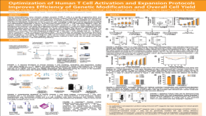 科学海报Optimization of Human T Cell Activation and Expansion Protocols Improves Efficiency of Genetic Modification and Overall Cell Yield
 沪公网安备31010102008431号