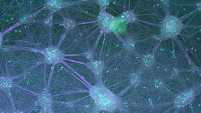
Characterization of enhancer activity in early human neurodevelopment using Massively Parallel Reporter Assay (MPRA) and forebrain organoids
Regulation of gene expression through enhancers is one of the major processes shaping the structure and function of the human brain during development. High-throughput assays have predicted thousands of enhancers involved in neurodevelopment,and confirming their activity through orthogonal functional assays is crucial. Here,we utilized Massively Parallel Reporter Assays (MPRAs) in stem cells and forebrain organoids to evaluate the activity of ~ 7000 gene-linked enhancers previously identified in human fetal tissues and brain organoids. We used a Gaussian mixture model to evaluate the contribution of background noise in the measured activity signal to confirm the activity of ~ 35% of the tested enhancers,with most showing temporal-specific activity,suggesting their evolving role in neurodevelopment. The temporal specificity was further supported by the correlation of activity with gene expression. Our findings provide a valuable gene regulatory resource to the scientific community.
View Publication
产品类型:
产品号#:
85850
85857
产品名:
mTeSR™1
mTeSR™1
(Jul 2025)
Frontiers in Pharmacology 16 3
Machine learning analysis of ARVC informed by sodium channel protein-based interactome networks
Arrhythmogenic right ventricular cardiomyopathy (ARVC) is an inherited cardiac disorder characterized by sodium channel dysfunction. However,the clinical management of ARVC remains challenging. Identifying novel compounds for the treatment of ARVC is crucial for advancing drug development.PurposeIn this study,we aim to identify novel compounds for treating ARVC.MethodsMachine learning (ML) models were constructed using proteins analyzed from the scRNA-seq data of ARVC rats and their corresponding protein-protein interaction (PPI) network to predict binding affinity (BA). To validate these predictions,a series of experiments in cardiac organoids were conducted,including Western blotting,ELISA,MEA,and Masson staining to assess the effects of these compounds.ResultsWe first discovered and identified SCN5A as the most significantly affected sodium channel protein in ARVC. ML models predicted that Kaempferol binds to SCN5A with high affinity. In vitro experiments further confirmed that Kaempferol exerted therapeutic effects in ARVC.ConclusionThis study presents a novel approach for identifying potential compounds to treat ARVC. By integrating ML modeling with organoid validation,our platform provides valuable support in addressing the public health challenges posed by ARVC,with broad application prospects. Kaempferol shows promise as a lead compound for ARVC treatment.
View Publication
产品类型:
产品号#:
100-0276
100-1130
产品名:
mTeSR™ Plus
mTeSR™ Plus
(Jul 2025)
bioRxiv 5 27
Robust Production of Parvalbumin Interneurons and Fast-Spiking Neurons from Human Medial Ganglionic Eminence Organoids
SummaryThe medial ganglionic eminence (MGE) gives rise to parvalbumin (PV)- and somatostatin (SST)-expressing cortical interneurons essential for regulating cortical excitability. Although PV interneurons are linked to various neurodevelopmental and neurodegenerative disorders,reliably generating them from human pluripotent stem cells (hPSCs) has been extremely challenging. We present a robust,reproducible protocol for generating single-rosette MGE organoids (MGEOs) from hPSCs. Transcriptomic analyses reveal that MGEOs exhibit MGE regional identity and faithfully model the developing human fetal MGE. As MGEOs mature,they generate abundant PV-expressing cortical interneurons,including putative basket and axoaxonic cells,at a scale not previously achieved in vitro. When fused with hPSC-derived cortical organoids,these interneurons rapidly migrate into cortical regions,integrate into excitatory networks,and contribute to complex electrophysiological patterns and the emergence of large numbers of fast-spiking neurons. MGEOs thus offer a powerful in vitro approach for probing human MGE-lineage cortical and subcortical GABAergic neuron development,modeling various neuropsychiatric disorders,and advancing cell-based therapies for neurodevelopmental and neurodegenerative disorders. Graphical abstract
View Publication
产品类型:
产品号#:
100-0276
100-1130
85850
85857
产品名:
mTeSR™ Plus
mTeSR™ Plus
mTeSR™1
mTeSR™1
(May 2024)
Nature Communications 15
mTORC1 regulates cell survival under glucose starvation through 4EBP1/2-mediated translational reprogramming of fatty acid metabolism
Energetic stress compels cells to evolve adaptive mechanisms to adjust their metabolism. Inhibition of mTOR kinase complex 1 (mTORC1) is essential for cell survival during glucose starvation. How mTORC1 controls cell viability during glucose starvation is not well understood. Here we show that the mTORC1 effectors eukaryotic initiation factor 4E binding proteins 1/2 (4EBP1/2) confer protection to mammalian cells and budding yeast under glucose starvation. Mechanistically,4EBP1/2 promote NADPH homeostasis by preventing NADPH-consuming fatty acid synthesis via translational repression of Acetyl-CoA Carboxylase 1 (ACC1),thereby mitigating oxidative stress. This has important relevance for cancer,as oncogene-transformed cells and glioma cells exploit the 4EBP1/2 regulation of ACC1 expression and redox balance to combat energetic stress,thereby supporting transformation and tumorigenicity in vitro and in vivo. Clinically,high EIF4EBP1 expression is associated with poor outcomes in several cancer types. Our data reveal that the mTORC1-4EBP1/2 axis provokes a metabolic switch essential for survival during glucose starvation which is exploited by transformed and tumor cells. How cells adapt to glucose starvation is still elusive. Here,Levy et al. show that the mTOR substrate 4EBP1 protects human,mouse,and yeast cells from glucose starvation and is exploited by cancer cells to promote tumorigenesis.
View Publication
产品类型:
产品号#:
85850
85857
产品名:
mTeSR™1
mTeSR™1
(Feb 2025)
Nucleic Acids Research 53 4
NEAT1-mediated regulation of proteostasis and mRNA localization impacts autophagy dysregulation in Rett syndrome
Rett syndrome (RTT) is a severe neurodevelopmental disorder primarily caused by loss-of-function mutations in the MECP2 gene,resulting in diverse cellular dysfunctions. Here,we investigated the role of the long noncoding RNA (lncRNA) NEAT1 in the context of MeCP2 deficiency using human neural cells and RTT patient samples. Through single-cell RNA sequencing and molecular analyses,we found that NEAT1 is markedly downregulated in MECP2 knockout (KO) cells at various stages of neural differentiation. NEAT1 downregulation correlated with aberrant activation of the mTOR pathway,abnormal protein metabolism,and dysregulated autophagy,contributing to the accumulation of protein aggregates and impaired mitochondrial function. Reactivation of NEAT1 in MECP2-KO cells rescued these phenotypes,indicating its critical role downstream of MECP2. Furthermore,direct RNA–RNA interaction was revealed as the key process for NEAT1 influence on autophagy genes,leading to altered subcellular localization of specific autophagy-related messenger RNAs and impaired biogenesis of autophagic complexes. Importantly,NEAT1 restoration rescued the morphological defects observed in MECP2-KO neurons,highlighting its crucial role in neuronal maturation. Overall,our findings elucidate lncRNA NEAT1 as a key mediator of MeCP2 function,regulating essential pathways involved in protein metabolism,autophagy,and neuronal morphology.
View Publication
产品类型:
产品号#:
08581
08582
85850
85857
产品名:
STEMdiff™SMADi神经诱导试剂盒
STEMdiff™SMADi神经诱导试剂盒,2套
mTeSR™1
mTeSR™1
(Jul 2025)
Cell Communication and Signaling : CCS 23 23
BackgroundTrichloroethylene (TCE) is a ubiquitous pollutant with potential capacity to induce congenital heart disease (CHD). However,the mechanisms underlying TCE-induced CHD are largely unraveled.MethodsWe exposed zebrafish embryos to TCE to investigate its cardiac development toxicity and related response factor through bulk RNA sequencing. We constructed transgenic fluorescent fish and employed the CRISPR/dCas9 system along with single-cell RNA sequencing to identify the genetic cause of TCE-induced CHD.ResultsWe found that early-stage exposure to TCE induced significant cardiac defects characterized by elongated SV-BA distance,thinned myocardium,and attenuated contractility. Gremlin1 encoding gene,grem1a,a putative target showing high expression at the beginning of cardiac development,was sharply down-regulated by TCE. Consistently,grem1a knockdown in zebrafish induced cardiac phenotypes generally like those of the TCE-treated group,accompanying the disarrangement of myofibril structure. Single-cell RNA-seq depicted that mitochondrial respiration in grem1a-repressed cardiomyocytes was greatly enhanced,ultimately leading to a branch from the normal trajectory of myocardial development. Accordingly,in vitro results demonstrated that GREM1 repression increased mitochondrial content,ATP production,mitochondrial reactive oxygen species,mitochondrial membrane potential,and disrupted myofibril expansion in hPSC-CMs.ConclusionsThese results suggested that TCE-induced gremlin1 repression could result in mitochondrial hyperfunction,thereby hampering cardiomyocyte development and causing cardiac defects in zebrafish embryos. This study not only provided a novel insight into the etiology for environmental stressor-caused cardiac development defects,but also offered a potential therapeutic and preventive target for TCE-induced CHD.Supplementary InformationThe online version contains supplementary material available at 10.1186/s12964-025-02314-9.
View Publication
产品类型:
产品号#:
05010
100-0483
100-0484
100-0276
100-1130
产品名:
STEMdiff™ 心室肌细胞分化试剂盒
Hausser Scientificᵀᴹ 明线血球计数板
ReLeSR™
mTeSR™ Plus
mTeSR™ Plus
L. Fang et al. (JUL 2018)
Cancer cell 34 1 103--118.e9
SET1A-Mediated Mono-Methylation at K342 Regulates YAP Activation by Blocking Its Nuclear Export and Promotes Tumorigenesis.
YAP,a key effector of Hippo pathway,is activated by its translocation from cytoplasm to nucleus to regulate gene expression and promote tumorigenesis. Although the mechanism by which YAP is suppressed in cytoplasm has been well-studied,how the activated YAP is sequestered in the nucleus remains unknown. Here,we demonstrate that YAP is a nucleocytoplasmic shuttling protein and its nuclear export is controlled by SET1A-mediated mono-methylation of YAP at K342,which disrupts the binding of YAP to CRM1. YAP mimetic methylation knockin mice are more susceptible to colorectal tumorigenesis. Clinically,YAP K342 methylation is reversely correlated with cancer survival. Collectively,our study identifies SET1A-mediated mono-methylation at K342 as an essential regulatory mechanism for regulating YAP activity and tumorigenesis.
View Publication
产品类型:
产品号#:
06005
产品名:
IntestiCult™ 类器官生长培养基 (小鼠)
S. Belluschi et al. ( 2018)
Nature communications 9 1 4100
Myelo-lymphoid lineage restriction occurs in the human haematopoietic stem cell compartment before lymphoid-primed multipotent progenitors.
Capturing where and how multipotency is lost is crucial to understand how blood formation is controlled. Blood lineage specification is currently thought to occur downstream of multipotent haematopoietic stem cells (HSC). Here we show that,in human,the first lineage restriction events occur within the CD19-CD34+CD38-CD45RA-CD49f+CD90+ (49f+) HSC compartment to generate myelo-lymphoid committed cells with no erythroid differentiation capacity. At single-cell resolution,we observe a continuous but polarised organisation of the 49f+ compartment,where transcriptional programmes and lineage potential progressively change along a gradient of opposing cell surface expression of CLEC9A and CD34. CLEC9AhiCD34lo cells contain long-term repopulating multipotent HSCs with slow quiescence exit kinetics,whereas CLEC9AloCD34hi cells are restricted to myelo-lymphoid differentiation and display infrequent but durable repopulation capacity. We thus propose that human HSCs gradually transition to a discrete lymphoid-primed state,distinct from lymphoid-primed multipotent progenitors,representing the earliest entry point into lymphoid commitment.
View Publication
产品类型:
产品号#:
22001
22005
22006
22007
22008
22009
22011
22012
22013
产品名:
STEMvision™ 人脐带血7-天CFU分析包
STEMvision™人脐带血14天CFU分析套装
STEMvision™人骨髓14天CFU分析套装
STEMvision™人动员外周血14天CFU分析套装
STEMvision™ 小鼠总CFU分析包
STEMvision™ 小鼠髓系CFU分析包
STEMvision™ 小鼠红系CFU分析包
STEMvision™小鼠CFU分析套装组合
B. Fregin et al. ( 2019)
Nature communications 10 1 415
High-throughput single-cell rheology in complex samples by dynamic real-time deformability cytometry.
In life sciences,the material properties of suspended cells have attained significance close to that of fluorescent markers but with the advantage of label-free and unbiased sample characterization. Until recently,cell rheological measurements were either limited by acquisition throughput,excessive post processing,or low-throughput real-time analysis. Real-time deformability cytometry expanded the application of mechanical cell assays to fast on-the-fly phenotyping of large sample sizes,but has been restricted to single material parameters as the Young's modulus. Here,we introduce dynamic real-time deformability cytometry for comprehensive cell rheological measurements at up to 100 cells per second. Utilizing Fourier decomposition,our microfluidic method is able to disentangle cell response to complex hydrodynamic stress distributions and to determine viscoelastic parameters independent of cell shape. We demonstrate the application of our technology for peripheral blood cells in whole blood samples including the discrimination of B- and CD4+ T-lymphocytes by cell rheological properties.
View Publication
产品类型:
产品号#:
19157
19157RF
19659
产品名:
EasySep™人记忆CD4+ T细胞富集试剂盒
RoboSep™ 人记忆CD4 T细胞富集试剂盒含滤芯吸头
EasySep™ Direct人Pan-粒细胞分选试剂盒
O. M. Omar et al. (nov 2018)
Molecular carcinogenesis 57 11 1577--1587
TFF1 antagonizes TIMP-1 mediated proliferative functions in gastric cancer.
Tissue inhibitor matrix metalloproteinase-1 (TIMP1) is one of four identified members of the TIMP family. We evaluated the role of TIMP1 in gastric cancer using human and mouse tissues along with gastric organoids and in vitro cell models. Using quantitative real-time RT-PCR,we detected significant overexpression of TIMP1 in the human gastric cancer samples,as compared to normal stomach samples (P {\textless} 0.01). We also detected overexpression of Timp1 in neoplastic gastric lesions of the Tff1-knockout (KO) mice,as compared to normal stomach tissues. Reconstitution of TFF1 in human gastric cancer cell lines led to a significant decrease in the mRNA expression level of TIMP1 (P {\textless} 0.05). In vitro analysis demonstrated that TIMP1 mRNA expression is induced by TNF-alpha and activation of NF-kappaB whereas inhibition of NF-kappaB using BAY11-7082 led to inhibition of NF-kappaB and downregulation of TIMP1. Western blot analysis confirmed the decrease in TIMP1 protein level following reconstitution of TFF1. By using immunofluorescence,we showed nuclear localization of NF-kappaB and expression of TIMP1 in gastric organoids established from the Tff1-KO stomach where reconstitution of Tff1 using recombinant protein led to a notable reduction in the expression of both NF-kappaB and TIMP1. Using EDU assay,as a measure of proliferating cells,we found that TIMP1 promotes cellular proliferation whereas TFF1 reconstitution leads to a significant decrease in cellular proliferation (P {\textless} 0.05). In summary,our findings demonstrate overexpression of TIMP1 in mouse and human gastric cancers through NF-kB-dependent mechanism. We also show that TFF1 suppresses NF-kappaB and inhibits TIMP1-mediated proliferative potential in gastric cancer.
View Publication
产品类型:
产品号#:
06005
产品名:
IntestiCult™ 类器官生长培养基 (小鼠)
K. E. Sivick et al. (dec 2018)
Cell reports 25 11 3074--3085.e5
Magnitude of Therapeutic STING Activation Determines CD8+ T Cell-Mediated Anti-tumor Immunity.
Intratumoral (IT) STING activation results in tumor regression in preclinical models,yet factors dictating the balance between innate and adaptive anti-tumor immunity are unclear. Here,clinical candidate STING agonist ADU-S100 (S100) is used in an IT dosing regimen optimized for adaptive immunity to uncover requirements for a T cell-driven response compatible with checkpoint inhibitors (CPIs). In contrast to high-dose tumor ablative regimens that result in systemic S100 distribution,low-dose immunogenic regimens induce local activation of tumor-specific CD8+ effector T cells that are responsible for durable anti-tumor immunity and can be enhanced with CPIs. Both hematopoietic cell STING expression and signaling through IFNAR are required for tumor-specific T cell activation,and in the context of optimized T cell responses,TNFalpha is dispensable for tumor control. In a poorly immunogenic model,S100 combined with CPIs generates a survival benefit and durable protection. These results provide fundamental mechanistic insights into STING-induced anti-tumor immunity.
View Publication

 EasySep™小鼠TIL(CD45)正选试剂盒


 沪公网安备31010102008431号