R. Listro et al. (Dec 2025)
International Journal of Molecular Sciences 27 1
HuR-Targeted Small Molecules Reduce Pseudomonas aeruginosa Adhesion in Cystic Fibrosis Airway Epithelial Cells
Antibiotic-resistant infections remain a major challenge in cystic fibrosis (CF),where chronic Pseudomonas aeruginosa colonization drives lung infection. The overexpression of adhesion-related proteins and extracellular matrix components,including fibronectin (Fn),facilitates bacterial colonization. Recent evidence identifies the RNA-binding protein Human Antigen R (HuR) as a key regulator of this process,as it stabilizes Vav3 mRNA,promoting Fn deposition and the formation of bacterial docking platforms. Here,we report the synthesis,optimization,and functional evaluation of the HuR-targeted small-molecule (2S,3S)-BOPC1. Functional assays in CF human airway epithelial cells demonstrated that (2S,3S)-BOPC1 significantly reduced P. aeruginosa adhesion in a dose-dependent manner without detectable cytotoxic effects. These findings provide the first evidence that targeting HuR can disrupt the HuR–Vav3–Fn axis,reducing bacterial attachment. This host-directed approach represents a promising strategy to prevent chronic infections in CF without promoting antibiotic resistance.
View Publication
产品类型:
产品号#:
05001
05021
05022
产品名:
PneumaCult™-ALI 培养基
PneumaCult™-ALI 培养基含12 mm Transwell®插件
PneumaCult™-ALI 培养基含6.5 mm Transwell®插件
S. Pisani et al. (mar 2020)
International journal of molecular sciences 21 5
Tissue Engineered Esophageal Patch by Mesenchymal Stromal Cells: Optimization of Electrospun Patch Engineering.
Aim of work was to locate a simple,reproducible protocol for uniform seeding and optimal cellularization of biodegradable patch minimizing the risk of structural damages of patch and its contamination in long-term culture. Two seeding procedures are exploited,namely static seeding procedures on biodegradable and biocompatible patches incubated as free floating (floating conditions) or supported by CellCrownTM insert (fixed conditions) and engineered by porcine bone marrow MSCs (p-MSCs). Scaffold prototypes having specific structural features with regard to pore size,pore orientation,porosity,and pore distribution were produced using two different techniques,such as temperature-induced precipitation method and electrospinning technology. The investigation on different prototypes allowed achieving several implementations in terms of cell distribution uniformity,seeding efficiency,and cellularization timing. The cell seeding protocol in stating conditions demonstrated to be the most suitable method,as these conditions successfully improved the cellularization of polymeric patches. Furthermore,the investigation provided interesting information on patches' stability in physiological simulating experimental conditions. Considering the in vitro results,it can be stated that the in vitro protocol proposed for patches cellularization is suitable to achieve homogeneous and complete cellularizations of patch. Moreover,the protocol turned out to be simple,repeatable,and reproducible.
View Publication
产品类型:
产品号#:
05402
产品名:
MesenCult™ MSC刺激添加物(人)
S. Song et al. (aug 2014)
Cancer research 74 15 4170--82
Hippo coactivator YAP1 upregulates SOX9 and endows esophageal cancer cells with stem-like properties.
Cancer stem cells (CSC) are purported to initiate and maintain tumor growth. Deregulation of normal stem cell signaling may lead to the generation of CSCs; however,the molecular determinants of this process remain poorly understood. Here we show that the transcriptional coactivator YAP1 is a major determinant of CSC properties in nontransformed cells and in esophageal cancer cells by direct upregulation of SOX9. YAP1 regulates the transcription of SOX9 through a conserved TEAD binding site in the SOX9 promoter. Expression of exogenous YAP1 in vitro or inhibition of its upstream negative regulators in vivo results in elevated SOX9 expression accompanied by the acquisition of CSC properties. Conversely,shRNA-mediated knockdown of YAP1 or SOX9 in transformed cells attenuates CSC phenotypes in vitro and tumorigenicity in vivo. The small-molecule inhibitor of YAP1,verteporfin,significantly blocks CSC properties in cells with high YAP1 and a high proportion of ALDH1(+). Our findings identify YAP1-driven SOX9 expression as a critical event in the acquisition of CSC properties,suggesting that YAP1 inhibition may offer an effective means of therapeutically targeting the CSC population.
View Publication
产品类型:
产品号#:
产品名:
S. Wang et al. ( 2020)
Scientific reports 10 1 12226
Label-free detection of rare circulating tumor cells by image analysis and machine learning.
Detection and characterization of rare circulating tumor cells (CTCs) in patients' blood is important for the diagnosis and monitoring of cancer. The traditional way of counting CTCs via fluorescent images requires a series of tedious experimental procedures and often impacts the viability of cells. Here we present a method for label-free detection of CTCs from patient blood samples,by taking advantage of data analysis of bright field microscopy images. The approach uses the convolutional neural network,a powerful image classification and machine learning algorithm to perform label-free classification of cells detected in microscopic images of patient blood samples containing white blood cells and CTCs. It requires minimal data pre-processing and has an easy experimental setup. Through our experiments,we show that our method can achieve high accuracy on the identification of rare CTCs without the need for advanced devices or expert users,thus providing a faster and simpler way for counting and identifying CTCs. With more data becoming available in the future,the machine learning model can be further improved and can serve as an accurate and easy-to-use tool for CTC analysis.
View Publication
E. Lucchinetti et al. (dec 2022)
The American journal of clinical nutrition 116 6 1805--1819
Novel lipid emulsion for total parenteral nutrition based on 18-carbon n-3 fatty acids elicits a superior immunometabolic phenotype in a murine model compared with standard lipid emulsions.
BACKGROUND While lipid emulsions in modern formulations for total parenteral nutrition (TPN) provide essential fatty acids and dense calories,they also promote inflammation and immunometabolic disruptions. OBJECTIVES We aimed to develop a novel lipid emulsion for TPN use with superior immunometabolic actions compared with available standard lipid emulsions. METHODS A novel lipid emulsion [Vegaven (VV)] containing 30% of 18-carbon n-3 fatty acids ($\alpha$-linolenic acid and stearidonic acid) was developed for TPN (VV-TPN) and compared with TPN containing soybean oil-based lipid emulsion (IL-TPN) and fish-oil-based lipid emulsion (OV-TPN). In vivo studies were performed in instrumented male C57BL/6 mice subjected to 7-d TPN prior to analysis of cytokines,indices of whole-body and hepatic glucose metabolism,immune cells,lipid mediators,and mucosal bowel microbiome. RESULTS IL-6 to IL-10 ratios were significantly lower in liver and skeletal muscle of VV-TPN mice when compared with IL-TPN or OV-TPN mice. VV-TPN and OV-TPN each increased hepatic insulin receptor abundance and resulted in similar HOMA-IR values,whereas only VV-TPN increased hepatic insulin receptor substrate 2 and maintained normal hepatic glycogen content,effects that were IL-10-dependent and mediated by glucokinase activation. The percentages of IFN-$\gamma$- and IL-17-expressing CD4+ T cells were increased in livers of VV-TPN mice,and liver macrophages exhibited primed phenotypes when compared with IL-TPN. This immunomodulation was associated with successful elimination of the microinvasive bacterium Akkermansia muciniphila from the bowel mucosa by VV-TPN as opposed to standard lipid emulsions. Assay of hepatic lipid mediators revealed a distinct profile with VV-TPN,including increases in 9(S)-hydroxy-octadecatrienoic acid. When co-administered with IL-TPN,hydroxy-octadecatrienoic acids mimicked the VV-TPN immunometabolic phenotype. CONCLUSIONS We here report the unique anti-inflammatory,insulin-sensitizing,and immunity-enhancing properties of a newly developed lipid emulsion designed for TPN use based on 18-carbon n-3 fatty acids.
View Publication
Sharifi K et al. (DEC 2013)
Cell and Tissue Research 354 3 683--695
Differential expression and regulatory roles of FABP5 and FABP7 in oligodendrocyte lineage cells
Fatty-acid-binding proteins (FABPs) are key intracellular molecules involved in the uptake,transportation and storage of fatty acids and in the mediation of signal transduction and gene transcription. However,little is known regarding their expression and function in the oligodendrocyte lineage. We evaluate the in vivo and in vitro expression of FABP5 and FABP7 in oligodendrocyte lineage cells in the cortex and corpus callosum of adult mice,mixed cortical culture and oligosphere culture by immunofluorescent counter-staining with major oligodendrocyte lineage markers. In all settings,FABP7 expression was detected in NG2(+)/PDGFRα(+) oligodendrocyte progenitor cells (OPCs) that did not express FABP5. FABP5 was detected in mature CC1(+)/MBP(+) oligodendrocytes that did not express FABP7. Analysis of cultured OPCs showed a significant decrease in the population of FABP7-knockout (KO) OPCs and their BrdU uptake compared with wild-type (WT) OPCs. Upon incubation of OPCs in oligodendrocyte differentiation medium,a significantly lower percentage of FABP7-KO OPCs differentiated into O4(+) oligodendrocytes. The percentage of mature MBP(+) oligodendrocytes relative to whole O4(+)/MBP(+) oligodendrocytes was significantly lower in FABP7-KO and FABP5-KO than in WT cell populations. The percentage of terminally mature oligodendrocytes with membrane sheet morphology was significantly lower in FABP5-KO compared with WT cell populations. Thus,FABP7 and FABP5 are differentially expressed in oligodendrocyte lineage cells and regulate their proliferation and/or differentiation. Our findings suggest the involvement of FABP7 and FABP5 in the pathophysiology of demyelinating disorders,neuropsychiatric disorder and glioma,conditions in which OPCs/oligodendrocytes play central roles.
View Publication
产品类型:
产品号#:
05707
产品名:
NeuroCult™化学解离试剂盒(小鼠)
Wei W et al. (APR 2013)
Proceedings of the National Academy of Sciences 110 15 E1352--E1360
Hypoxia induces a phase transition within a kinase signaling network in cancer cells
Hypoxia is a near-universal feature of cancer,promoting glycolysis,cellular proliferation,and angiogenesis. The molecular mechanisms of hypoxic signaling have been intensively studied,but the impact of changes in oxygen partial pressure (pO2) on the state of signaling networks is less clear. In a glioblastoma multiforme (GBM) cancer cell model,we examined the response of signaling networks to targeted pathway inhibition between 21% and 1% pO2. We used a microchip technology that facilitates quantification of a panel of functional proteins from statistical numbers of single cells. We find that near 1.5% pO2,the signaling network associated with mammalian target of rapamycin (mTOR) complex 1 (mTORC1)--a critical component of hypoxic signaling and a compelling cancer drug target--is deregulated in a manner such that it will be unresponsive to mTOR kinase inhibitors near 1.5% pO2,but will respond at higher or lower pO2 values. These predictions were validated through experiments on bulk GBM cell line cultures and on neurosphere cultures of a human-origin GBM xenograft tumor. We attempt to understand this behavior through the use of a quantitative version of Le Chatelier's principle,as well as through a steady-state kinetic model of protein interactions,both of which indicate that hypoxia can influence mTORC1 signaling as a switch. The Le Chatelier approach also indicates that this switch may be thought of as a type of phase transition. Our analysis indicates that certain biologically complex cell behaviors may be understood using fundamental,thermodynamics-motivated principles.
View Publication
产品类型:
产品号#:
05761
产品名:
用于小鼠和大鼠神经干细胞和祖细胞分化培养的试剂盒
Beer PA et al. (JAN 2015)
Blood 125 3 504--15
Disruption of IKAROS activity in primitive chronic-phase CML cells mimics myeloid disease progression.
Without effective therapy,chronic-phase chronic myeloid leukemia (CP-CML) evolves into an acute leukemia (blast crisis [BC]) that displays either myeloid or B-lymphoid characteristics. This transition is often preceded by a clinically recognized,but biologically poorly characterized,accelerated phase (AP). Here,we report that IKAROS protein is absent or reduced in bone marrow blasts from most CML patients with advanced myeloid disease (AP or BC). This contrasts with primitive CP-CML cells and BCR-ABL1-negative acute myeloid leukemia blasts,which express readily detectable IKAROS. To investigate whether loss of IKAROS contributes to myeloid disease progression in CP-CML,we examined the effects of forced expression of a dominant-negative isoform of IKAROS (IK6) in CP-CML patients' CD34(+) cells. We confirmed that IK6 disrupts IKAROS activity in transduced CP-CML cells and showed that it confers on them features of AP-CML,including a prolonged increased output in vitro and in xenografted mice of primitive cells with an enhanced ability to differentiate into basophils. Expression of IK6 in CD34(+) CP-CML cells also led to activation of signal transducer and activator of transcription 5 and transcriptional repression of its negative regulators. These findings implicate loss of IKAROS as a frequent step and potential diagnostic harbinger of progressive myeloid disease in CML patients.
View Publication
产品类型:
产品号#:
18056
18056RF
产品名:
Ware CB et al. (APR 2009)
Cell stem cell 4 4 359--69
Histone deacetylase inhibition elicits an evolutionarily conserved self-renewal program in embryonic stem cells.
Recent evidence indicates that mouse and human embryonic stem cells (ESCs) are fixed at different developmental stages,with the former positioned earlier. We show that a narrow concentration of the naturally occurring short-chain fatty acid,sodium butyrate,supports the extensive self-renewal of mouse and human ESCs,while promoting their convergence toward an intermediate stem cell state. In response to butyrate,human ESCs regress to an earlier developmental stage characterized by a gene expression profile resembling that of mouse ESCs,preventing precocious Xist expression while retaining the ability to form complex teratomas in vivo. Other histone deacetylase inhibitors (HDACi) also support human ESC self-renewal. Our results indicate that HDACi can promote ESC self-renewal across species,and demonstrate that ESCs can toggle between alternative states in response to environmental factors.
View Publication

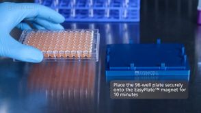
 EasySep™小鼠TIL(CD45)正选试剂盒


 沪公网安备31010102008431号