Rawat VPS et al. (SEP 2010)
Proceedings of the National Academy of Sciences of the United States of America 107 39 16946--51
The vent-like homeobox gene VENTX promotes human myeloid differentiation and is highly expressed in acute myeloid leukemia.
Recent data indicate that a variety of regulatory molecules active in embryonic development may also play a role in the regulation of early hematopoiesis. Here we report that the human Vent-like homeobox gene VENTX,a putative homolog of the Xenopus xvent2 gene,is a unique regulatory hematopoietic gene that is aberrantly expressed in CD34(+) leukemic stem-cell candidates in human acute myeloid leukemia (AML). Quantitative RT-PCR documented expression of the gene in lineage positive hematopoietic subpopulations,with the highest expression in CD33(+) myeloid cells. Notably,expression levels of VENTX were negligible in normal CD34(+)/CD38(-) or CD34(+) human progenitor cells. In contrast to this,leukemic CD34(+)/CD38(-) cells from AML patients with translocation t(8,21) and normal karyotype displayed aberrantly high expression of VENTX. Gene expression and pathway analysis demonstrated that in normal CD34(+) cells enforced expression of VENTX initiates genes associated with myeloid development and down-regulates genes involved in early lymphoid development. Functional analyses confirmed that aberrant expression of VENTX in normal CD34(+) human progenitor cells perturbs normal hematopoietic development,promoting generation of myeloid cells and impairing generation of lymphoid cells in vitro and in vivo. Stable knockdown of VENTX expression inhibited the proliferation of human AML cell lines. Taken together,these data extend our insights into the function of embryonic mesodermal factors in human postnatal hematopoiesis and indicate a role for VENTX in normal and malignant myelopoiesis.
View Publication
产品类型:
产品号#:
04434
04444
产品名:
MethoCult™ H4434 Classic
MethoCult™ H4434 Classic
Kozikowski AP et al. (AUG 2008)
Journal of medicinal chemistry 51 15 4370--3
Use of the nitrile oxide cycloaddition (NOC) reaction for molecular probe generation: a new class of enzyme selective histone deacetylase inhibitors (HDACIs) showing picomolar activity at HDAC6.
A series of hydroxamate based HDAC inhibitors containing a phenylisoxazole as the CAP group has been synthesized using nitrile oxide cycloaddition chemistry. An HDAC6 selective inhibitor having a potency of approximately 2 picomolar was identified. Some of the compounds were examined for their ability to block pancreatic cancer cell growth and found to be about 10-fold more potent than SAHA. This research provides valuable,new molecular probes for use in exploring HDAC biology.
View Publication
Roybal KT et al. (SEP 2016)
Cell 167 2 419--432.e16
Engineering T Cells with Customized Therapeutic Response Programs Using Synthetic Notch Receptors
Redirecting T cells to attack cancer using engineered chimeric receptors provides powerful new therapeutic capabilities. However,the effectiveness of therapeutic T cells is constrained by the endogenous T cell response: certain facets of natural response programs can be toxic,whereas other responses,such as the ability to overcome tumor immunosuppression,are absent. Thus,the efficacy and safety of therapeutic cells could be improved if we could custom sculpt immune cell responses. Synthetic Notch (synNotch) receptors induce transcriptional activation in response to recognition of user-specified antigens. We show that synNotch receptors can be used to sculpt custom response programs in primary T cells: they can drive a la carte cytokine secretion profiles,biased T cell differentiation,and local delivery of non-native therapeutic payloads,such as antibodies,in response to antigen. SynNotch T cells can thus be used as a general platform to recognize and remodel local microenvironments associated with diverse diseases.
View Publication
产品类型:
产品号#:
15022
15062
15023
15063
产品名:
RosetteSep™人CD4+ T细胞富集抗体混合物
RosetteSep™人CD4+ T细胞富集抗体混合物
RosetteSep™人CD8+ T细胞富集抗体混合物
RosetteSep™人CD8+ T细胞富集抗体混合物
Raju R et al. (FEB 2017)
Stem cells and development 26 4 274--284
Cell Expansion During Directed Differentiation of Stem Cells Toward the Hepatic Lineage.
The differentiation of human pluripotent stem cells toward the hepatocyte lineage can potentially provide an unlimited source of functional hepatocytes for transplantation and extracorporeal bioartificial liver applications. It is anticipated that the quantities of cells needed for these applications will be in the order of 10(9)-10(10) cells,because of the size of the liver. An ideal differentiation protocol would be to enable directed differentiation to the hepatocyte lineage with simultaneous cell expansion. We introduced a cell expansion stage after the commitment of human embryonic stem cells to the endodermal lineage,to allow for at least an eightfold increase in cell number,with continuation of cell maturation toward the hepatocyte lineage. The progressive changes in the transcriptome were measured by expression array,and the expression dynamics of certain lineage markers was measured by mass cytometry during the differentiation and expansion process. The findings revealed that while cells were expanding they were also capable of progressing in their differentiation toward the hepatocyte lineage. In addition,our transcriptome,protein and functional studies,including albumin secretion,drug-induced CYP450 expression and urea production,all indicated that the hepatocyte-like cells obtained with or without cell expansion are very similar. This method of simultaneous cell expansion and hepatocyte differentiation should facilitate obtaining large quantities of cells for liver cell applications.
View Publication
Effects of sodium butyrate, a new pharmacological agent, on cells in culture.
Sodium butyrate,at millimolar concentrations,when added to cell cultures produces many morphological and biochemical modifications in a reversible manner. Some of them occur in all cell lines. They concern regulatory mechanisms of gene expression and cell growth: an hyperacetylation of histone resulting from an inhibition of histone deacetylase and an arrest of cell proliferation are almost constantly observed. Some other modifications vary from one cell type to another: induction of proteins,including enzymes,hormones,hemoglobin,inhibition of cell differentiation,reversion of transformed characteristics of cells to normal morphological and biochemical pattern,increase in interferon antiviral efficiency and induction of integrated viruses. Most if not all these effects of butyrate could result from histone hyperacetylation,from changes in chromatin structures as measured by accessibility to DNases and from modifications in cytoskeleton assembly. We do not know at the present time whether butyrate acts on a very specific target site in cell or if it acts on several cell components.
View Publication
产品类型:
产品号#:
72242
产品名:
丁酸钠(Sodium Butyrate)
Simõ et al. (AUG 2011)
Breast cancer research and treatment 129 1 23--35
Effects of estrogen on the proportion of stem cells in the breast.
There is increasing evidence that breast cancers contain tumor-initiating cells with stem cell properties. The importance of estrogen in the development of the mammary gland and in breast cancer is well known,but the influence of estrogen on the stem cell population has not been assessed. We show that estrogen reduces the proportion of stem cells in the normal human mammary gland and in breast cancer cells. The embryonic stem cell genes NANOG,OCT4,and SOX2 are expressed in normal breast stem cells and at higher levels in breast tumor cells and their expression decreases upon differentiation. Overexpression of each stem cell gene reduces estrogen receptor (ER) expression,and increases the number of stem cells and their capacity for invasion,properties associated with tumorigenesis and poor prognosis. These results indicate that estrogen reduces the size of the human breast stem cell pool and may provide an explanation for the better prognosis of ER-positive tumors.
View Publication
产品类型:
产品号#:
01700
01705
01702
产品名:
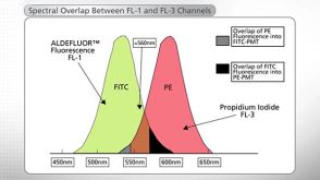
ALDEFLUOR™ 试剂盒
ALDEFLUOR™ DEAB试剂, 1.5 mM, 1 mL
ALDEFLUOR™检测缓冲液
Guan H et al. (JUL 2007)
Journal of immunology (Baltimore,Md. : 1950) 179 1 590--6
NK cells enhance dendritic cell response against parasite antigens via NKG2D pathway.
Recent studies have shown that NK-dendritic cell (DC) interaction plays an important role in the induction of immune response against tumors and certain viruses. Although the effect of this interaction is bidirectional,the mechanism or molecules involved in this cross-talk have not been identified. In this study,we report that coculture with NK cells causes several fold increase in IL-12 production by Toxoplasma gondii lysate Ag-pulsed DC. This interaction also leads to stronger priming of Ag-specific CD8+ T cell response by these cells. In vitro blockade of NKG2D,a molecule present on human and murine NK cells,neutralizes the NK cell-induced up-regulation of DC response. Moreover,treatment of infected animals with Ab to NKG2D receptor compromises the development of Ag-specific CD8+ T cell immunity and reduces their ability to clear parasites. These studies emphasize the critical role played by NKG2D in the NK-DC interaction,which apparently is important for the generation of robust CD8+ T cell immunity against intracellular pathogens. To the best of our knowledge,this is the first work that describes in vivo importance of NKG2D during natural infection.
View Publication
产品类型:
产品号#:
18556
18556RF
产品名:
Y. Dieudonn\'e et al. (may 2019)
Journal of autoimmunity
Transitional B cells in quiescent SLE: An early checkpoint imprinted by IFN.
Systemic lupus (SLE) is characterized by a break of B cell tolerance that plays a central role in disease pathophysiology. An early checkpoint defect occurs at the transitional stage leading to the survival of autoreactive B cells and consequently the production of pathogenic autoantibodies. The main purpose of our work was to determine whether transitional B cells,as the most immature na{\{i}}ve B cell subset upstream of pathogenic B cells display specific features compared to healthy non SLE subjects. Through extensive analysis of transitional B cells from untreated or low treated mostly Caucasian SLE patients we demonstrated that transitional (T1 and T2) B cell frequencies were increased in SLE and positively correlated with disease activity. SLE transitional B cells displayed defects in two closely inter-related molecules (i.e. TLR9 defective responses and CD19 downregulation). RNA sequencing of sorted transitional B cells from untreated patients revealed a predominant overexpression of interferon stimulated genes (ISGs) even out of flares. In addition early transitional B cells from the bone marrow displayed the highest interferon score reflecting a B cell interferon burden of central origin. Hence the IFN signature in transitional B cells is not confined to African American SLE patients and exists in quiescent disease since the medullary stage. These results suggest that in SLE these 3 factors (i.e. IFN imprintment CD19 downregulation and TLR9 responses impairment) could take part at the early transitional B cell stage in B cell tolerance by-pass ultimately leading in periphery to the expansion of autoantibodies-secreting cells."
View Publication
产品类型:
产品号#:
17954
17954RF
100-0971
产品名:
EasySep™人B细胞分选试剂盒
RoboSep™ 人B细胞分选试剂盒
EasySep™人B细胞分离试剂盒
Cho SK et al. (AUG 1999)
Proceedings of the National Academy of Sciences of the United States of America 96 17 9797--802
Functional characterization of B lymphocytes generated in vitro from embryonic stem cells.
To study molecular events involved in B lymphocyte development and V(D)J rearrangement,we have established an efficient system for the differentiation of embryonic stem (ES) cells into mature Ig-secreting B lymphocytes. Here,we show that B lineage cells generated in vitro from ES cells are functionally analogous to normal fetal liver-derived or bone marrow-derived B lineage cells at three important developmental stages: first,they respond to Flt-3 ligand during an early lymphopoietic progenitor stage; second,they become targets for Abelson murine leukemia virus (A-MuLV) infection at a pre-B cell stage; third,they secrete Ig upon stimulation with lipopolysaccharide at a mature mitogen-responsive stage. Moreover,the ES cell-derived A-MuLV-transformed pre-B (EAB) cells are phenotypically and functionally indistinguishable from standard A-MuLV-transformed pre-B cells derived from infection of mouse fetal liver or bone marrow. Notably,EAB cells possess functional V(D)J recombinase activity. In particular,the generation of A-MuLV transformants from ES cells will provide an advantageous system to investigate genetic modifications that will help to elucidate molecular mechanisms in V(D)J recombination and in A-MuLV-mediated transformation.
View Publication
产品类型:
产品号#:
06902
06952
00321
00322
00323
00324
00325
产品名:
Niwa H et al. (APR 2000)
Nature genetics 24 4 372--6
Quantitative expression of Oct-3/4 defines differentiation, dedifferentiation or self-renewal of ES cells.
Cell fate during development is defined by transcription factors that act as molecular switches to activate or repress specific gene expression programmes. The POU transcription factor Oct-3/4 (encoded by Pou5f1) is a candidate regulator in pluripotent and germline cells and is essential for the initial formation of a pluripotent founder cell population in the mammalian embryo. Here we use conditional expression and repression in embryonic stem (ES) cells to determine requirements for Oct-3/4 in the maintenance of developmental potency. Although transcriptional determination has usually been considered as a binary on-off control system,we found that the precise level of Oct-3/4 governs three distinct fates of ES cells. A less than twofold increase in expression causes differentiation into primitive endoderm and mesoderm. In contrast,repression of Oct-3/4 induces loss of pluripotency and dedifferentiation to trophectoderm. Thus a critical amount of Oct-3/4 is required to sustain stem-cell self-renewal,and up- or downregulation induce divergent developmental programmes. Our findings establish a role for Oct-3/4 as a master regulator of pluripotency that controls lineage commitment and illustrate the sophistication of critical transcriptional regulators and the consequent importance of quantitative analyses.
View Publication
产品类型:
产品号#:
72602
产品名:
OAC1
Dioum EM et al. ( 2011)
Proceedings of the National Academy of Sciences of the United States of America 108 51 20713--20718
A small molecule differentiation inducer increases insulin production by pancreatic β cells.
New drugs for preserving and restoring pancreatic β-cell function are critically needed for the worldwide epidemic of type 2 diabetes and the cure for type 1 diabetes. We previously identified a family of neurogenic 3,5-disubstituted isoxazoles (Isx) that increased expression of neurogenic differentiation 1 (NeuroD1,also known as BETA2); this transcription factor functions in neuronal and pancreatic β-cell differentiation and is essential for insulin gene transcription. Here,we probed effects of Isx on human cadaveric islets and MIN6 pancreatic β cells. Isx increased the expression and secretion of insulin in islets that made little insulin after prolonged ex vivo culture and increased expression of neurogenic differentiation 1 and other regulators of islet differentiation and insulin gene transcription. Within the first few hours of exposure,Isx caused biphasic activation of ERK1/2 and increased bulk histone acetylation. Although there was little effect on histone deacetylase activity,Isx increased histone acetyl transferase activity in nuclear extracts. Reconstitution assays indicated that Isx increased the activity of the histone acetyl transferase p300 through an ERK1/2-dependent mechanism. In summary,we have identified a small molecule with antidiabetic activity,providing a tool for exploring islet function and a possible lead for therapeutic intervention in diabetes.
View Publication

 EasySep™小鼠TIL(CD45)正选试剂盒


 沪公网安备31010102008431号