Mary V et al. (DEC 2012)
Toxicology 302 2-3 299--307
Reactive oxygen species sources and biomolecular oxidative damage induced by aflatoxin B1 and fumonisin B1 in rat spleen mononuclear cells.
Aflatoxin B1 (AFB(1)) and fumonisin B1 (FB(1)) are mycotoxins widely found as cereal contaminants. Their immunotoxicities predispose to infectious diseases and may alter the tumor immunosurveillance of human and animals,but the mechanisms underlying have not been fully elucidated,and the induction of oxidative stress has been proposed as a probable mechanism. This work was aimed at evaluating in spleen mononuclear cells (SMC) from Wistar rats the effects of the exposure,in vitro for up to 48 h,to 20 μM AFB(1),10 μM FB(1) and AFB(1)-FB(1) mixture (MIX),over cellular oxidative status,as well as at elucidating the contribution of different reactive oxygen species (ROS) to biomolecular oxidative damage,the biochemical pathways involved,and the probable interaction of both toxins to induce oxidative stress. All the treatments increased total ROS and oxidation of biomolecules,with MIX having the greatest effects. However,only MIX increased superoxide anion radical. The main ROS involved in oxidation of proteins,lipids and DNA appear to be hydrogen peroxide and hydroxyl radical. The mitochondrial complex I and CYP450 were involved in the ROS generation induced by all treatments. The NADPH oxidase system was induced by FB1 and MIX. The arachidonic acid metabolism contributed to the ROS formation induced by AFB(1) and MIX. These results demonstrate that an interaction between AFB(1) and FB(1) occur in the oxidative stress induction,and show the biochemical pathways involved in ROS generation in SMC. The oxidative stress could mediate the AFB(1) and FB(1) individual and combined immunotoxicities.
View Publication
产品类型:
产品号#:
73682
73684
产品名:
Fumonisin B1
Fumonisin B1
C. A. Clough et al. (Jun 2025)
Leukemia 39 8
Characterization of E1 enzyme dependencies in mutant- UBA1 human cells reveals UBA6 as a novel therapeutic target in VEXAS syndrome
VEXAS syndrome is a clonal hematopoietic disorder characterized by hyperinflammation,bone marrow failure,and high mortality. The molecular hallmark of VEXAS is somatic mutations at methionine 41 (M41) in the E1 ubiquitin enzyme,UBA1. These mutations induce a protein isoform switch,but the mechanisms underlying disease pathogenesis remain unclear. Here,we developed a human cell model of VEXAS syndrome by engineering the male monocytic THP1 cell line to express the common UBA1 M41V mutation. We found that mutant UBA1 M41V cells exhibit aberrant UBA1 isoform expression,increased vacuolization,and upregulation of the unfolded protein response,recapitulating key features of VEXAS. Moreover,proteomic analyses revealed dysregulated ubiquitination and proteotoxic stress in UBA1 M41V cells,with alterations in inflammatory and stress-response pathways. Functional studies demonstrated that UBA1 M41V cells were highly sensitive to genetic or pharmacological inhibition of E1 ubiquitin enzymes. Treatment with the E1 enzyme inhibitor TAK-243 preferentially suppressed colony formation of UBA1 M41V cells as compared to WT cells. Moreover,UBA1 M41V cells exhibited greater sensitivity to TAK-243 in competition assays and showed increased apoptosis. Interestingly,TAK-243 preferentially inhibited UBA6 activity over UBA1,suggesting that UBA6 may compensate for UBA1 dysfunction in UBA1 M41V cells. Targeting UBA6 using shRNA or the UBA6-specific inhibitor phytic acid further revealed an acquired dependency on UBA6 in UBA1 M41V cells. Phytic acid selectively impaired growth and colony formation in UBA1 M41V cells while sparing WT cells,highlighting a potential therapeutic vulnerability. Together,these findings establish a novel human model of VEXAS syndrome,identify key roles for UBA1 and UBA6 in disease pathogenesis,and demonstrate that UBA6 inhibition represents a promising therapeutic strategy for selectively targeting UBA1 mutant clones. Subject terms: Haematological cancer,Cell signalling
View Publication
S. Gupta et al. ( 2018)
Immunity & ageing : I & A 15 2
Molecular changes associated with increased TNF-?-induced apoptotis in naive (TN) and central memory (TCM) CD8+ T cells in aged humans.
Background Progressive T cell decline in aged humans is associated with a deficiency of naive (TN) and central memory (TCM) T cells. We have previously reported increased tumor necrosis factor-? (TNF-?)-induced apoptosis in TN and TCM T cells in aged humans; however,the molecular basis of increased apoptosis remains to be defined. Since expression of TNF receptors (TNFRs) was reported to be comparable in young and aged,we investigated signaling events downstream of TNFRs to understand the molecular basis of increased TNF-?-induced apoptosis in aged TN and TCM CD8+ cells. Results The expression of TRAF-2 and RIP,phosphorylation of JNK,IKK?/?,and I?B?,and activation of NF-?B activation were significantly decreased in TN and TCM CD8+ cells from aged subjects as compared to young controls. Furthermore,expression of A20,Bcl-xL,cIAP1,and FLIP-L and FLIP-S was significantly decreased in TN and TCM CD8+ cells from aged subjects. Conclusions These data demonstrate that an impaired expression/function of molecules downstream TNFR signaling pathway that confer survival signals contribute to increased apoptosis of TN and TCM CD8+ cells in aged humans.
View Publication
产品类型:
产品号#:
17654
19053
19053RF
产品名:
EasySep™ Release人PE正选试剂盒
EasySep™人CD8+ T细胞富集试剂盒
RoboSep™ 人CD8+ T细胞富集试剂盒含滤芯吸头
Liu CC et al. (JUN 2016)
Stem cells (Dayton,Ohio) 1--19
Distinct Responses of stem Cells to Telomere Uncapping - a Potential Strategy to Improve the Safety Of Cell Therapy.
In most human somatic cells,the lack of telomerase activity results in progressive telomere shortening during each cell division. Eventually,DNA damage responses triggered by critically short telomeres induce an irreversible cell cycle arrest termed replicative senescence. However,the cellular responses of human pluripotent stem cells to telomere uncapping remain unknown. We generated telomerase knockout human embryonic stem (ES) cells through gene targeting. Telomerase inactivation in ES cells results in progressive telomere shortening. Telomere DNA damage in ES cells and neural progenitor cells induces rapid apoptosis when telomeres are uncapped,in contrast to fibroblast cells that enter a state of replicative senescence. Significantly,telomerase inactivation limits the proliferation capacity of human ES cells without affecting their pluripotency. By targeting telomerase activity,we can functionally separate the two unique properties of human pluripotent stem cells,namely unlimited self-renewal and pluripotency. We show that the potential of ES cells to form teratomas in vivo is dictated by their telomere length. By controlling telomere length of ES cells through telomerase inactivation,we can inhibit teratoma formation and potentially improve the safety of cell therapies involving terminally differentiated cells as well as specific progenitor cells that do not require sustained cellular proliferation in vivo,and thus sustained telomerase activity. This article is protected by copyright. All rights reserved.
View Publication
产品类型:
产品号#:
05850
05857
05870
05875
85850
85857
85870
85875
产品名:
mTeSR™1
mTeSR™1
Pettinato G et al. (SEP 2016)
Scientific reports 6 32888
Scalable Differentiation of Human iPSCs in a Multicellular Spheroid-based 3D Culture into Hepatocyte-like Cells through Direct Wnt/β-catenin Pathway Inhibition.
Treatment of acute liver failure by cell transplantation is hindered by a shortage of human hepatocytes. Current protocols for hepatic differentiation of human induced pluripotent stem cells (hiPSCs) result in low yields,cellular heterogeneity,and limited scalability. In the present study,we have developed a novel multicellular spheroid-based hepatic differentiation protocol starting from embryoid bodies of hiPSCs (hiPSC-EBs) for robust mass production of human hepatocyte-like cells (HLCs) using two novel inhibitors of the Wnt pathway. The resultant hiPSC-EB-HLCs expressed liver-specific genes,secreted hepatic proteins such as Albumin,Alpha Fetoprotein,and Fibrinogen,metabolized ammonia,and displayed cytochrome P450 activities and functional activities typical of mature primary hepatocytes,such as LDL storage and uptake,ICG uptake and release,and glycogen storage. Cell transplantation of hiPSC-EB-HLC in a rat model of acute liver failure significantly prolonged the mean survival time and resolved the liver injury when compared to the no-transplantation control animals. The transplanted hiPSC-EB-HLCs secreted human albumin into the host plasma throughout the examination period (2 weeks). Transplantation successfully bridged the animals through the critical period for survival after acute liver failure,providing promising clues of integration and full in vivo functionality of these cells after treatment with WIF-1 and DKK-1.
View Publication
产品类型:
产品号#:
05850
05857
05870
05875
85850
85857
85870
85875
产品名:
mTeSR™1
mTeSR™1
Li R et al. (NOV 2016)
Cancer research
Macrophage-secreted TNFα and TGFβ1 Influence Migration Speed and Persistence of Cancer Cells in 3D Tissue Culture via Independent Pathways.
The ability of a cancer cell to migrate through the dense extracellular matrix (ECM) within and surrounding the solid tumor is a critical determinant of metastasis. Macrophages enhance invasion and metastasis in the tumor microenvironment but the basis for their effects are not fully understood. Using a microfluidic 3D cell migration assay,we found that the presence of macrophages enhanced the speed and persistence of cancer cell migration through a 3D extracellular matrix in a matrix metalloproteinases (MMP)-dependent fashion. Mechanistic investigations revealed that macrophage-released TNFα and TGFβ1 mediated the observed behaviors by two distinct pathways. These factors synergistically enhanced migration persistence through a synergistic induction of NF-κB-dependent MMP1 expression in cancer cells. In contrast,macrophage-released TGFβ1 enhanced migration speed primarily by inducing MT1-MMP expression. Taken together,our results reveal new insights into how macrophages enhance cancer cell metastasis,and they identify TNFα and TGFβ1 dual blockade as an anti-metastatic strategy in solid tumors.
View Publication
产品类型:
产品号#:
19059
19059RF
产品名:
EasySep™人单核细胞富集试剂盒
RoboSep™ 人单核细胞富集试剂盒含滤芯吸头
Katzenellenbogen BS et al. (JAN 1984)
Cancer research 44 1 112--9
Bioactivities, estrogen receptor interactions, and plasminogen activator-inducing activities of tamoxifen and hydroxy-tamoxifen isomers in MCF-7 human breast cancer cells.
Tamoxifen is used widely in the treatment of endocrine-responsive breast cancers in humans. Studies were undertaken to examine the biological character (estrogenic-antiestrogenic properties) and estrogen receptor (ER) interaction of the cis- and trans-isomers of tamoxifen and hydroxytamoxifen in MCF-7 human breast cancer cells. For each compound,the following parameters were monitored: affinity for ER and effects on cellular ER levels; stimulation-inhibition of cell growth,plasminogen activator activity,and cellular progesterone receptor levels; and isomer interconversion and metabolism in vitro. The relative binding affinities of the compounds cis-tamoxifen,trans-tamoxifen,cis-hydroxytamoxifen,and trans-hydroxytamoxifen for cytosol ER were 0.3,2.5,1.8,and 310%,respectively,in which the affinity of estradiol is considered 100%. cis-Tamoxifen behaved as a weak estrogen agonist in all assays,while trans-tamoxifen was an effective estrogen antagonist. cis-Tamoxifen behaved like estradiol in stimulating MCF-7 cell growth and increasing plasminogen activator activity and cellular progesterone receptor content,although very much higher concentrations of cis-tamoxifen (10(-6) M) were needed to achieve the levels of stimulation observed with 10(-10) M estradiol. trans-Tamoxifen and trans-hydroxytamoxifen suppressed cell growth,inhibited plasminogen activator activity of control cells,and suppressed estradiol-stimulation of plasminogen activator activity,and they evoked minimal increases in cellular progesterone receptor levels. trans-Hydroxytamoxifen had a 100-fold increased affinity for ER and was approximately 100-times more potent than was trans-tamoxifen in suppressing cell growth and plasminogen activator activity. cis-Hydroxytamoxifen behaved as an estrogen antagonist,suppressing cell growth and plasminogen activator activity,and it elicited submaximal increases in progesterone receptor levels. This apparently paradoxical behavior of cis-hydroxytamoxifen was shown to be due to the fact that the cis- and trans-hydroxytamoxifens readily undergo isomeric interconversion upon exposure to our cell culture conditions,resulting in substantial accumulation of the higher-affinity trans-hydroxytamoxifen in the nuclear ER fraction of cells. In contrast to the facile interconversion of the hydroxytamoxifen isomers,there is no metabolism or interconversion of the parent compounds cis- and trans-tamoxifen in vitro. Hence,by the criteria we have used,the biological characters of trans-tamoxifen and trans-hydroxytamoxifen are similar,the major difference being the approximately 100-fold enhanced potency of the hydroxylated form. In contrast,cis-t
View Publication
产品类型:
产品号#:
72662
产品名:
他莫昔芬(Tamoxifen)
Lichterfeld M et al. (SEP 2004)
The Journal of experimental medicine 200 6 701--12
Loss of HIV-1-specific CD8+ T cell proliferation after acute HIV-1 infection and restoration by vaccine-induced HIV-1-specific CD4+ T cells.
Virus-specific CD8(+) T cells are associated with declining viremia in acute human immunodeficiency virus (HIV)1 infection,but do not correlate with control of viremia in chronic infection,suggesting a progressive functional defect not measured by interferon gamma assays presently used. Here,we demonstrate that HIV-1-specific CD8(+) T cells proliferate rapidly upon encounter with cognate antigen in acute infection,but lose this capacity with ongoing viral replication. This functional defect can be induced in vitro by depletion of CD4(+) T cells or addition of interleukin 2-neutralizing antibodies,and can be corrected in chronic infection in vitro by addition of autologous CD4(+) T cells isolated during acute infection and in vivo by vaccine-mediated induction of HIV-1-specific CD4(+) T helper cell responses. These data demonstrate a loss of HIV-1-specific CD8(+) T cell function that not only correlates with progressive infection,but also can be restored in chronic infection by augmentation of HIV-1-specific T helper cell function. This identification of a reversible defect in cell-mediated immunity in chronic HIV-1 infection has important implications for immunotherapeutic interventions.
View Publication
产品类型:
产品号#:
15023
15063
产品名:
RosetteSep™人CD8+ T细胞富集抗体混合物
RosetteSep™人CD8+ T细胞富集抗体混合物
Arbab AS et al. (MAR 2006)
Stem cells (Dayton,Ohio) 24 3 671--8
Magnetic resonance imaging and confocal microscopy studies of magnetically labeled endothelial progenitor cells trafficking to sites of tumor angiogenesis.
UNLABELLED: AC133 cells,a subpopulation of CD34+ hematopoietic stem cells,can transform into endothelial cells that may integrate into the neovasculature of tumors or ischemic tissue. Most current imaging modalities do not allow monitoring of early migration and incorporation of endothelial progenitor cells (EPCs) into tumor neovasculature. The goals of this study were to use magnetic resonance imaging (MRI) to track the migration and incorporation of intravenously injected,magnetically labeled EPCs into the blood vessels in a rapidly growing flank tumor model and to determine whether the pattern of EPC incorporation is related to the time of injection or tumor size. MATERIALS AND METHODS: EPCs labeled with ferumoxide-protamine sulfate (FePro) complexes were injected into mice bearing xenografted glioma,and MRI was obtained at different stages of tumor development and size. RESULTS: Migration and incorporation of labeled EPCs into tumor neovasculature were detected as low signal intensity on MRI at the tumor periphery as early as 3 days after EPC administration in preformed tumors. However,low signal intensities were not observed in tumors implanted at the time of EPC administration until tumor size reached 1 cm at 12 to 14 days. Prussian blue staining showed iron-positive cells at the sites corresponding to low signal intensity on MRI. Confocal microscopy showed incorporation into the neovasculature,and immunohistochemistry clearly demonstrated the transformation of the administered EPCs into endothelial cells. CONCLUSION: MRI demonstrated the incorporation of FePro-labeled human CD34+/AC133+ EPCs into the neovasculature of implanted flank tumors.
View Publication
产品类型:
产品号#:
09850
产品名:
Thum T et al. (NOV 2005)
Journal of the American College of Cardiology 46 9 1693--701
Suppression of endothelial progenitor cells in human coronary artery disease by the endogenous nitric oxide synthase inhibitor asymmetric dimethylarginine.
OBJECTIVES: We tested the hypothesis that asymmetric dimethylarginine (ADMA) may be an endogenous inhibitor of endothelial progenitor cells (EPCs). BACKGROUND: Endothelial progenitor cells play a pivotal role in regeneration of injured endothelium,thereby limiting the formation of atherosclerotic lesions. Reduced numbers of EPCs may affect progression of coronary artery disease. Regulation of EPC mobilization and function is mediated in part by nitric oxide (NO). Endogenous inhibitors of NO synthases,such as ADMA,contribute to endothelial dysfunction and injury. METHODS: We used flow cytometry and in vitro assays to investigate the relationship between EPC number and function with ADMA plasma levels in patients with stable angina. RESULTS: The plasma concentration of ADMA was related to the severity of coronary artery disease and correlated inversely with the number of circulating CD34+/CD133+ progenitor cells (r = -0.69; p textless 0.0001) and endothelial colony forming units (CFUs) (r = -0.75; p textless 0.0001). Adjusting for all patient characteristics,we confirmed these findings in multivariate regression analyses. In vitro differentiation of EPCs was repressed by ADMA in a concentration-dependent manner. Compared with untreated cells,ADMA reduced EPC incorporation into endothelial tube-like structures to 27 +/- 11% (p textless 0.001). Asymmetric dimethylarginine repressed the formation of CFUs from cultured peripheral blood mononuclear cells to 35 +/- 7% (p textless 0.001). Asymmetric dimethylarginine decreased endothelial nitric oxide synthase activity in EPCs to 64 +/- 6% (p textless 0.05) when compared with controls. Co-incubation with the hydroxymethyl glutaryl coenzyme A reductase inhibitor rosuvastatin abolished the detrimental effects of ADMA. CONCLUSIONS: Asymmetric dimethylarginine is an endogenous inhibitor of mobilization,differentiation,and function of EPCs. This contributes to the cardiovascular risk in patients with high ADMA levels and may explain low numbers and function of EPCs in patients with coronary artery disease.
View Publication
产品类型:
产品号#:
05900
05950
产品名:
Windmolders S et al. (JAN 2014)
Journal of molecular and cellular cardiology 66 177--188
Mesenchymal stem cell secreted platelet derived growth factor exerts a pro-migratory effect on resident Cardiac Atrial appendage Stem Cells.
Mesenchymal stem cells (MSCs) modulate cardiac healing after myocardial injury through the release of paracrine factors,but the exact mechanisms are still unknown. One possible mechanism is through mobilization of endogenous cardiac stem cells (CSCs). This study aimed to test the pro-migratory effect of MSC conditioned medium (MSC-CM) on endogenous CSCs from human cardiac tissue. By using a three-dimensional collagen assay,we found that MSC-CM improved migration of cells from human cardiac tissue. Cell counts,perimeter and area measurements were utilized to quantify migration effects. To examine whether resident stem cells were among the migrating cells,specific stem cell properties were investigated. The migrating cells displayed strong similarities with resident Cardiac Atrial appendage Stem Cells (CASCs),including a clonogenic potential of ˜21.5% and expression of pluripotency associated genes like Oct-4,Nanog,c-Myc and Klf-4. Similar to CASCs,migrating cells demonstrated high aldehyde dehydrogenase activity and were able to differentiate towards cardiomyocytes. Receptor tyrosine kinase analysis and collagen assays performed with recombinant platelet derived growth factor (PDGF)-AA and Imatinib Mesylate,a PDGF receptor inhibitor,suggested a role for the PDGF-AA/PDGF receptor $$ axis in enhancing the migration process of CASCs. In conclusion,our findings demonstrate that factors present in MSC-CM improve migration of resident stem cells from human cardiac tissue. These data open doors towards future therapies in which MSC secreted factors,like PDGF-AA,can be utilized to enhance the recruitment of CASCs towards the site of myocardial injury.
View Publication


 EasySep™小鼠TIL(CD45)正选试剂盒





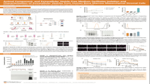 科学海报Animal Component- and Extracellular Vesicle-Free Medium Facilitates Isolation and Characterization of Extracellular Vesicles from Human Bone Marrow Mesenchymal Stromal Cells
 沪公网安备31010102008431号