Lack of expression of Thy-1 (CD90) on acute myeloid leukemia cells with long-term proliferative ability in vitro and in vivo.
Acute myeloid leukaemia (AML) is thought to be maintained by a small population of leukemic progenitor cells. To define the phenotype of such cells with long-term proliferative capacity in vitro and in vivo,we have used the production of leukemic clonogenic cells (CFU) after 2 to 8 weeks in suspension culture as a measure of these cells in vitro and compared their phenotype with that of cells capable of engrafting nonobese diabetic severe combined immune deficient (NOD/SCID) mice. Leukemic blast peripheral blood cells were evaluated for expression of CD34 and Thy-1 (CD90) antigens. The majority of AML blast cells at diagnosis lacked expression of Thy-1. Most primary CFU-blast and the CFU detected at up to 8 weeks from suspension cultures were CD34+/Thy-1-. AML cells that were capable of engrafting NOD/SCID mice were also found to have the CD34+/Thy-1- phenotype. However,significant engraftment was achieved using both CD34+/Thy-1- and CD34- subfractions from one AML M5 patient. These results suggest that while heterogeneity exists between individual patients,the leukemic progenitor cells that are capable of maintaining the disease in vitro and in vivo differ from normal hematopoietic progenitor cells in their lack of expression of Thy-1.
View Publication
产品类型:
产品号#:
02690
02696
02697
09300
09500
09600
09650
09850
产品名:
StemSpan™ CC100
StemSpan™巨核细胞扩增添加物 (100X)
StemSpan™ CC110
含有10% 牛血清白蛋白(BSA)的 Iscove's MDM
BIT 9500血清替代物
StemSpan™ SFEM
StemSpan™ SFEM
Daynac M et al. (FEB 2016)
Scientific reports 6 21505
Age-related neurogenesis decline in the subventricular zone is associated with specific cell cycle regulation changes in activated neural stem cells.
Although neural stem cells (NSCs) sustain continuous neurogenesis throughout the adult lifespan of mammals,they progressively exhibit proliferation defects that contribute to a sharp reduction in subventricular neurogenesis during aging. However,little is known regarding the early age-related events in neurogenic niches. Using a fluorescence-activated cell sorting technique that allows for the prospective purification of the main neurogenic populations from the subventricular zone (SVZ),we demonstrated an early decline in adult neurogenesis with a dramatic loss of progenitor cells in 4 month-old young adult mice. Whereas the activated and quiescent NSC pools remained stable up to 12 months,the proliferative status of activated NSCs was already altered by 6 months,with an overall extension of the cell cycle resulting from a specific lengthening of G1. Whole genome analysis of activated NSCs from 2- and 6-month-old mice further revealed distinct transcriptomic and molecular signatures,as well as a modulation of the TGFβ signalling pathway. Our microarray study constitutes a cogent identification of new molecular players and signalling pathways regulating adult neurogenesis and its early modifications.
View Publication
产品类型:
产品号#:
05700
05701
05702
产品名:
NeuroCult™ 基础培养基(小鼠和大鼠)
NeuroCult™ 扩增添加物(小鼠和大鼠)
NeuroCult™扩增试剂盒(小鼠和大鼠)
Verstovsek S et al. ( 2005)
Cancer 104 6 1230--1236
AMN107, a novel aminopyrimidine inhibitor of p190 Bcr-Abl activation and of in vitro proliferation of Philadelphia-positive acute lymphoblastic leukemia cells.
BACKGROUND: Previous studies have shown that patients with Bcr-Abl-positive acute lymphoblastic leukemia (ALL) either have primary disease that is refractory to imatinib mesylate or develop disease recurrence after an initial response. METHODS: The authors investigated the effects of a newly designed Bcr-Abl inhibitor,AMN107,by comparing its in vitro inhibitory potency on p190 Bcr-Abl ALL cell lines with that of imatinib. RESULTS: In two Philadelphia (Ph)-positive ALL cell lines,AMN107 was found to be 30-40 times more potent than imatinib in inhibiting cellular proliferation. AMN107 was also more effective than imatinib in inhibiting phosphorylation of p190 Bcr-Abl tyrosine kinase in cell lines and primary ALL cells. The inhibition of cellular proliferation was associated with the induction of apoptosis in only one of the cell lines. No activity was observed in cell lines lacking the BCR-ABL genotype. CONCLUSIONS: The results of the current study suggest the superior potency of AMN107 compared with imatinib in Ph-positive ALL and support clinical trials of AMN107 in patients with Ph-positive ALL.
View Publication
产品类型:
产品号#:
73302
73304
产品名:
Nilotinib
Nilotinib
Corti S et al. (JAN 2006)
Human molecular genetics 15 2 167--87
Transplanted ALDHhiSSClo neural stem cells generate motor neurons and delay disease progression of nmd mice, an animal model of SMARD1.
Spinal muscular atrophy with respiratory distress type 1 (SMARD1) is an infantile autosomal-recessive motor neuron disease caused by mutations in the immunoglobulin micro-binding protein 2. We investigated the potential of a spinal cord neural stem cell population isolated on the basis of aldehyde dehydrogenase (ALDH) activity to modify disease progression of nmd mice,an animal model of SMARD1. ALDH(hi)SSC(lo) stem cells are self-renewing and multipotent and when intrathecally transplanted in nmd mice generate motor neurons properly localized in the spinal cord ventral horns. Transplanted nmd animals presented delayed disease progression,sparing of motor neurons and ventral root axons and increased lifespan. To further investigate the molecular events responsible for these differences,microarray and real-time reverse transcription-polymerase chain reaction analyses of wild-type,mutated and transplanted nmd spinal cord were undertaken. We demonstrated a down-regulation of genes involved in excitatory amino acid toxicity and oxidative stress handling,as well as an up-regulation of genes related to the chromatin organization in nmd compared with wild-type mice,suggesting that they may play a role in SMARD1 pathogenesis. Spinal cord of nmd-transplanted mice expressed high transcript levels for genes related to neurogenesis such as doublecortin (DCX),LIS1 and drebrin. The presence of DCX-expressing cells in adult nmd spinal cord suggests that both exogenous and endogenous neurogeneses may contribute to the observed nmd phenotype amelioration.
View Publication
产品类型:
产品号#:
01700
01705
01701
01702
产品名:
ALDEFLUOR™ 试剂盒
ALDEFLUOR™ DEAB试剂, 1.5 mM, 1 mL
ALDEFLUOR™检测缓冲液
S. L. Locatelli et al. (OCT 2018)
Clinical cancer research : an official journal of the American Association for Cancer Research
Targeting cancer cells and tumor microenvironment in preclinical and clinical models of Hodgkin lymphoma using the dual PI3K$\delta$/$\gamma$ inhibitor RP6530.
PURPOSE Tumor-associated macrophages (TAMs) and the hyperactivation of phosphoinositide 3-kinase(PI3K)/AKT pathway are involved in the pathogenesis of Hodgkin lymphoma (HL) and affect disease outcome. Since the $\delta$ and $\gamma$ isoforms of PI3K are overexpressed in Hodgkin/Reed-Sternberg (HRS) cells and the tumor microenvironment (TME),we propose that the PI3K$\delta$/$\gamma$ inhibitor RP6530 might affect both HRS cells and TME,ultimately leading to an enhanced antitumor response. EXPERIMENTAL DESIGN HL cell lines (L-540,KM-H2 and L-428) and primary human macrophages were used to investigate the activity of RP6530 in vitro and in vivo in HL cell line xenografts. RESULTS In vitro,RP6530 besides killing and inhibiting the proliferation of HL cells,downregulated lactic acid metabolism,switching the activation of macrophages from an immunosuppressive M2-like phenotype to a more inflammatory M1-like state. By RNA sequencing,we define tumor glycolysis as a specific PI3K$\delta$/$\gamma$-dependent pathway implicated in the metabolic reprogramming of cancer cells. We identify the metabolic regulator Pyruvate Kinase M2 (PKM2) as the main mediator of tumor-induced immunosuppressive phenotype of macrophages. Furthermore,we show in human tumor xenografts that RP6530 repolarizes TAMs into pro-inflammatory macrophages and inhibits tumor vasculature,leading to tumor regression. Interestingly,HL patients experiencing objective responses (CR and PR) in a phase 1 trial using RP6530 showed a significant inhibition of circulating MDSCs and an average mean reduction in serum TARC levels of 40{\%} (range,4-76{\%}). CONCLUSIONS Our results support PI3K$\delta$/$\gamma$ inhibition as a novel therapeutic strategy that targets both malignant cells and the TME to treat HL patients.
View Publication
Vazin T et al. (FEB 2014)
Neurobiology of Disease 62 62--72
Efficient derivation of cortical glutamatergic neurons from human pluripotent stem cells: a model system to study neurotoxicity in Alzheimer's disease.
Alzheimer's disease (AD) is among the most prevalent forms of dementia affecting the aging population,and pharmacological therapies to date have not been successful in preventing disease progression. Future therapeutic efforts may benefit from the development of models that enable basic investigation of early disease pathology. In particular,disease-relevant models based on human pluripotent stem cells (hPSCs) may be promising approaches to assess the impact of neurotoxic agents in AD on specific neuronal populations and thereby facilitate the development of novel interventions to avert early disease mechanisms. We implemented an efficient paradigm to convert hPSCs into enriched populations of cortical glutamatergic neurons emerging from dorsal forebrain neural progenitors,aided by modulating Sonic hedgehog (Shh) signaling. Since AD is generally known to be toxic to glutamatergic circuits,we exposed glutamatergic neurons derived from hESCs to an oligomeric pre-fibrillar forms of Aβ known as globulomers"�
View Publication
产品类型:
产品号#:
05850
05857
05870
05875
85850
85857
85870
85875
产品名:
mTeSR™1
mTeSR™1
Obakan P et al. (JAN 2014)
Molecular biology reports 41 1 145--54
Purvalanol A is a strong apoptotic inducer via activating polyamine catabolic pathway in MCF-7 estrogen receptor positive breast cancer cells.
Purvalanol A is a specific CDK inhibitor which triggers apoptosis by causing cell cycle arrest in cancer cells. Although it has strong apoptotic potential,the mechanistic action of Purvalanol A on significant cell signaling targets has not been clarified yet. Polyamines are crucial metabolic regulators affected by CDK inhibition because of their role in cell cycle progress as well. In addition,malignant cells possess impaired polyamine homeostasis with high level of intracellular polyamines. Especially induction of polyamine catabolic enzymes spermidine/spermine N1-acetyltransferase (SSAT),polyamine oxidase (PAO) and spermine oxidase (SMO) induced toxic by-products in correlation with the induction of apoptosis in cancer cells. In this study,we showed that Purvalanol A induced apoptosis in caspase- dependent manner in MCF-7 ER(+) cells,while MDA-MB-231 (ER-) cells were less sensitive against drug. In addition Bcl-2 is a critical target for Purvalanol A,since Bcl-2 overexpressed cells are more resistant to Purvalanol A-mediated apoptosis. Furthermore,exposure of MCF-7 cells to Purvalanol A triggered SSAT and PAO upregulation and the presence of PAO/SMO inhibitor,MDL 72,527 prevented Purvalanol A-induced apoptosis.
View Publication
产品类型:
产品号#:
73772
73774
产品名:
ndrea de Oliveira Georges JA et al. (AUG 2014)
Stem cell reviews 10 4 472--479
Aberrant patterns of X chromosome inactivation in a new line of human embryonic stem cells established in physiological oxygen concentrations
One of the differences between murine and human embryonic stem cells (ESCs) is the epigenetic state of the X chromosomes in female lines. Murine ESCs (mESCs) present two transcriptionally active Xs that will undergo the dosage compensation process of XCI upon differentiation,whereas most human ESCs (hESCs) spontaneously inactivate one X while keeping their pluripotency. Whether this reflects differences in embryonic development of mice and humans,or distinct culture requirements for the two kinds of pluripotent cells is not known. Recently it has been shown that hESCs established in physiological oxygen levels are in a stable pre-XCI state equivalent to that of mESCs,suggesting that culture in low oxygen concentration is enough to preserve that epigenetic state of the X chromosomes. Here we describe the establishment of two new lines of hESCs under physiological oxygen level and the characterization of the XCI state in the 46,XX line BR-5. We show that a fraction of undifferentiated cells present XIST RNA accumulation and single H3K27me foci,characteristic of the inactive X. Moreover,analysis of allele specific gene expression suggests that pluripotent BR-5 cells present completely skewed XCI. Our data indicate that physiological levels of oxygen are not sufficient for the stabilization of the pre-XCI state in hESCs.
View Publication
产品类型:
产品号#:
05850
05857
05870
05875
85850
85857
85870
85875
产品名:
mTeSR™1
mTeSR™1
Kallas A et al. (FEB 2014)
Stem Cells International 2014 298163
SOX2 is regulated differently from NANOG and OCT4 in human embryonic stem cells during early differentiation initiated with sodium butyrate
Transcription factors NANOG,OCT4,and SOX2 regulate self-renewal and pluripotency in human embryonic stem (hES) cells; however,their expression profiles during early differentiation of hES cells are unclear. In this study,we used multiparameter flow cytometric assay to detect all three transcription factors (NANOG,OCT4,and SOX2) simultaneously at single cell level and monitored the changes in their expression during early differentiation towards endodermal lineage (induced by sodium butyrate). We observed at least four distinct populations of hES cells,characterized by specific expression patterns of NANOG,OCT4,and SOX2 and differentiation markers. Our results show that a single cell can express both differentiation and pluripotency markers at the same time,indicating a gradual mode of developmental transition in these cells. Notably,distinct regulation of SOX2 during early differentiation events was detected,highlighting the potential importance of this transcription factor for self-renewal of hES cells during differentiation.
View Publication
产品类型:
产品号#:
05850
05857
05870
05875
85850
85857
85870
85875
产品名:
mTeSR™1
mTeSR™1
Dang LTH et al. (SEP 2014)
Biomaterials 35 27 7786--7799
Inhibition of apoptosis in human induced pluripotent stem cells during expansion in a defined culture using angiopoietin-1 derived peptide QHREDGS
Adhesion molecule signaling is critical to human pluripotent stem cell (hPSC) survival,self-renewal,and differentiation. Thus,hPSCs are grown as clumps of cells on feeder cell layers or poorly defined extracellular matrices such as Matrigel. We sought to define a small molecule that would initiate adhesion-based signaling to serve as a basis for a defined substrate for hPSC culture. Soluble angiopoeitin-1 (Ang-1)-derived peptide QHREDGS added to defined serum-free media increased hPSC colony cell number and size during long- and short-term culture when grown on feeder cell layers or Matrigel,i.e. on standard substrates,without affecting hPSC morphology,growth rate or the ability to differentiate into multiple lineages both invitro and invivo. Importantly,QHREDGS treatment decreased hPSC apoptosis during routine passaging and single-cell dissociation. Mechanistically,the interaction of QHREDGS with ??1-integrins increased expression of integrin-linked kinase (ILK),increased expression and activation of extracellular signal-regulated kinases 1/2 (ERK1/2),and decreased caspase-3/7 activity. QHREDGS immobilization to polyethylene glycol hydrogels significantly increased cell adhesion in a dose-dependent manner. We propose QHREDGS as a small molecule inhibitor of hPSC apoptosis and the basis of an affordable defined substrate for hPSC maintenance. ?? 2014 Elsevier Ltd.
View Publication
产品类型:
产品号#:
05850
05857
05870
05875
85850
85857
85870
85875
产品名:
mTeSR™1
mTeSR™1
Opyrchal M et al. ( 2014)
International journal of oncology 45 3 1193--1199
Inhibition of Cdk2 kinase activity selectively targets the CD44�?�/CD24�?�/Low stem-like subpopulation and restores chemosensitivity of SUM149PT triple-negative breast cancer cells.
Inflammatory breast cancer (IBC) is an angioinvasive and most aggressive type of advanced breast cancer characterized by rapid proliferation,chemoresistance,early metastatic development and poor prognosis. IBC tumors display a triple-negative breast cancer (TNBC) phenotype characterized by centrosome amplification,high grade of chromosomal instability (CIN) and low levels of expression of estrogen receptor α (ERα),progesterone receptor (PR) and HER-2 tyrosine kinase receptor. Since the TNBC cells lack these receptors necessary to promote tumor growth,common treatments such as endocrine therapy and molecular targeting of HER-2 receptor are ineffective for this subtype of breast cancer. To date,not a single targeted therapy has been approved for non-inflammatory and inflammatory TNBC tumors and combination of conventional cytotoxic chemotherapeutic agents remains the standard therapy. IBC tumors generally display activation of epithelial to mesenchymal transition (EMT) that is functionally linked to a CD44+/CD24-/Low stem-like phenotype. Development of EMT and consequent activation of stemness programming is responsible for invasion,tumor self-renewal and drug resistance leading to breast cancer progression,distant metastases and poor prognosis. In this study,we employed the luminal ER+ MCF-7 and the IBC SUM149PT breast cancer cell lines to establish the extent to which high grade of CIN and chemoresistance were mechanistically linked to the enrichment of CD44+/CD24low/- CSCs. Here,we demonstrate that SUM149PT cells displayed higher CIN than MCF-7 cells characterized by higher percentage of structural and numerical chromosomal aberrations. Moreover,centrosome amplification,cyclin E overexpression and phosphorylation of retinoblastoma (Rb) were restricted to the stem-like CD44+/CD24-/Low subpopulation isolated from SUM149PT cells. Significantly,CD44+/CD24-/Low CSCs displayed resistance to conventional chemotherapy but higher sensitivity to SU9516,a specific cyclin-dependent kinase 2 (Cdk2) inhibitor,demonstrating that aberrant activation of cyclin E/Cdk2 oncogenic signaling is essential for the maintenance and expansion of CD44+/CD24-/Low CSC subpopulation in IBC. In conclusion,our findings propose a novel therapeutic approach to restore chemosensitivity and delay recurrence of IBC tumors based on the combination of conventional chemotherapy with small molecule inhibitors of the Cdk2 cell cycle kinase.
View Publication

 EasySep™小鼠TIL(CD45)正选试剂盒


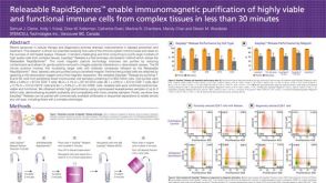 科学海报Releasable RapidSpheres Enable Immunomagnetic Purification of Highly Viable and Functional Immune Cells from Complex Tissues in Less Than 30 Minutes
 沪公网安备31010102008431号