Sandt C et al. (JAN 2013)
Journal of Biophotonics 6 1 60--72
Profiling pluripotent stem cells and organelles using synchrotron radiation infrared microspectroscopy
FTIR micro-spectroscopy is a sensitive,non-destructive and label-free method offering diffraction-limited resolution with high signal-to-noise ratios when combined with a synchrotron radiation source. The vibrational signature of individual cells was used to validate an alternative strategy for reprogramming induced pluripotent stem cells generated from amniocytes. The iPSC lines PB09 and PB10,were reprogrammed from the same amniocyte cell line using respectively the Oct54,Sox2,Lin28,and Nanog and the Oct4 and Sox2 transcription factor cocktail. We show that cells reprogrammed by the two different sets of transfection factors have similar spectral signatures after reprogramming,except for a small subpopulation of cells in one of the cell lines. Mapping HeLa cells at subcellular resolution,we show that the Golgi apparatus,the cytoplasm and the nucleus have a specific spectral signature. The CH(3):CH(2) ratio is the highest in the nucleus and the lowest in the Golgi apparatus/endoplasmic reticulum,in agreement with the membrane composition of these organelles. This is confirmed by specific staining of the organelles with fluorescent dyes. Subcellular differentiation of cell compartments is also demonstrated in living cells.
View Publication
产品类型:
产品号#:
05850
05857
05870
05875
85850
85857
85870
85875
产品名:
mTeSR™1
mTeSR™1
S. K. Atkins et al. ( 2022)
Frontiers in cardiovascular medicine 9 925777
Induced pluripotent stem cell-derived smooth muscle cells to study cardiovascular calcification.
Cardiovascular calcification is the lead predictor of cardiovascular events and the top cause of morbidity and mortality worldwide. To date,only invasive surgical options are available to treat cardiovascular calcification despite the growing understanding of underlying pathological mechanisms. Key players in vascular calcification are vascular smooth muscle cells (SMCs),which transform into calcifying SMCs and secrete mineralizing extracellular vesicles that form microcalcifications,subsequently increasing plaque instability and consequential plaque rupture. There is an increasing,practical need for a large scale and inexhaustible source of functional SMCs. Here we describe an induced pluripotent stem cell (iPSC)-derived model of SMCs by differentiating iPSCs toward SMCs to study the pathogenesis of vascular calcification. Specifically,we characterize the proteome during iPSC differentiation to better understand the cellular dynamics during this process. First,we differentiated human iPSCs toward an induced-SMC (iSMC) phenotype in a 10-day protocol. The success of iSMC differentiation was demonstrated through morphological analysis,immunofluorescent staining,flow cytometry,and proteomics characterization. Proteomics was performed throughout the entire differentiation time course to provide a robust,well-defined starting and ending cell population. Proteomics data verified iPSC differentiation to iSMCs,and functional enrichment of proteins on different days showed the key pathways changing during iSMC development. Proteomics comparison with primary human SMCs showed a high correlation with iSMCs. After iSMC differentiation,we initiated calcification in the iSMCs by culturing the cells in osteogenic media for 17 days. Calcification was verified using Alizarin Red S staining and proteomics data analysis. This study presents an inexhaustible source of functional vascular SMCs and calcifying vascular SMCs to create an in vitro model of vascular calcification in osteogenic conditions,with high potential for future applications in cardiovascular calcification research.
View Publication
Phospho-seq: integrated, multi-modal profiling of intracellular protein dynamics in single cells
Cell signaling plays a critical role in neurodevelopment,regulating cellular behavior and fate. While multimodal single-cell sequencing technologies are rapidly advancing,scalable and flexible profiling of cell signaling states alongside other molecular modalities remains challenging. Here we present Phospho-seq,an integrated approach that aims to quantify cytoplasmic and nuclear proteins,including those with post-translational modifications,and to connect their activity with cis-regulatory elements and transcriptional targets. We utilize a simplified benchtop antibody conjugation method to create large custom neuro-focused antibody panels for simultaneous protein and scATAC-seq profiling on whole cells,alongside both experimental and computational strategies to incorporate transcriptomic measurements. We apply our workflow to cell lines,induced pluripotent stem cells,and months-old retinal and brain organoids to demonstrate its broad applicability. We show that Phospho-seq can provide insights into cellular states and trajectories,shed light on gene regulatory relationships,and help explore the causes and effects of diverse cell signaling in neurodevelopment. Here,the authors demonstrate Phospho-seq,a single-cell multiomics method capable of quantifying chromatin accessibility alongside intracellular proteins,including post-translationally modified proteins. Then,they apply Phospho-seq to organoid models of neurodevelopment.
View Publication
Hypoimmune gene edited human pluripotent stem cells (hPSCs) are a promising platform for developing reparative cellular therapies that evade immune rejection. Existing first-generation hypoimmune strategies have used CRISPR/Cas9 editing to modulate genes associated with adaptive (e.g.,T cell) immune responses,but have largely not addressed the innate immune cells (e.g.,monocytes,neutrophils) that mediate inflammation and rejection processes occurring early after graft transplantation. We identified the adhesion molecule ICAM-1 as a novel hypoimmune target that plays multiple critical roles in both adaptive and innate immune responses post-transplantation. In a series of studies,we found that ICAM-1 blocking or knock-out (KO) in hPSC-derived cardiovascular therapies imparted significantly diminished binding of multiple immune cell types. ICAM-1 KO resulted in diminished T cell proliferation responses in vitro and in longer in vivo retention/protection of KO grafts following immune cell encounter in NeoThy humanized mice. The ICAM-1 KO edit was also introduced into existing first-generation hypoimmune hPSCs and prevented immune cell binding,thereby enhancing the overall hypoimmune capacity of the cells. This novel hypoimmune editing strategy has the potential to improve the long-term efficacy and safety profiles of regenerative therapies for cardiovascular pathologies and a number of other diseases. Graphical Abstract ICAM-1 Knock-out in Transendothelial Migration and at the Immune Synapse. Abbreviations: PSC-EC - pluripotent stem cell-derived endothelial cells; KO – knock-out; dSMAC – distal supramolecular activation complex; pSMAC – peripheral supramolecular activation complex; cSMAC – central supramolecular activation complex.
View Publication
产品类型:
产品号#:
19666
18002
18000
100-0404
产品名:
EasySep™ Direct人中性粒细胞分选试剂盒
Easy50 EasySep™磁极
EasySep™磁极
RoboSep™ 人中性粒细胞分选试剂盒
M. Dobersberger et al. (Mar 2024)
Cell Reports Methods 4 4
An engineering strategy to target activated EGFR with CAR T cells
Chimeric antigen receptor (CAR) T cells have shown remarkable response rates in hematological malignancies. In contrast,CAR T cell treatment of solid tumors is associated with several challenges,in particular the expression of most tumor-associated antigens at lower levels in vital organs,resulting in on-target/off-tumor toxicities. Thus,innovative approaches to improve the tumor specificity of CAR T cells are urgently needed. Based on the observation that many human solid tumors activate epidermal growth factor receptor (EGFR) on their surface through secretion of EGFR ligands,we developed an engineering strategy for CAR-binding domains specifically directed against the ligand-activated conformation of EGFR. We show,in several experimental systems,that the generated binding domains indeed enable CAR T cells to distinguish between active and inactive EGFR. We anticipate that this engineering concept will be an important step forward to improve the tumor specificity of CAR T cells directed against EGFR-positive solid cancers.
View Publication
产品类型:
产品号#:
15021
15061
产品名:
RosetteSep™人T细胞富集抗体混合物
RosetteSep™人T细胞富集抗体混合物
Q. Guo et al. (Sep 2024)
Journal of Translational Medicine 22 10060
The SIX2/PFN2 feedback loop promotes the stemness of gastric cancer cells
The roles of the transcriptional factor SIX2 have been identified in several tumors. However,its roles in gastric cancer (GC) progression have not yet been revealed. Our objective is to explore the impact and underlying mechanisms of SIX2 on the stemness of GC cells. Lentivirus infection was employed to establish stable expression SIX2 or PFN2 in GC cells. Gain- and loss-of-function experiments were conducted to detect changes of stemness markers,flow cytometry profiles,tumor spheroid formation,and tumor-initiating ability. ChIP,RNA-sequencing,tissue microarray,and bioinformatics analysis were performed to reveal the correlation between SIX2 and PFN2. The mechanisms underlying the SIX2/PFN2 loop-mediated effects were elucidated through tissue microarray analysis,RNA stability assay,IP-MS,Co-Immunoprecipitation,and inhibition of the JNK signaling pathway. The stemness of GC cells was enhanced by SIX2. Mechanistically,SIX2 directly bound to PFN2’s promoter and promoted PFN2 activity. PFN2,in turn,promoted the mRNA stability of SIX2 by recruiting RNA binding protein YBX-1,subsequently activating the downstream MAPK/JNK pathway. This study unveils the roles of SIX2 in governing GC cell stemness,defining a novel SIX2/PFN2 regulatory loop responsible for this regulation. This suggests the potential of targeting the SIX2/PFN2 loop for GC treatment (Graphical Abstracts). The online version contains supplementary material available at 10.1186/s12967-024-05618-5.
View Publication
Kim MY et al. (MAR 2017)
Oncology letters 13 3 1767--1774
Accumulation of low-dose BIX01294 promotes metastatic potential of U251 glioblastoma cells.
BIX01294 (Bix) is known to be a euchromatic histone-lysine N-methyltransferase 2 inhibitor and treatment with Bix suppresses cancer cell survival and proliferation. In the present study,it was observed that sequential treatment with low-dose Bix notably increases glioblastoma cell migration and metastasis. It was demonstrated that U251 cells sequentially treated with low-dose Bix exhibited induced characteristic changes in critical epithelial-mesenchymal transition (EMT) markers,including E-cadherin,N-cadherin,β-catenin and zinc finger protein SNAI2. Notably,sequential treatment with Bix also increased the expression of cancer stem cell-associated markers,including sex determining region Y-box 2,octamer-binding transcription factor 4 and cluster of differentiation 133. Neurosphere formation was significantly enhanced in cells sequentially treated with Bix,compared with control cells (control: P=0.011; single treatment of Bix,P=0.045). The results of the present study suggest that accumulation of low-dose Bix enhanced the migration and metastatic potential of glioblastoma cells by regulating EMT-associated gene expression,which may be the cause of the altered properties of glioblastoma stem cells.
View Publication
产品类型:
产品号#:
05750
产品名:
NeuroCult™ NS-A 基础培养基(人)
Stipcevic T et al. (DEC 2013)
Acta Neurologica Belgica 113 4 501--506
Stimulation of adult neural stem cells with a novel glycolipid biosurfactant
Glycolipids are amphipathic molecules which are highly expressed on cell membranes in skin and brain where they mediate several key cellular processes. Neural stem cells are defined as undifferentiated,proliferative,multipotential cells with extensive self-renewal and are responsive to brain injury. Di-rhamnolipid: α-L-rhamnopyranosyl-(1-2)α-L-rhamnopyranosyl-3-hydroxydecanoyl-3-hydroxydecanoic acid,also referred to as di-rhamnolipid BAC-3,is a glycolipid isolated from the bacteria Pseudomonas aeruginosa. In the previous studies,di-rhamnolipid enhanced dermal tissue healing and regeneration. The present study provides the first assessment of di-rhamnolipid,and glycolipid biosurfactants in general,on the nervous system. Treatment of neural stem cells isolated from the lateral ventricle of adult mice and cultured in defined media containing growth factors at 0.5 and 1 μg/ml of di-rhamnolipid increased the number of neurospheres (2.7- and 2.8-fold,respectively) compared to controls and this effect remained even after passaging in the absence of di-rhamnolipid. In addition,neural stem cells treated with di-rhamnolipid at 50 and 100 μg/ml in defined media supplemented with fetal calf serum and without growth factors exhibited increased cell viability,indicating an interaction between di-rhamnolipid and serum components in the regulation of neural stem cells and neuroprogenitors. Intracerebroventricular administration of di-rhamnolipid at 300 and 120 ng/day increased the number of neurospheres (1.3- and 1.63-fold,respectively) that could be derived from the anterior lateral ventricles of adult mice. These results indicate that di-rhamnolipid stimulates proliferation of neural stem cells and increases their endogenous pools which may have therapeutic potential in managing neurodegenerative or neuropsychiatric disorders and promoting nervous tissue regeneration following injury.
View Publication
产品类型:
产品号#:
05700
产品名:
NeuroCult™ 基础培养基(小鼠和大鼠)
Baud A et al. (FEB 2017)
Analytical chemistry 89 4 2440--2448
Induced pluripotent stem cells have great potential as a human model system in regenerative medicine,disease modeling,and drug screening. However,their use in medical research is hampered by laborious reprogramming procedures that yield low numbers of induced pluripotent stem cells. For further applications in research,only the best,competent clones should be used. The standard assays for pluripotency are based on genomic approaches,which take up to 1 week to perform and incur significant cost. Therefore,there is a need for a rapid and cost-effective assay able to distinguish between pluripotent and nonpluripotent cells. Here,we describe a novel multiplexed,high-throughput,and sensitive peptide-based multiple reaction monitoring mass spectrometry assay,allowing for the identification and absolute quantitation of multiple core transcription factors and pluripotency markers. This assay provides simpler and high-throughput classification into either pluripotent or nonpluripotent cells in 7 min analysis while being more cost-effective than conventional genomic tests.
View Publication
产品类型:
产品号#:
85850
85857
85870
85875
产品名:
mTeSR™1
mTeSR™1
Carrera Silva EA et al. ( 2017)
Blood 130 17 1898--1902
CD207+CD1a+ cells circulate in pediatric patients with active Langerhans cell histiocytosis.
Langerhans cell histiocytosis (LCH) is a rare disease with an unknown etiology characterized by heterogeneous lesions containing CD207+CD1a+ cells that can arise in almost any tissue and cause significant morbidity and mortality. Precursors of pathological Langerhans cells have yet to be defined. Our aim was to identify circulating CD207+CD1a+ cells and their inducers in LCH. Expression of CD207 and CD1a in the blood myeloid compartment as well as thymic stromal lymphopoietin (TSLP) and transforming growth factor β (TGF-β) plasma levels were measured in 22 pediatric patients with active disease (AD) or nonactive disease (NAD). In patients with AD vs those with NAD,the myeloid compartment showed an increased CD11b (CD11bhigh plus CD11b+) fraction (39.7 ± 3.6 vs 18.6 ± 1.9),a higher percentage of circulating CD11bhighCD11c+CD207+ cells (44.5 ± 11.3 vs 3.2 ± 0.5),and the presence of CD11chighCD207+CD1a+ cells (25.0 ± 9.1 vs 2.3 ± 0.5). Blood CD207+CD1a+ cells were not observed in adult controls or umbilical cord. Increased TSLP and TGF-β levels were detected in patients with AD. Interestingly,plasma from patients with AD induces CD207 expression on CD14+ monocytes. We conclude that CD207+CD1a+ cells are circulating in patients with active LCH,and TSLP and TGF-β are potential drivers of Langerhans-like cells in vivo.
View Publication


 EasySep™小鼠TIL(CD45)正选试剂盒





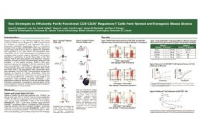 科学海报Cell Isolation of Functional CD4+CD25+ Regulatory T Cells from Mouse Strains
 沪公网安备31010102008431号