Vector-Free and Transgene-Free Human iPS Cells Differentiate into Functional Neurons and Enhance Functional Recovery after Ischemic Stroke in Mice
Stroke is a leading cause of human death and disability in the adult population in the United States and around the world. While stroke treatment is limited,stem cell transplantation has emerged as a promising regenerative therapy to replace or repair damaged tissues and enhance functional recovery after stroke. Recently,the creation of induced pluripotent stem (iPS) cells through reprogramming of somatic cells has revolutionized cell therapy by providing an unlimited source of autologous cells for transplantation. In addition,the creation of vector-free and transgene-free human iPS (hiPS) cells provides a new generation of stem cells with a reduced risk of tumor formation that was associated with the random integration of viral vectors seen with previous techniques. However,the potential use of these cells in the treatment of ischemic stroke has not been explored. In the present investigation,we examined the neuronal differentiation of vector-free and transgene-free hiPS cells and the transplantation of hiPS cell-derived neural progenitor cells (hiPS-NPCs) in an ischemic stroke model in mice. Vector-free hiPS cells were maintained in feeder-free and serum-free conditions and differentiated into functional neurons in vitro using a newly developed differentiation protocol. Twenty eight days after transplantation in stroke mice,hiPS-NPCs showed mature neuronal markers in vivo. No tumor formation was seen up to 12 months after transplantation. Transplantation of hiPS-NPCs restored neurovascular coupling,increased trophic support and promoted behavioral recovery after stroke. These data suggest that using vector-free and transgene-free hiPS cells in stem cell therapy are safe and efficacious in enhancing recovery after focal ischemic stroke in mice.
View Publication
产品类型:
产品号#:
05850
05857
05870
05875
85850
85857
85870
85875
产品名:
mTeSR™1
mTeSR™1
Wang H et al. (APR 2016)
The Journal of biological chemistry 291 16 8644--8652
Germ Cell Nuclear Factor (GCNF) Represses Oct4 Expression and Globally Modulates Gene Expression in Human Embryonic Stem (hES) Cells.
Oct4 is considered a key transcription factor for pluripotent stem cell self-renewal. It binds to specific regions within target genes to regulate their expression and is downregulated upon induction of differentiation of pluripotent stem cells; however,the mechanisms that regulate the levels of human Oct4 expression remain poorly understood. Here we show that expression of human Oct4 is directly repressed by germ cell nuclear factor (GCNF),an orphan nuclear receptor,in hES cells. Knockdown of GCNF by siRNA resulted in maintenance of Oct4 expression during RA-induced hES cell differentiation. While overexpression of GCNF promoted repression of Oct4 expression in both undifferentiated and differentiated hES cells. The level of Oct4 repression was dependent on the level of GCNF expression in a dose-dependent manner. mRNA microarray analysis demonstrated that overexpression of GCNF globally regulates gene expression in undifferentiated and differentiated hES cells. Within the group of altered genes,GCNF down-regulated 36% of the genes,and up-regulated 64% in undifferentiated hES cells. In addition,GCNF also showed a regulatory gene pattern that is different from RA treatment during hES cell differentiation. These findings increase our understanding of the mechanisms that maintain hES cell pluripotency and regulate gene expression during the differentiation process.
View Publication
T. Ito-Kureha et al. (aug 2022)
Nature immunology 23 8 1208--1221
The function of Wtap in N6-adenosine methylation of mRNAs controls T cell receptor signaling and survival of T cells.
T cell antigen-receptor (TCR) signaling controls the development,activation and survival of T cells by involving several layers and numerous mechanisms of gene regulation. N6-methyladenosine (m6A) is the most prevalent messenger RNA modification affecting splicing,translation and stability of transcripts. In the present study,we describe the Wtap protein as essential for m6A methyltransferase complex function and reveal its crucial role in TCR signaling in mouse T cells. Wtap and m6A methyltransferase functions were required for the differentiation of thymocytes,control of activation-induced death of peripheral T cells and prevention of colitis by enabling gut ROR?t+ regulatory T cell function. Transcriptome and epitranscriptomic analyses reveal that m6A modification destabilizes Orai1 and Ripk1 mRNAs. Lack of post-transcriptional repression of the encoded proteins correlated with increased store-operated calcium entry activity and diminished survival of T cells with conditional genetic inactivation of Wtap. These findings uncover how m6A modification impacts on TCR signal transduction and determines activation and survival of T cells.
View Publication
产品类型:
产品号#:
19852
19852RF
产品名:
EasySep™小鼠CD4+ T细胞分选试剂盒
RoboSep™ 小鼠CD4+ T细胞分选试剂盒
L. Wang et al. (nov 2019)
European journal of pharmacology 863 172676
Decitabine promotes apoptosis in mesenchymal stromal cells isolated from patients with myelodysplastic syndromes by inducing reactive oxygen species generation.
Myelodysplastic syndromes (MDSs) are a group of clonal disorders of hematopoietic stem cells,resulting in ineffective hematopoiesis. Previous studies have reported that decitabine (DAC) plays an essential role in cell cycle arrest and cell death induction in multiple cell types. Nevertheless,the effect of decitabine on mesenchymal stromal cells derived from bone marrow of patients with MDSs is not completely clarified. Here,we explored the apoptotic and anti-proliferative effect of DAC on MSCs isolated from patients with MDSs. Treatment with DAC inhibited cell growth in a concentration- and time-dependent manner by inducing apoptosis. We found a positive relationship between cell death triggered by DAC in MSCs and the death receptor family members Fas and FasL mRNA and protein levels (***P {\textless} 0.00085),cleaved caspase (-3,-8,and -9) activity,and mitochondrial membrane potential reduction. Additionally,DAC-induced apoptosis was inhibited by Kp7-6,a FasL/Fas antagonist,indicating a crucial role of FasL/Fas,a cell death receptor,in mediating the apoptotic effect of DAC. DAC also induced reactive oxygen species (ROS) generation in MSCs derived from MDSs patients (*P = 0.038). Furthermore,N-acetyl-L-cysteine (NAC),a widely accepted ROS scavenger,efficiently reversed DAC-induced apoptosis by inhibiting ROS generation (***P {\textless} 0.00051) in mitochondria and restoring mitochondrial membrane potential. Furthermore,ROS production was found to be a consequence of caspase activation via caspases inhibition. Our data imply that DAC triggers ROS production in human MSCs,which serves as a crucial factor for mitochondrial membrane potential reduction,and DAC induces cell death prior to FasL/Fas stimulation.
View Publication
产品类型:
产品号#:
85450
85460
产品名:
SepMate™-50 (IVD)
SepMate™-50 (IVD)
(Oct 2024)
BMC Psychiatry 24 1
Patient iPSC-derived neural progenitor cells display aberrant cell cycle control, p53, and DNA damage response protein expression in schizophrenia
BackgroundSchizophrenia (SCZ) is a severe psychiatric disorder associated with alterations in early brain development. Details of underlying pathomechanisms remain unclear,despite genome and transcriptome studies providing evidence for aberrant cellular phenotypes and pathway deregulation in developing neuronal cells. However,mechanistic insight at the protein level is limited.MethodsHere,we investigate SCZ-specific protein expression signatures of neuronal progenitor cells (NPC) derived from patient iPSC in comparison to healthy controls using high-throughput Western Blotting (DigiWest) in a targeted proteomics approach.ResultsSCZ neural progenitors displayed altered expression and phosphorylation patterns related to Wnt and MAPK signaling,protein synthesis,cell cycle regulation and DNA damage response. Consistent with impaired cell cycle control,SCZ NPCs also showed accumulation in the G2/M cell phase and reduced differentiation capacity. Furthermore,we correlated these findings with elevated p53 expression and phosphorylation levels in SCZ patient-derived cells,indicating a potential implication of p53 in hampering cell cycle progression and efficient neurodevelopment in SCZ.ConclusionsThrough targeted proteomics we demonstrate that SCZ NPC display coherent mechanistic alterations in regulation of DNA damage response,cell cycle control and p53 expression. These findings highlight the suitability of iPSC-based approaches for modeling psychiatric disorders and contribute to a better understanding of the disease mechanisms underlying SCZ,particularly during early development.Supplementary InformationThe online version contains supplementary material available at 10.1186/s12888-024-06127-x.
View Publication
产品类型:
产品号#:
05833
08581
08582
100-0276
100-1130
产品名:
STEMdiff™神经前体细胞培养基
STEMdiff™SMADi神经诱导试剂盒
STEMdiff™SMADi神经诱导试剂盒,2套
mTeSR™ Plus
mTeSR™ Plus
(Sep 2024)
Nature Cancer 5 10
ISB 2001 trispecific T cell engager shows strong tumor cytotoxicity and overcomes immune escape mechanisms of multiple myeloma cells
Despite recent advances in immunotherapies targeting single tumor-associated antigens,patients with multiple myeloma eventually relapse. ISB 2001 is a CD3+ T cell engager (TCE) co-targeting BCMA and CD38 designed to improve cytotoxicity against multiple myeloma. Targeting of two tumor-associated antigens by a single TCE resulted in superior cytotoxic potency across a variable range of BCMA and CD38 tumor expression profiles mimicking natural tumor heterogeneity,improved resistance to competing soluble factors and exhibited superior cytotoxic potency on patient-derived samples and in mouse models. Despite the broad expression of CD38 across human tissues,ISB 2001 demonstrated a reduced T cell activation profile in the absence of tumor cells when compared to TCEs targeting CD38 only. To determine an optimal first-in-human dose for the ongoing clinical trial (NCT05862012),we developed an innovative quantitative systems pharmacology model leveraging preclinical data,using a minimum pharmacologically active dose approach,therefore reducing patient exposure to subefficacious doses of therapies. Perro and colleagues develop a CD3+ T cell engager co-targeting BCMA and CD38 to improve immunotherapy for multiple myeloma,demonstrate cytotoxicity in patient-derived samples and murine models and develop a quantitative systems pharmacology model.
View Publication
产品类型:
产品号#:
17951
100-0695
17951RF
产品名:
EasySep™人T细胞分选试剂盒
EasySep™人T细胞分选试剂盒
RoboSep™ 人T细胞分选试剂盒
(Jun 2025)
Frontiers in Medicine 12
The microbial metabolite butyrate enhances the effector and memory functions of murine CD8+ T cells and improves anti-tumor activity
IntroductionCD8+ T cells are vital in the immune control of cancer and a key player in cell-based cancer immunotherapy. Recent studies have shown that microbial short-chain fatty acids (SCFA) can promote both effector and memory phenotypes in CD8+ T cells and may thereby enhance protection against cancer.MethodsIn this study,we determined the effect of SCFA butyrate on mouse CD8+ T cell function in vitro and in vivo,using the OT-I model.ResultsButyrate co-culture with anti-CD3 + anti-CD28 activated T cells in vitro enhanced the frequency of effector CD8+ IFN-γ-producing cells,and the amount of cytokine produced per cell. Culture with butyrate also enhanced the activation,TCR expression,and levels of phosphorylated mTOR proteins within CD8+ T cells but reduced proliferation rate and increased apoptosis. Butyrate-treated activated cells conferred tumor protection after adoptive transfer. Butyrate-treated cells were present at higher frequencies within the tumor compared to non-butyrate treated cells,and expressed IFN-γ. When analyzed using high dimensional cytometry,the tumors of mice that received butyrate-treated cells were enriched in clusters displaying an effector memory phenotype with high expression of IL-15Rβ and T-bet.DiscussionOur findings show that butyrate promotes the effector activity of CD8+ T cells in culture,which can persist in vivo while also stimulating memory phenotypes. Consequently,butyrate treatment may have strong application in T cell-based immunotherapies to improve protective cell functions and patient outcomes.
View Publication
Schmidt K et al. (MAR 2009)
Journal of leukocyte biology 85 3 563--73
Histone deacetylase inhibition improves differentiation of dendritic cells from leukemic blasts of patients with TEL/AML1-positive acute lymphoblastic leukemia.
Histone deacetylase inhibitors (HdI) could potentially improve the differentiation of leukemic dendritic cells (DC). Therefore,bone marrow samples from 100 children with acute lymphoblastic leukemia (ALL) were cultured in the cytokines TNF-alpha,GM-CSF,c-kit ligand,and fetal liver tyrosine kinase 3 ligand,with or without IL-3 and -4 and after administration of HdI valproic acid (VAL),suberoylanilide hydroxamic acid (SAHA),isobutyramid,or trichostatin A. Among the tested samples,25 were positive for the chromosomal translocation t(12;21),encoding the fusion gene translocation ETS-like leukemia/acute myeloid leukemia 1 (TEL/AML1). SAHA increased CD83 expression of TEL/AML1-positive blasts in conditions without ILs,and SAHA and VAL increased the number of CD86(+)80(-) cells in the presence of ILs. VAL and isobutyramid supported the allostimulatory capacities of TEL/AML1-positive,leukemic DC; VAL and SAHA reduced those of TEL/AML1-negative DC. Cytotoxic T cells sensitized with leukemic DC produced more IFN-gamma and TNF-alpha upon presentation of the TEL/AML1 peptide. They also induced the cytotoxic lysis of nondifferentiated blasts,which was enhanced when TEL/AML1-positive DC had developed after addition of VAL or SAHA. Therefore,the use of HdI in the differentiation of leukemic DC from patients with TEL/AML1-positive ALL is recommended.
View Publication
产品类型:
产品号#:
09850
产品名:
Burger R et al. ( 2009)
Molecular Cancer Therapeutics 8 1 26--35
Janus kinase inhibitor INCB20 has antiproliferative and apoptotic effects on human myeloma cells in vitro and in vivo
Protein tyrosine kinases of the Janus kinase (JAK) family are associated with many cytokine receptors,which,on ligand binding,regulate important cellular functions such as proliferation,survival,and differentiation. In multiple myeloma,JAKs may be persistently activated due to a constant stimulation by interleukin (IL)-6,which is produced in the bone marrow environment. INCB20 is a synthetic molecule that potently inhibits all members of the JAK family with a 100- to 1,000-fold selectivity for JAKs over textgreater70 other kinases. Treatment of multiple myeloma cell lines and patient tumor cells with INCB20 resulted in a significant and dose-dependent inhibition of spontaneous as well as IL-6-induced cell growth. Importantly,multiple myeloma cell growth was inhibited in the presence of bone marrow stromal cells. The IL-6 dependent cell line INA-6 was particularly sensitive to the drug (IC50textless1 micromol/L). Growth suppression of INA-6 correlated with an increase in the percentage of apoptotic cells and inhibition of signal transducer and activator of transcription 3 phosphorylation. INCB20 also abrogated the protective effect of IL-6 against dexamethasone by blocking phosphorylation of SHP-2 and AKT. In contrast,AKT phosphorylation induced by insulin-like growth factor-I remained unchanged,showing selectivity of the compound. In a s.c. severe combined immunodeficient mouse model with INA-6,INCB20 significantly delayed INA-6 tumor growth. Our studies show that disruption of JAKs and downstream signaling pathways may both inhibit multiple myeloma cell growth and survival and overcome cytokine-mediated drug resistance,thereby providing the preclinical rationale for the use of JAK inhibitors as a novel therapeutic approach in multiple myeloma.
View Publication
产品类型:
产品号#:
72932
72934
产品名:
AG - 490
Eberhard Y et al. (OCT 2009)
Blood 114 14 3064--73
Chelation of intracellular iron with the antifungal agent ciclopirox olamine induces cell death in leukemia and myeloma cells.
Off-patent drugs with previously unrecognized anticancer activity could be rapidly repurposed for this new indication. To identify such compounds,we conducted 2 independent cell-based chemical screens and identified the antimicrobial ciclopirox olamine (CPX) in both screens. CPX decreased cell growth and viability of malignant leukemia,myeloma,and solid tumor cell lines as well as primary AML patient samples at low-micromolar concentrations that appear pharmacologically achievable. Furthermore,oral CPX decreased tumor weight and volume in 3 mouse models of leukemia by up to 65% compared with control without evidence of weight loss or gross organ toxicity. In addition,oral CPX prevented the engraftment of primary AML cells in nonobese diabetic/severe combined immunodeficiency mouse models,thereby establishing its ability to target leukemia stem cells. Mechanistically,CPX bound intracellular iron,and this intracellular iron chelation was functionally important for its cytotoxicity. By electron paramagnetic resonance,CPX inhibited the iron-dependent enzyme ribonucleotide reductase at concentrations associated with cell death. Thus,in summary,CPX has previously unrecognized anticancer activity at concentrations that are pharmacologically achievable. Therefore,CPX could be rapidly repurposed for the treatment of malignancies,including leukemia and myeloma.
View Publication

 EasySep™小鼠TIL(CD45)正选试剂盒


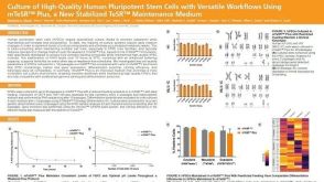 科学海报Culture of High-Quality Human Pluripotent Stem Cells with Versatile Workflows Using mTeSR™ Plus, a New Stabilized TeSR™ Maintenance Medium
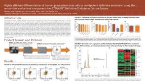 科学海报Highly Efficient Differentiation of Human Pluripotent Stem Cells to Multipotent Definitive Endoderm Using STEMdiff™ Definitive Endoderm Culture System
 沪公网安备31010102008431号