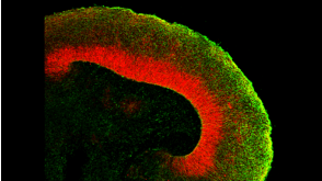
Laumont C et al. (JAN 2016)
Nature Communications 7 10238
Global proteogenomic analysis of human MHC class I-associated peptides derived from non-canonical reading frames.
In view of recent reports documenting pervasive translation outside of canonical protein-coding sequences,we wished to determine the proportion of major histocompatibility complex (MHC) class I-associated peptides (MAPs) derived from non-canonical reading frames. Here we perform proteogenomic analyses of MAPs eluted from human B cells using high-throughput mass spectrometry to probe the six-frame translation of the B-cell transcriptome. We report that ∼ 10% of MAPs originate from allegedly noncoding genomic sequences or exonic out-of-frame translation. The biogenesis and properties of these 'cryptic MAPs' differ from those of conventional MAPs. Cryptic MAPs come from very short proteins with atypical C termini,and are coded by transcripts bearing long 3'UTRs enriched in destabilizing elements. Relative to conventional MAPs,cryptic MAPs display different MHC class I-binding preferences and harbour more genomic polymorphisms,some of which are immunogenic. Cryptic MAPs increase the complexity of the MAP repertoire and enhance the scope of CD8 T-cell immunosurveillance.
View Publication
产品类型:
产品号#:
产品名:
Di Cello F et al. (APR 2013)
Biochemical and biophysical research communications 434 1 70--74
Knockdown of HMGA1 inhibits human breast cancer cell growth and metastasis in immunodeficient mice.
The high mobility group A1 gene (HMGA1) has been previously implicated in breast carcinogenesis,and is considered an attractive target for therapeutic intervention because its expression is virtually absent in normal adult tissue. Other studies have shown that knockdown of HMGA1 reduces the tumorigenic potential of breast cancer cells in vitro. Therefore,we sought to determine if silencing HMGA1 can affect breast cancer development and metastatic progression in vivo. We silenced HMGA1 expression in the human breast cancer cell line MDA-MB-231 using an RNA interference vector,and observed a significant reduction in anchorage-independent growth and tumorsphere formation,which respectively indicate loss of tumorigenesis and self-renewal ability. Moreover,silencing HMGA1 significantly impaired xenograft growth in immunodeficient mice,and while control cells metastasized extensively to the lungs and lymph nodes,HMGA1-silenced cells generated only a few small metastases. Thus,our results show that interfering with HMGA1 expression reduces the tumorigenic and metastatic potential of breast cancer cells in vivo,and lend further support to investigations into targeting HMGA1 as a potential treatment for breast cancer.
View Publication
产品类型:
产品号#:
05620
产品名:
MammoCult™ 人源培养基套装
D. G. Belair et al. (jul 2020)
Toxicology in vitro : an international journal published in association with BIBRA 68 104928
Human ileal organoid model recapitulates clinical incidence of diarrhea associated with small molecule drugs.
Drug-induced gastrointestinal toxicity (GIT) is a common treatment-emergent adverse event that can negatively impact dosing,thereby limiting efficacy and treatment options for patients. An in vitro assay of GIT is needed to address patient variability,mimic the microphysiology of the gut,and accurately predict drug-induced GIT. Primary human ileal organoids (termed 'enteroids') have proven useful for stimulating intestinal stem cell proliferation and differentiation to multiple cell types present in the gut epithelium. Enteroids have enabled characterization of gut biology and the signaling involved in the pathogenesis of disease. Here,enteroids were differentiated from four healthy human donors and assessed for culture duration-dependent differentiation status by immunostaining for gut epithelial markers lysozyme,chromogranin A,mucin,and sucrase isomaltase. Differentiated enteroids were evaluated with a reference set of 31 drugs exhibiting varying degrees of clinical incidence of diarrhea,a common manifestation of GIT that can be caused by drug-induced thinning of the gut epithelium. An assay examining enteroid viability in response to drug treatment demonstrated 90{\%} accuracy for recapitulating the incidence of drug-induced diarrhea. The human enteroid viability assay developed here presents a promising in vitro model for evaluating drug-induced diarrhea.
View Publication
产品类型:
产品号#:
06010
产品名:
IntestiCult™ 类器官生长培养基 (人)
A. Nemoto et al. (Oct 2025)
Nature Communications 16
Rescue of imprinted genes by epigenome editing in human cellular models of Prader-Willi syndrome
Prader-Willi syndrome (PWS) is a genomic imprinting disorder caused by the loss of function of the paternal chromosome 15q11-13,resulting in a spectrum of symptoms associated with hypothalamic dysfunction. PWS patients lack the expression of paternally expressed genes (PEGs) in the 15q11-13 locus but possess an epigenetically silenced set of these genes in the maternal allele. Thus,activation of these silenced genes can serve as a therapeutic target for PWS. Here,we leverage CRISPR-based epigenome editing system to modulate the DNA methylation status of the PWS imprinting control region (PWS-ICR) in induced pluripotent stem cells (iPSCs) derived from PWS patients. Successful demethylation in the PWS-ICR restores the PEG expression from the maternal allele and reorganizes the methylation patterns in other PWS-associated imprinted regions beyond the PWS-ICR. Remarkably,these corrected epigenomic patterns and PEG expression are maintained following the differentiation of these cells into hypothalamic organoids. Finally,the single-cell transcriptomic analysis of epigenome-edited organoids demonstrates a partial restoration of the transcriptomic dysregulation observed in PWS. This study highlights the utility of epigenome editing technology as a therapeutic approach in addressing PWS and potentially other imprinting disorders. The authors develop CRISPR-based epigenome editing strategy to reactivate silenced maternally inherited genes for Prader-Willi syndrome in human iPSC and hypothalamic organoid models,highlighting its potential for treating imprinting disorders.
View Publication
Konorov SO et al. (JUL 2010)
Applied spectroscopy 64 7 767--74
Lorentzian amplitude and phase pulse shaping for nonresonant background suppression and enhanced spectral resolution in coherent anti-Stokes Raman scattering spectroscopy and microscopy.
Femtosecond coherent anti-Stokes Raman scattering (CARS) spectroscopy offers several advantages over spontaneous Raman spectroscopy due to the inherently high sensitivity and low average power deposition in the sample. Femtosecond CARS can be implemented in a collinear pump/probe beam configuration for microspectroscopy applications and has emerged as a powerful technique for chemical imaging of biological specimens. However,one serious limitation of this approach is the presence of a high nonresonant background component that often obscures the resonant signals of interest. We report here an innovative pulse-shaping method based on Lorentzian amplitude and phase spectral modulation of a broadband femtosecond probe pulse that yields spectra with both high spectral resolution and no nonresonant background. No further mathematical analysis is needed to extract Raman spectra. The utility of the proposed method for CARS microscopy is demonstrated using a mixture of polystyrene and latex beads,as well as dry-fixed embryonic stem cells.
View Publication
产品类型:
产品号#:
05850
05857
05870
05875
85850
85857
85870
85875
产品名:
mTeSR™1
mTeSR™1
Matsumoto K et al. (JAN 2000)
Stem cells (Dayton,Ohio) 18 3 196--203
In vitro proliferation potential of AC133 positive cells in peripheral blood.
AC133 antigen is a novel marker for human hematopoietic stem/progenitor cells. In this study,we examined the expression and proliferation potential of AC133(+) cells obtained from steady-state peripheral blood (PB). The proportion of AC133(+) cells in the CD34(+) subpopulation of steady-state PB was significantly lower than that of cord blood (CB),although that of cytokine-mobilized PB was higher than that of CB. The proliferation potential of AC133(+)CD34(+) and AC133(-)CD34(+) cells was examined by colony-forming analysis and analysis of long-term culture-initiating cells (LTC-IC). Although the total number of colony-forming cells was essentially the same in the AC133(+)CD34(+) fraction as in the AC133(-)CD34(+) fraction,the proportion of LTC-IC was much higher in the AC133(+)CD34(+) fraction. Virtually no LTC-IC were detected in the AC133(-)CD34(+) fraction. In addition,the features of the colonies grown from these two fractions were quite different. Approximately 70% of the colonies derived from the AC133(+)CD34(+) fraction were granulocyte-macrophage colonies,whereas more than 90% of the colonies derived from the AC133(-)CD34(+) fraction were erythroid colonies. Furthermore,an ex vivo expansion study observed expansion of colony-forming cells only in the AC133(+)CD34(+) population,and not in the AC133(-)CD34(+) population. These findings suggest that to isolate primitive hematopoietic cells from steady-state PB,selection by AC133 expression is better than selection by CD34 expression.
View Publication
产品类型:
产品号#:
04034
04044
产品名:
MethoCult™ H4034 Optimum
MethoCult™ H4034 Optimum
Schwieger M et al. (SEP 2009)
Blood 114 12 2476--88
Homing and invasiveness of MLL/ENL leukemic cells is regulated by MEF2C.
Acute myelogenous leukemia is driven by leukemic stem cells (LSCs) generated by mutations that confer (or maintain) self-renewal potential coupled to an aberrant differentiation program. Using retroviral mutagenesis,we identified genes that generate LSCs in collaboration with genetic disruption of the gene encoding interferon response factor 8 (Irf8),which induces a myeloproliferation in vivo. Among the targeted genes,we identified Mef2c,encoding a MCM1-agamous-deficiens-serum response factor transcription factor,and confirmed that overexpression induced a myelomonocytic leukemia in cooperation with Irf8 deficiency. Strikingly,several of the genes identified in our screen have been reported to be up-regulated in the mixed-lineage leukemia (MLL) subtype. High MEF2C expression levels were confirmed in acute myelogenous leukemia patient samples with MLL gene disruptions,prompting an investigation of the causal interplay. Using a conditional mouse strain,we demonstrated that Mef2c deficiency does not impair the establishment or maintenance of LSCs generated in vitro by MLL/ENL fusion proteins; however,its loss led to compromised homing and invasiveness of the tumor cells. Mef2c-dependent targets included several genes encoding matrix metalloproteinases and chemokine ligands and receptors,providing a mechanistic link to increased homing and motility. Thus,MEF2C up-regulation may be responsible for the aggressive nature of this leukemia subtype.
View Publication
产品类型:
产品号#:
03434
03444
09600
09650
产品名:
MethoCult™ GF M3434
MethoCult™ GF M3434
StemSpan™ SFEM
StemSpan™ SFEM
Eguchi M et al. (JAN 2005)
Proceedings of the National Academy of Sciences of the United States of America 102 4 1133--8
Directing oncogenic fusion genes into stem cells via an SCL enhancer.
TEL-TRKC is a fusion gene generated by chromosomal translocation and encodes an activated tyrosine kinase. Uniquely,it is found in both solid tumors and leukemia. However,a single exon difference (in TEL) in TEL-TRKC fusions is associated with the two sets of cancer phenotypes. We expressed the two TEL-TRKC variants in vivo by using the 3' regulatory element of SCL that is selectively active in a subset of mesodermal cell lineages,including endothelial and hematopoietic stem cells and progenitors. The leukemia form of TEL-TRKC (-exon 5 of TEL) enhanced hematopoietic stem cell renewal and initiated leukemia. In contrast,the TEL-TRKC solid tumor variant (+ TEL exon 5) elicited an embryonic lethal phenotype with impairment of both angiogenesis and hematopoiesis indicative of an effect at the level of the hemangioblasts. The ability of TEL-TRKC to repress expression of Flk1,a critical regulator of early endothelial and hematopoietic cells,depended on TEL exon 5. These data indicate that related oncogenic fusion proteins similarly expressed in a hierarchy of early stem cells can have selective,cell type-specific developmental impacts.
View Publication
产品类型:
产品号#:
03231
产品名:
MethoCult™ M3231
Nayak RC et al. (AUG 2015)
The Journal of clinical investigation 125 8 3103--3116
Pathogenesis of ELANE-mutant severe neutropenia revealed by induced pluripotent stem cells.
Severe congenital neutropenia (SCN) is often associated with inherited heterozygous point mutations in ELANE,which encodes neutrophil elastase (NE). However,a lack of appropriate models to recapitulate SCN has substantially hampered the understanding of the genetic etiology and pathobiology of this disease. To this end,we generated both normal and SCN patient-derived induced pluripotent stem cells (iPSCs),and performed genome editing and differentiation protocols that recapitulate the major features of granulopoiesis. Pathogenesis of ELANE point mutations was the result of promyelocyte death and differentiation arrest,and was associated with NE mislocalization and activation of the unfolded protein response/ER stress (UPR/ER stress). Similarly,high-dose G-CSF (or downstream signaling through AKT/BCL2) rescues the dysgranulopoietic defect in SCN patient-derived iPSCs through C/EBP$$-dependent emergency granulopoiesis. In contrast,sivelestat,an NE-specific small-molecule inhibitor,corrected dysgranulopoiesis by restoring normal intracellular NE localization in primary granules; ameliorating UPR/ER stress; increasing expression of CEBPA,but not CEBPB; and promoting promyelocyte survival and differentiation. Together,these data suggest that SCN disease pathogenesis includes NE mislocalization,which in turn triggers dysfunctional survival signaling and UPR/ER stress. This paradigm has the potential to be clinically exploited to achieve therapeutic responses using lower doses of G-CSF combined with targeting to correct NE mislocalization.
View Publication
产品类型:
产品号#:
05850
05857
05870
05875
04034
04044
85850
85857
85870
85875
产品名:
MethoCult™ H4034 Optimum
MethoCult™ H4034 Optimum
mTeSR™1
mTeSR™1
Tamaki T et al. (MAY 2002)
The Journal of cell biology 157 4 571--7
Identification of myogenic-endothelial progenitor cells in the interstitial spaces of skeletal muscle.
Putative myogenic and endothelial (myo-endothelial) cell progenitors were identified in the interstitial spaces of murine skeletal muscle by immunohistochemistry and immunoelectron microscopy using CD34 antigen. Enzymatically isolated cells were characterized by fluorescence-activated cell sorting on the basis of cell surface antigen expression,and were sorted as a CD34+ and CD45- fraction. Cells in this fraction were approximately 94% positive for Sca-1,and mostly negative (textless3% positive) for CD14,31,49,144,c-kit,and FLK-1. The CD34+/45- cells formed colonies in clonal cell cultures and colony-forming units displayed the potential to differentiate into adipocytes,endothelial,and myogenic cells. The CD34+/45- cells fully differentiated into vascular endothelial cells and skeletal muscle fibers in vivo after transplantation. Immediately after sorting,CD34+/45- cells expressed only c-met mRNA,and did not express any other myogenic cell-related markers such as MyoD,myf-5,myf-6,myogenin,M-cadherin,Pax-3,and Pax-7. However,after 3 d of culture,these cells expressed mRNA for all myogenic markers. CD34+/45- cells were distinct from satellite cells,as they expressed Bcrp1/ABCG2 gene mRNA (Zhou et al.,2001). These findings suggest that myo-endothelial progenitors reside in the interstitial spaces of mammalian skeletal muscles,and that they can potentially contribute to postnatal skeletal muscle growth.
View Publication
产品类型:
产品号#:
04034
04044
产品名:
MethoCult™ H4034 Optimum
MethoCult™ H4034 Optimum
Dotsenko O et al. (DEC 2010)
The Annals of thoracic surgery 90 6 1944--51
Bone marrow resident and circulating progenitor cells in patients undergoing cardiac surgery.
BACKGROUND: Vascular trauma induced by surgical revascularization stimulates mobilization of hematopoietic and nonhematopoietic progenitor cells. However,it is not clear whether mobilized progenitors are functionally active and participate in peripheral homing. We have found no clinical studies available regarding the reaction of bone marrow to surgical revascularization. METHODS: This was an observational prospective study of 76 patients undergoing elective coronary artery bypass graft surgery. Bone marrow aspirates and blood samples were collected at baseline,at the end of surgery,and 24 hours postoperatively (blood samples only). The CD34+,CD34+CD133+,and CD34+CXCR4+ progenitor cell counts,CXCR4+ mononuclear cell counts,and CXCR4 expression on CD34+ cells were measured by flow cytometry. Progenitor cell functions were studied in vitro by clonogenic and migration assays. RESULTS: In response to coronary revascularization there was mobilization of CD34+ progenitors,having increased migratory and clonogenic function. The CD34+CXCR4+ subsets and CXCR4 expression on CD34+ cells in peripheral blood increased significantly 24 hours postoperatively. The CXCR4 expression on mobilized progenitors at the end of surgery was independently related to baseline CXCR4 expression on bone marrow resident CD34+ cells and duration of cardiopulmonary bypass in a multivariate model. At the end of surgery there was a significant fall in the expression of CXCR4 on CD34+ bone marrow cells,suggesting egress into peripheral circulation of the most active CXCR4-expressing progenitors. CONCLUSIONS: Coronary artery bypass graft surgery is associated with bone marrow release of functionally active progenitor cells. Further studies are needed to verify whether mobilized progenitors participate in regeneration of injured tissues.
View Publication

 EasySep™小鼠TIL(CD45)正选试剂盒


 沪公网安备31010102008431号