Yan Z et al. (AUG 2017)
Human gene therapy 28 8 612--625
Human Bocavirus Type-1 Capsid Facilitates the Transduction of Ferret Airways by Adeno-Associated Virus Genomes.
Human bocavirus type-1 (HBoV1) has a high tropism for the apical membrane of human airway epithelia. The packaging of a recombinant adeno-associated virus 2 (rAAV2) genome into HBoV1 capsid produces a chimeric vector (rAAV2/HBoV1) that also efficiently transduces human airway epithelia. As such,this vector is attractive for use in gene therapies to treat lung diseases such as cystic fibrosis. However,preclinical development of rAAV2/HBoV1 vectors has been hindered by the fact that humans are the only known host for HBoV1 infection. This study reports that rAAV2/HBoV1 vector is capable of efficiently transducing the lungs of both newborn (3- to 7-day-old) and juvenile (29-day-old) ferrets,predominantly in the distal airways. Analyses of in vivo,ex vivo,and in vitro models of the ferret proximal airway demonstrate that infection of this particular region is less effective than it is in humans. Studies of vector binding and endocytosis in polarized ferret proximal airway epithelial cultures revealed that a lack of effective vector endocytosis is the main cause of inefficient transduction in vitro. While transgene expression declined proportionally with growth of the ferrets following infection at 7 days of age,reinfection of ferrets with rAAV2/HBoV1 at 29 days gave rise to approximately 5-fold higher levels of transduction than observed in naive infected 29-day-old animals. The findings presented here lay the foundation for clinical development of HBoV1 capsid-based vectors for lung gene therapy in cystic fibrosis using ferret models.
View Publication
Pig Induced Pluripotent Stem Cell-Derived Neural Rosettes Parallel Human Differentiation Into Sensory Neural Subtypes.
The pig is the large animal model of choice for study of nerve regeneration and wound repair. Availability of porcine sensory neural cells would conceptually allow for analogous cell-based peripheral nerve regeneration in porcine injuries of similar severity and size to those found in humans. After recently reporting that porcine (or pig) induced pluripotent stem cells (piPSCs) differentiate into neural rosette (NR) structures similar to human NRs,here we demonstrate that pig NR cells could differentiate into neural crest cells and other peripheral nervous system-relevant cell types. Treatment with either bone morphogenetic protein 4 or fetal bovine serum led to differentiation into BRN3A-positive sensory cells and increased expression of sensory neuron TRK receptor gene family: TRKA,TRKB,and TRKC. Porcine sensory neural cells would allow determination of parallels between human and porcine cells in response to noxious stimuli,analgesics,and reparative mechanisms. In vitro differentiation of pig sensory neurons provides a novel model system for neural cell subtype specification and would provide a novel platform for the study of regenerative therapeutics by elucidating the requirements for innervation following injury and axonal survival.
View Publication
Induction of a human pluripotent state with distinct regulatory circuitry that resembles preimplantation epiblast
Human embryonic stem cells (hESCs) are derived from the inner cell mass of the blastocyst. Despite sharing the common property of pluripotency,hESCs are notably distinct from epiblast cells of the preimplantation blastocyst. Here we use a combination of three small-molecule inhibitors to sustain hESCs in a LIF signaling-dependent hESC state (3iL hESCs) with elevated expression of NANOG and epiblast-enriched genes such as KLF4,DPPA3,and TBX3. Genome-wide transcriptome analysis confirms that the expression signature of 3iL hESCs shares similarities with native preimplantation epiblast cells. We also show that 3iL hESCs have a distinct epigenetic landscape,characterized by derepression of preimplantation epiblast genes. Using genome-wide binding profiles of NANOG and OCT4,we identify enhancers that contribute to rewiring of the regulatory circuitry. In summary,our study identifies a distinct hESC state with defined regulatory circuitry that will facilitate future analysis of human preimplantation embryogenesis and pluripotency.
View Publication
产品类型:
产品号#:
05850
05857
05870
05875
85850
85857
85870
85875
产品名:
mTeSR™1
mTeSR™1
Hur J et al. (AUG 2014)
Molecular therapy : the journal of the American Society of Gene Therapy 22 8 1518--29
Human podoplanin-positive monocytes and platelets enhance lymphangiogenesis through the activation of the podoplanin/CLEC-2 axis.
Emerging studies suggested that murine podoplanin-positive monocytes (PPMs) are involved in lymphangiogenesis. The goal of this study was to demonstrate the therapeutic lymphangiogenesis of human PPMs by the interaction with platelets. Aggregation culture of human peripheral blood mononuclear cells (PBMCs) resulted in cellular aggregates termed hematospheres. During 5-day culture,PPMs expanded exponentially and expressed several lymphatic endothelial cell-specific markers including vascular endothelial growth factor receptor (VEGFR)-3 and well-established lymphangiogenic transcription factors. Next,we investigated the potential interaction of PPMs with platelets that had C-type lectin-like receptor-2 (CLEC-2),a receptor of podoplanin. In vitro coculture of PPMs and platelets stimulated PPMs to strongly express lymphatic endothelial markers and upregulate lymphangiogenic cytokines. Recombinant human CLEC-2 also stimulated PPMs through Akt and Erk signaling. Likewise,platelets in coculture with PPMs augmented secretion of a lymphangiogenic cytokine,interleukin (IL)-1-β,via podoplanin/CLEC-2 axis. The supernatant obtained from coculture was able to enhance the migration,viability,and proliferation of lymphatic endothelial cell. Local injection of hematospheres with platelets significantly increased lymphatic neovascularization and facilitated wound healing in the full-thickness skin wounds of nude mice. Cotreatment with PPMs and platelets augments lymphangiogenesis through podoplanin/CLEC-2 axis,which thus would be a promising novel strategy of cell therapy to treat human lymphatic vessel disease.
View Publication
The primate-specific noncoding RNA HPAT5 regulates pluripotency during human preimplantation development and nuclear reprogramming.
Long intergenic noncoding RNAs (lincRNAs) are derived from thousands of loci in mammalian genomes and are frequently enriched in transposable elements (TEs). Although families of TE-derived lincRNAs have recently been implicated in the regulation of pluripotency,little is known of the specific functions of individual family members. Here we characterize three new individual TE-derived human lincRNAs,human pluripotency-associated transcripts 2,3 and 5 (HPAT2,HPAT3 and HPAT5). Loss-of-function experiments indicate that HPAT2,HPAT3 and HPAT5 function in preimplantation embryo development to modulate the acquisition of pluripotency and the formation of the inner cell mass. CRISPR-mediated disruption of the genes for these lincRNAs in pluripotent stem cells,followed by whole-transcriptome analysis,identifies HPAT5 as a key component of the pluripotency network. Protein binding and reporter-based assays further demonstrate that HPAT5 interacts with the let-7 microRNA family. Our results indicate that unique individual members of large primate-specific lincRNA families modulate gene expression during development and differentiation to reinforce cell fate.
View Publication
产品类型:
产品号#:
05850
05857
05870
05875
85850
85857
85870
85875
产品名:
mTeSR™1
mTeSR™1
Joanne P et al. (FEB 2016)
Biomaterials 80 157--168
Nanofibrous clinical-grade collagen scaffolds seeded with human cardiomyocytes induces cardiac remodeling in dilated cardiomyopathy.
Limited data are available on the effects of stem cells in non-ischemic dilated cardiomyopathy (DCM). Since the diffuse nature of the disease calls for a broad distribution of cells,this study investigated the scaffold-based delivery of human induced pluripotent stem cell-derived cardiomyocytes (hiPS-CM) in a mouse model of DCM. Nanofibrous scaffolds were produced using a clinical grade atelocollagen which was electrospun and cross-linked under different conditions. As assessed by scanning electron microscopy and shearwave elastography,the optimum crosslinking conditions for hiPS-CM colonization proved to be a 10% concentration of citric acid crosslinking agent and 150 min of post-electrospinning baking. Acellular collagen scaffolds were first implanted in both healthy mice and those with induced DCM by a cardiac-specific invalidation of serum response factor (SRF). Seven and fourteen days after implantation,the safety of the scaffold was demonstrated by echocardiography and histological assessments. The subsequent step of implantation of the scaffolds seeded with hiPS-CM in DCM induced mice,using cell-free scaffolds as controls,revealed that after fourteen days heart function decreased in controls while it remained stable in the treated mice. This pattern was associated with an increased number of endothelial cells,in line with the greater vascularity of the scaffold. Moreover,a lesser degree of fibrosis consistent with the upregulation of several genes involved in extracellular matrix remodeling was observed. These results support the interest of the proposed hiPS-CM seeded electrospun scaffold for the stabilization of the DCM outcome with potential for its clinical use in the future.
View Publication
产品类型:
产品号#:
05850
05857
05870
05875
85850
85857
85870
85875
产品名:
mTeSR™1
mTeSR™1
B. S. Panchatcharam et al. (jul 2020)
International forum of allergy {\&} rhinology 10 7 871--883
Staphylococcus aureus biofilm exoproteins are cytotoxic to human nasal epithelial barrier in chronic rhinosinusitis.
BACKGROUND Chronic rhinosinusitis patients (CRS) suffer from chronic inflammation of the sinus mucosa associated with chronic relapsing infections. Mucosal biofilms,associated with Staphylococcus aureus,have been implicated as a cause. We compared the effect of exoproteins secreted from clinical isolates of S aureus from CRS patients in planktonic and biofilm form on the nasal epithelial barrier. METHODS Clinical S aureus isolates from 39 CRS patients were grown in planktonic and biofilm forms and their exoproteins concentrated. These were applied to primary human nasal epithelial cells grown at the air-liquid interface. Transepithelial electrical resistance,permeability of flourescein isothiocyanate-dextrans,and cytotoxicity were measured. Structure and expression of tight junctions zona occludens-1,and claudin-1 proteins were assessed by electron microscopy and immunofluorescence. The Wilcoxon signed rank test was used for statistical analyses. RESULTS S aureus biofilm exoproteins showed dose- and time-dependent reduction of transepithelial electrical resistance,increased cell toxicity,and increased permeability (p {\textless} 0.001) compared with equal concentrations of planktonic cultures. Discontinuity in zona occludens-1 and claudin-1 immunofluorescence was confirmed as disrupted tight junctions on electron microscopy. CONCLUSION S aureus biofilm exoproteins disrupt the mucosal barrier structure in a time- and dose-dependent manner and are toxic. Damage to the mucosal barrier by S aureus biofilm exoproteins may play a major role in CRS etiopathogenesis.
View Publication
产品类型:
产品号#:
05001
05022
05021
产品名:
PneumaCult™-ALI 培养基
PneumaCult™-ALI 培养基含6.5 mm Transwell®插件
PneumaCult™-ALI 培养基含12 mm Transwell®插件
C. Bouffi et al. (Jun 2023)
Nature biotechnology 41 824-831
In vivo development of immune tissue in human intestinal organoids transplanted into humanized mice.
Human intestinal organoids (HIOs) derived from pluripotent stem cells provide a valuable model for investigating human intestinal organogenesis and physiology,but they lack the immune components required to fully recapitulate the complexity of human intestinal biology and diseases. To address this issue and to begin to decipher human intestinal-immune crosstalk during development,we generated HIOs containing immune cells by transplanting HIOs under the kidney capsule of mice with a humanized immune system. We found that human immune cells temporally migrate to the mucosa and form cellular aggregates that resemble human intestinal lymphoid follicles. Moreover,after microbial exposure,epithelial microfold cells are increased in number,leading to immune cell activation determined by the secretion of IgA antibodies in the HIO lumen. This in vivo HIO system with human immune cells provides a framework for future studies on infection- or allergen-driven intestinal diseases.
View Publication
Study protocol for the ROLEX-DUO randomised placebo-controlled trial: ROmosozumab Loaded with EXercise – DUal effects on bone and muscle in postmenopausal Osteoporosis and Osteopenia
AbstractIntroductionNovel strategies are needed to address the rising burden of osteoporosis and fragility fractures. High-intensity resistance and impact (HiRIT) exercise has shown benefit in improving bone density in postmenopausal women with osteoporosis/osteopenia. Whether HiRIT can enhance the therapeutic effects of osteoporosis pharmacotherapy has not been established. ROLEX-DUO is a randomised controlled trial designed to assess the efficacy of romosozumab on various bone and muscle outcomes in combination with different exercise interventions in women with postmenopausal osteoporosis/osteopenia.Methods and analysisROLEX-DUO is an 8-month randomised placebo-controlled trial conducted at two tertiary referral centres for patients with osteoporosis/osteopenia in Sydney,New South Wales,Australia. The study is implementing the combination of romosozumab or placebo with different forms of exercise in postmenopausal women with osteoporosis/osteopenia without recent fragility fracture (n=102). Eligible women will be randomised 1:1:1 into one of three groups: (1) romosozumab with supervised HiRIT,(2) romosozumab with unsupervised low-intensity exercise or (3) placebo with unsupervised low-intensity exercise. Co-primary outcomes are the mean percentage change in lumbar spine bone mineral density (BMD),and mean change in five times sit-to-stand test performance (seconds) at 8 months. Secondary/exploratory outcomes include BMD changes at the femoral neck,total hip and distal radius,three-dimensional dual-energy X-ray absorptiometry (DXA) hip outcomes,DXA-derived lean and fat mass,serum markers of bone turnover (procollagen type 1 peptide,C-telopeptide of type 1 collagen) and bone biomarkers (dickkopf-1),serum extracellular vesicle analyses,36-Item Short Form Survey (SF-36) quality-of-life scores,Menopause-Specific Quality Of Life (MENQOL) Questionnaire menopause symptom burden scores,number of falls and fractures. Mixed-effects models will be performed to compare longitudinal outcome results between groups using intention-to-treat analysis.Ethics and disseminationThe trial was approved by the Northern Sydney Local Health District Human Research Ethics Committee (2022/ETH01794,protocol V.8,dated 03 July 2024). Participants will provide written informed consent prior to inclusion. Findings will be disseminated via peer-reviewed journals,scientific conferences and summary reports to funding bodies.Trial registration numberACTRN12623000867695.
View Publication
产品类型:
产品号#:
17891
产品名:
EasySep™人总细胞外囊泡正选试剂盒
L. Hang et al. (apr 2019)
Journal of immunology (Baltimore,Md. : 1950) 202 8 2473--2481
Heligmosomoides polygyrus bakeri Infection Decreases Smad7 Expression in Intestinal CD4+ T Cells, Which Allows TGF-beta to Induce IL-10-Producing Regulatory T Cells That Block Colitis.
Helminthic infections modulate host immunity and may protect their hosts from developing immunological diseases like inflammatory bowel disease. Induction of regulatory T cells (Tregs) may be an important part of this protective process. Heligmosomoides polygyrus bakeri infection also promotes the production of the regulatory cytokines TGF-beta and IL-10 in the gut. In the intestines,TGF-beta helps induce regulatory T cells. This study used Foxp3/IL-10 double reporter mice to investigate the effect of TGF-beta on the differentiation of colon and mesenteric lymph node-derived murine Foxp3- IL-10- CD4+ T cells into their regulatory phenotypes. Foxp3- IL-10- CD4+ T cells from H. polygyrus bakeri-infected mice,as opposed to T cells from uninfected animals,cultured in vitro with TGF-beta and anti-CD3/CD28 mAb differentiated into Foxp3+ and/or IL-10+ T cells. The IL-10-producing T cells nearly all displayed CD25. Smad7 is a natural inhibitor of TGF-beta signaling. In contrast to gut T cells from uninfected mice,Foxp3- IL10- CD4+ T cells from H. polygyrus bakeri-infected mice displayed reduced Smad7 expression and responded to TGF-beta with Smad2/3 phosphorylation. The TGF-beta-induced Tregs that express IL-10 blocked colitis when transferred into the Rag/CD25- CD4+ T cell transfer model of inflammatory bowel disease. TGF-beta had a greatly diminished capacity to induce Tregs in H. polygyrus bakeri-infected transgenic mice with constitutively high T cell-specific Smad7 expression. Thus,infection with H. polygyrus bakeri causes down-modulation in Smad7 expression in intestinal CD4+ T cells,which allows the TGF-beta produced in response to the infection to induce the Tregs that prevent colitis.
View Publication


 EasySep™小鼠TIL(CD45)正选试剂盒





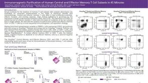 科学海报Immunomagnetic Purification of Human Central and Effector Memory T Cell Subsets in 45 Minutes
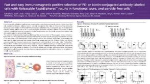 科学海报Fast and Easy Immunomagnetic Positive Selection of PE- or Biotin-Conjugated Antibody Labeled Cells with Releasable RapidSpheres™ Results in Functional, Pure and Particle-Free Cells
 沪公网安备31010102008431号