Y. Kim et al. (MAY 2018)
Cell reports 23 9 2550--2558
Mitochondrial Aging Defects Emerge in Directly Reprogrammed Human Neurons due to Their Metabolic Profile.
Mitochondria are a major target for aging and are instrumental in the age-dependent deterioration of the human brain,but studying mitochondria in aging human neurons has been challenging. Direct fibroblast-to-induced neuron (iN) conversion yields functional neurons that retain important signs of aging,in contrast to iPSC differentiation. Here,we analyzed mitochondrial features in iNs from individuals of different ages. iNs from old donors display decreased oxidative phosphorylation (OXPHOS)-related gene expression,impaired axonal mitochondrial morphologies,lower mitochondrial membrane potentials,reduced energy production,and increased oxidized proteins levels. In contrast,the fibroblasts from which iNs were generated show only mild age-dependent changes,consistent with a metabolic shift from glycolysis-dependent fibroblasts to OXPHOS-dependent iNs. Indeed,OXPHOS-induced old fibroblasts show increased mitochondrial aging features similar to iNs. Our data indicate that iNs are a valuable tool for studying mitochondrial aging and support a bioenergetic explanation for the high susceptibility of the brain to aging.
View Publication
产品类型:
产品号#:
05790
05792
05793
05794
05795
产品名:
BrainPhys™神经元培养基
BrainPhys™神经元培养基和SM1试剂盒
BrainPhys™ 神经元培养基N2-A和SM1试剂盒
BrainPhys™原代神经元试剂盒
BrainPhys™ hPSC 神经元试剂盒
(Jan 2025)
Cells 14 2
Derivation and Characterization of Isogenic OPA1 Mutant and Control Human Pluripotent Stem Cell Lines
Dominant optic atrophy (DOA) is the most commonly inherited optic neuropathy. The majority of DOA is caused by mutations in the OPA1 gene,which encodes a dynamin-related GTPase located to the mitochondrion. OPA1 has been shown to regulate mitochondrial dynamics and promote fusion. Within the mitochondrion,proteolytically processed OPA1 proteins form complexes to maintain membrane integrity and the respiratory chain complexity. Although OPA1 is broadly expressed,human OPA1 mutations predominantly affect retinal ganglion cells (RGCs) that are responsible for transmitting visual information from the retina to the brain. Due to the scarcity of human RGCs,DOA has not been studied in depth using the disease affected neurons. To enable studies of DOA using stem-cell-derived human RGCs,we performed CRISPR-Cas9 gene editing to generate OPA1 mutant pluripotent stem cell (PSC) lines with corresponding isogenic controls. CRISPR-Cas9 gene editing yielded both OPA1 homozygous and heterozygous mutant ESC lines from a parental control ESC line. In addition,CRISPR-mediated homology-directed repair (HDR) successfully corrected the OPA1 mutation in a DOA patient’s iPSCs. In comparison to the isogenic controls,the heterozygous mutant PSCs expressed the same OPA1 protein isoforms but at reduced levels; whereas the homozygous mutant PSCs showed a loss of OPA1 protein and altered mitochondrial morphology. Furthermore,OPA1 mutant PSCs exhibited reduced rates of oxygen consumption and ATP production associated with mitochondria. These isogenic PSC lines will be valuable tools for establishing OPA1-DOA disease models in vitro and developing treatments for mitochondrial deficiency associated neurodegeneration.
View Publication
产品类型:
产品号#:
100-0276
100-1130
产品名:
mTeSR™ Plus
mTeSR™ Plus
(Jan 2025)
Nature Communications 16
A cell atlas of the human fallopian tube throughout the menstrual cycle and menopause
The fallopian tube undergoes extensive molecular changes during the menstrual cycle and menopause. We use single-cell RNA and ATAC sequencing to construct a comprehensive cell atlas of healthy human fallopian tubes during the menstrual cycle and menopause. Our scRNA-seq comparison of 85,107 pre- and 46,111 post-menopausal fallopian tube cells reveals substantial shifts in cell type frequencies,gene expression,transcription factor activity,and cell-to-cell communications during menopause and menstrual cycle. Menstrual cycle dependent hormonal changes regulate distinct molecular states in fallopian tube secretory epithelial cells. Postmenopausal fallopian tubes show high chromatin accessibility in transcription factors associated with aging such as Jun,Fos,and BACH1/2,while hormone receptors were generally downregulated,a small proportion of secretory epithelial cells had high expression of ESR2,IGF1R,and LEPR. While a pre-menopausal secretory epithelial gene cluster is enriched in the immunoreactive molecular subtype,a subset of genes expressed in post-menopausal secretory epithelial cells show enrichment in the mesenchymal molecular type of high-grade serous ovarian cancer. The fallopian tube undergoes extensive cellular and molecular changes during the menstrual cycle and aging. Here,Weigert et al. present a single-cell atlas of the normal human fallopian tube revealing the transition of secretory epithelial cells throughout the menstrual cycle and menopause.
View Publication
E. Y. Flores et al. (Nov 2025)
PLOS Pathogens 21 11
Filovirus infection disrupts epithelial barrier function and ion transport in human iPSC-derived gut organoids
Gastrointestinal (GI) dysfunction,characterized by severe diarrhea and dehydration,is a central contributor to morbidity and mortality in filovirus disease in patients,yet the role of the epithelium in this clinical outcome remains poorly defined. Here,we employ induced pluripotent stem cell (iPSC)-derived human intestinal (HIOs) and colonic organoids (HCOs) to model Ebola virus (EBOV) and Marburg virus (MARV) infection. These organoids are permissive to filovirus infection and support viral replication. Bulk RNA sequencing revealed distinct intestinal and colonic epithelial responses,including apical and junctional disruption and a delayed virus-specific induction of interferon-stimulated genes. Moreover,infection impaired adenylate cyclase signaling and CFTR-mediated ion transport,providing mechanistic insight into virus-induced secretory diarrhea. This platform recapitulates key features of human GI pathology in filoviral disease and serves as a powerful system to dissect host-pathogen interactions and identify therapeutic targets. Author summaryEbola virus (EBOV) and Marburg virus (MARV) are among the most lethal viruses known. Infection with these viruses leads to severe disease and death. One of their most harmful effects is damage to the gastrointestinal tract,causing intense diarrhea and life-threatening dehydration. Yet,how these viruses affect the gut remains poorly understood. In this study,we used human mini-guts—small,three-dimensional tissues grown from stem cells that mimic the human intestinal and colonic epithelium—to investigate how these viruses interact with gut epithelial cells. We found that both EBOV and MARV infect and replicate in these tissues,disrupt key barrier structures,and interfere with the cells’ ability to regulate fluid secretion. These effects mirror the severe symptoms seen in patients. Our study provides new insight into how EBOV and MARV damage the gut and identifies specific cellular pathways that may be targeted for treatment. This research not only improves our understanding of EBOV and MARV infections but also offers new infection platforms for testing therapies aimed at protecting the gastrointestinal system during filovirus outbreaks.
View Publication
产品类型:
产品号#:
05110
100-0483
100-0484
85850
85857
产品名:
STEMdiff™定型内胚层检测试剂盒
Hausser Scientificᵀᴹ 明线血球计数板
ReLeSR™
mTeSR™1
mTeSR™1
I. Miralda et al. ( 2020)
Frontiers in immunology 11 497
Whole Transcriptome Analysis Reveals That Filifactor alocis Modulates TNF$\alpha$-Stimulated MAPK Activation in Human Neutrophils.
Periodontitis is an irreversible,bacteria-induced,chronic inflammatory disease that compromises the integrity of tooth-supporting tissues and adversely affects systemic health. As the immune system's first line of defense against bacteria,neutrophils use their microbicidal functions in the oral cavity to protect the host against periodontal disease. However,periodontal pathogens have adapted to resist neutrophil microbicidal mechanisms while still propagating inflammation,which provides essential nutrients for the bacteria to proliferate and cause disease. Advances in sequencing technologies have recognized several newly appreciated bacteria associated with periodontal lesions such as the Gram-positive anaerobic rod,Filifactor alocis. With the discovery of these oral bacterial species,there is also a growing need to assess their pathogenic potential and determine their contribution to disease progression. Currently,few studies have addressed the pathogenic mechanisms used by oral bacteria to manipulate the neutrophil functional responses at the level of the transcriptome. Thus,this study aims to characterize the global changes at the gene expression level in human neutrophils during infection with F. alocis. Our results indicate that the challenge of human neutrophils with F. alocis results in the differential expression of genes involved in multiple neutrophil effector functions such as chemotaxis,cytokine and chemokine signaling pathways,and apoptosis. Moreover,F. alocis challenges affected the expression of components from the TNF and MAPK kinase signaling pathways. This resulted in transient,dampened p38 MAPK activation by secondary stimuli TNF$\alpha$ but not by fMLF. Functionally,the F. alocis-mediated inhibition of p38 activation by TNF$\alpha$ resulted in decreased cytokine production but had no effect on the priming of the respiratory burst response or the delay of apoptosis by TNF$\alpha$. Since the modulatory effect was characteristic of viable F. alocis only,we propose this as one of F. alocis' mechanisms to control neutrophils and their functional responses.
View Publication
T. Shibata et al. (Apr 2026)
Signal Transduction and Targeted Therapy 11
Bioengineered iPSC-derived human macrophages with increased angiotensin-converting enzyme (ACE) expression suppress solid tumor growth
The potential of the immune system to decrease cancer progression is widely recognized and has led to the development of innovative anti-cancer immunotherapies. Here,we studied human macrophages derived from genetically engineered iPSCs (iMac) with angiotensin-converting enzyme (ACE) expression regulatable by a doxycycline (dox)-inducible promoter as a novel anti-cancer immunotherapy. Increased ACE expression in iMac (cells now termed ACE-iMac) augments polarization towards an M1 macrophage phenotype characterized by increased production of proinflammatory cytokines,reactive oxygen species,nitric oxide,and an RNA profile indicating an aggressive immune response. ACE-iMac kills tumor cells in vitro significantly better than iMac. In vivo,studies using tumor xenografts for melanoma,breast cancer,and head and neck squamous cell carcinoma (HNSCC) showed a highly significant 3.4- to 7.2-fold reduction in solid tumor size following ACE-expressing ACE-iMac immunotherapy as compared to results with iMac. To further investigate the impact of ACE on human anti-tumor responses,we developed a humanized BLT-NSG mouse model with a fully functional adaptive immune system. Here,ACE-iMac treatment significantly reduced the growth of human melanoma xenografts by enhancing the activation of human T cells and NK cells. In conclusion,enhancing ACE expression in human-derived macrophages (ACE-iMac) greatly amplifies their anti-cancer phenotype,offering a compelling new therapeutic strategy with the potential to improve clinical outcomes for cancer patients.
View Publication
产品类型:
产品号#:
100-0276
100-1130
产品名:
mTeSR™ Plus
mTeSR™ Plus
Elkabetz Y et al. (JAN 2008)
Genes & development 22 2 152--65
Human ES cell-derived neural rosettes reveal a functionally distinct early neural stem cell stage.
Neural stem cells (NSCs) yield both neuronal and glial progeny,but their differentiation potential toward multiple region-specific neuron types remains remarkably poor. In contrast,embryonic stem cell (ESC) progeny readily yield region-specific neuronal fates in response to appropriate developmental signals. Here we demonstrate prospective and clonal isolation of neural rosette cells (termed R-NSCs),a novel NSC type with broad differentiation potential toward CNS and PNS fates and capable of in vivo engraftment. R-NSCs can be derived from human and mouse ESCs or from neural plate stage embryos. While R-NSCs express markers classically associated with NSC fate,we identified a set of genes that specifically mark the R-NSC state. Maintenance of R-NSCs is promoted by activation of SHH and Notch pathways. In the absence of these signals,R-NSCs rapidly lose rosette organization and progress to a more restricted NSC stage. We propose that R-NSCs represent the first characterized NSC stage capable of responding to patterning cues that direct differentiation toward region-specific neuronal fates. In addition,the R-NSC-specific genetic markers presented here offer new tools for harnessing the differentiation potential of human ESCs.
View Publication
产品类型:
产品号#:
72082
产品名:
DAPT
Bauwens C et al. (SEP 2008)
Stem cells (Dayton,Ohio) 26 9 2300--10
Control of human embryonic stem cell colony and aggregate size heterogeneity influences differentiation trajectories.
To better understand endogenous parameters that influence pluripotent cell differentiation we used human embryonic stem cells (hESCs) as a model system. We demonstrate that differentiation trajectories in aggregate (embryoid body [EB])-induced differentiation,a common approach to mimic some of the spatial and temporal aspects of in vivo development,are affected by three factors: input hESC composition,input hESC colony size,and EB size. Using a microcontact printing approach,size-specified hESC colonies were formed by plating single-cell suspensions onto micropatterned (MP) extracellular matrix islands. Subsequently,size-controlled EBs were formed by transferring entire colonies into suspension culture enabling the independent investigation of colony and aggregate size effects on differentiation induction. Gene and protein expression analysis of MP-hESC populations revealed that the ratio of Gata6 (endoderm-associated marker) to Pax6 (neural-associated marker) expression increased with decreasing colony size. Moreover,upon forming EBs from these MP-hESCs,we observed that differentiation trajectories were affected by both colony and EB size-influenced parameters. In MP-EBs generated from endoderm-biased (high Gata6/Pax6) input hESCs,higher mesoderm and cardiac induction was observed at larger EB sizes. Conversely,neural-biased (low Gata6/Pax6) input hESCs generated MP-EBs that exhibited higher cardiac induction in smaller EBs. Our analysis demonstrates that heterogeneity in hESC colony and aggregate size,typical in most differentiation strategies,produces subsets of appropriate conditions for differentiation into specific cell types. Moreover,our findings suggest that the local microenvironment modulates endogenous parameters that can be used to influence pluripotent cell differentiation trajectories.
View Publication
产品类型:
产品号#:
产品名:
Graham JD et al. (JUL 2009)
Endocrinology 150 7 3318--26
DNA replication licensing and progenitor numbers are increased by progesterone in normal human breast.
Proliferation in the nonpregnant human breast is highest in the luteal phase of the menstrual cycle when serum progesterone levels are high,and exposure to progesterone analogues in hormone replacement therapy is known to elevate breast cancer risk,yet the proliferative effects of progesterone in the human breast are poorly understood. In a model of normal human breast,we have shown that progesterone increased incorporation of 5-bromo-2'-deoxyuridine and increased cell numbers by activation of pathways involved in DNA replication licensing,including E2F transcription factors,chromatin licensing and DNA replication factor 1 (Cdt1),and the minichromosome maintenance proteins and by increased expression of proteins involved in kinetochore formation including Ras-related nuclear protein (Ran) and regulation of chromosome condensation 1 (RCC1). Progenitor cells competent to give rise to both myoepithelial and luminal epithelial cells were increased by progesterone,showing that progesterone influences epithelial cell lineage differentiation. Therefore,we have demonstrated that progesterone augments proliferation of normal human breast cells by both activating DNA replication licensing and kinetochore formation and increasing bipotent progenitor numbers.
View Publication
产品类型:
产品号#:
01700
01705
01702
产品名:
ALDEFLUOR™ 试剂盒
ALDEFLUOR™ DEAB试剂, 1.5 mM, 1 mL
ALDEFLUOR™检测缓冲液
Avery S et al. (MAY 2010)
Stem Cells 28 5 863--73
The role of SMAD4 in human embryonic stem cell self-renewal and stem cell fate.
Transforming growth factor (TGF)-beta superfamily proteins play a key role in the regulation of human embryonic stem cells (hESCs). Those of the TGFbeta/activin/nodal branch seem to support self-renewal and pluripotency,whereas those of the bone morphogenic protein (BMP) branch induce differentiation. In contrast to this generalization,we found that hESC remained undifferentiated after knockdown of SMAD4 with inducible short hairpin RNA interference,although the knockdown inhibited TGFbeta signaling and rendered the cells nonresponsive to BMP-induced differentiation. Moreover,the rapid differentiation of hESC after pharmacological inhibition of TGFbeta/activin/nodal receptor signaling was restricted after SMAD4 knockdown. These results suggest that TGFbeta/activin/nodal signaling supports the undifferentiated phenotype of hESC by suppressing BMP activity. During long-term culture,SMAD4 knockdown cell populations became less stable and more permissive to neural induction,a situation that was rescued by re-establishment of SMAD4 expression. These results suggest that SMAD4 is not required for maintenance of the undifferentiated state of hESC,but rather to stabilize that state.
View Publication

 EasySep™小鼠TIL(CD45)正选试剂盒


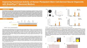 科学海报Improving Functional Activity of Human Pluripotent Stem Cell-Derived Neural Organoids with BrainPhys Neuronal Medium

 沪公网安备31010102008431号