Protocol for CRISPR-mediated deletion of cis-regulatory element in murine Th17 cells for in vivo assessment of effector function
SummaryStudying the cis-regulatory elements (CREs) of genes in Th17 cells during autoimmune disease progression,such as experimental autoimmune encephalomyelitis (EAE),is often limited by the availability of gene-edited mice. Here,we present a protocol for CRISPR-mediated deletion of a CRE in murine Th17 cells for in vivo assessment of effector function in EAE. We describe steps for dual U6gRNA construction,preparation of retroviruses,viral delivery,and Th17 differentiation. We then detail procedures for in vivo functionality analysis.For complete details on the use and execution of this protocol,please refer to Zhong et al.1,2 Graphical abstract Highlights•Steps for designing and cloning dual U6gRNA cassettes to delete a specific CRE•Instructions for optimized retrovirus production and transduction into CD4+ T cells•Guidance on Th17 differentiation and confirmation of CRE deletion in cultured T cells•Procedures for adoptive transfer of CRISPR-edited Th17 cells to assess in vivo function Publisher’s note: Undertaking any experimental protocol requires adherence to local institutional guidelines for laboratory safety and ethics. Studying the cis-regulatory elements (CREs) of genes in Th17 cells during autoimmune disease progression,such as experimental autoimmune encephalomyelitis (EAE),is often limited by the availability of gene-edited mice. Here,we present a protocol for CRISPR-mediated deletion of a CRE in murine Th17 cells for in vivo assessment of effector function in EAE. We describe steps for dual U6gRNA construction,preparation of retroviruses,viral delivery,and Th17 differentiation. We then detail procedures for in vivo functionality analysis.
View Publication
Hu Y-L et al. (SEP 2010)
Nucleic acids research 38 16 5472--8
HOXA9 regulates miR-155 in hematopoietic cells.
HOXA9-mediated up-regulation of miR-155 was noted during an array-based analysis of microRNA expression in Hoxa9(-/-)bone marrow (BM) cells. HOXA9 induction of miR-155 was confirmed in these samples,as well as in wild-type versus Hoxa9-deficient marrow,using northern analysis and qRT-PCR. Infection of wild-type BM with HOXA9 expressing or GFP(+) control virus further confirmed HOXA9-mediated regulation of miR-155. miR-155 expression paralleled Hoxa9 mRNA expression in fractionated BM progenitors,being highest in the stem cell enriched pools. HOXA9 capacity to induce myeloid colony formation was blunted in miR-155-deficient BM cells,indicating that miR-155 is a downstream mediator of HOXA9 function in blood cells. Pu.1,an important regulator of myelopoiesis,was identified as a putative down stream target for miR-155. Although miR-155 was shown to down-regulate the Pu.1 protein,HOXA9 did not appear to modulate Pu.1 expression in murine BM cells.
View Publication
产品类型:
产品号#:
03434
03444
产品名:
MethoCult™ GF M3434
MethoCult™ GF M3434
Chase LG and Firpo MT (AUG 2007)
Current opinion in chemical biology 11 4 367--72
Development of serum-free culture systems for human embryonic stem cells.
Human embryonic stem cells,because of their unique combination of long-term self-renewal properties and pluripotency,are providing new avenues of investigation of stem cell biology and human development and show promise in providing a new source of human cells for transplantation therapies and pharmaceutical testing. Current methods of propagating these cells using combinations of mouse fibroblast feeder cultures and bovine serum components are inexpensive and,in general,useful. However,the systematic investigation of the regulation of self-renewal and the production of safer sources of cells for transplantation depends on the elimination of animal products and the use of defined culture conditions. Both goals are served by the development of serum-free culture methods for human embryonic stem cells.
View Publication
产品类型:
产品号#:
05850
05857
05870
05875
85850
85857
85870
85875
产品名:
mTeSR™1
mTeSR™1
Sun Y et al. (MAR 2010)
Blood 115 9 1709--17
Slug deficiency enhances self-renewal of hematopoietic stem cells during hematopoietic regeneration.
Both extrinsic and intrinsic mechanisms tightly govern hematopoietic stem cell (HSC) decisions of self-renewal and differentiation. However,transcription factors that can selectively regulate HSC self-renewal division after stress remain to be identified. Slug is an evolutionarily conserved zinc-finger transcription factor that is highly expressed in primitive hematopoietic cells and is critical for the radioprotection of these key cells. We studied the effect of Slug in the regulation of HSCs in Slug-deficient mice under normal and stress conditions using serial functional assays. Here,we show that Slug deficiency does not disturb hematopoiesis or alter HSC homeostasis and differentiation in bone marrow but increases the numbers of primitive hematopoietic cells in the extramedullary spleen site. Deletion of Slug enhances HSC repopulating potential but not its homing and differentiation ability. Furthermore,Slug deficiency increases HSC proliferation and repopulating potential in vivo after myelosuppression and accelerates HSC expansion during in vitro culture. Therefore,we propose that Slug is essential for controlling the transition of HSCs from relative quiescence under steady-state condition to rapid proliferation under stress conditions. Our data suggest that inhibition of Slug in HSCs may present a novel strategy for accelerating hematopoietic recovery,thus providing therapeutic benefits for patients after clinical myelosuppressive treatment.
View Publication
Domashenko AD et al. (OCT 2010)
Blood 116 15 2676--83
TAT-mediated transduction of NF-Ya peptide induces the ex vivo proliferation and engraftment potential of human hematopoietic progenitor cells.
Retroviral overexpression of NF-Ya,the regulatory subunit of the transcription factor NF-Y,activates the transcription of multiple genes implicated in hematopoietic stem cell (HSC) self-renewal and differentiation and directs HSCs toward self-renewal. We asked whether TAT-NF-Ya fusion protein could be used to transduce human CD34(+) cells as a safer,more regulated alternative approach to gene therapy. Here we show that externally added recombinant protein was able to enter the cell nucleus and activate HOXB4,a target gene of NF-Ya,using real-time polymerase chain reaction RNA and luciferase-based protein assays. After TAT-NF-Ya transduction,the proliferation of human CD34(+) cells in the presence of myeloid cytokines was increased 4-fold. Moreover,TAT-NF-Ya-treated human primary bone marrow cells showed a 4-fold increase in the percentage of huCD45(+) cells recovered from the bone marrow of sublethally irradiated,transplanted NOD-Scid IL2Rγ(null) mice. These data demonstrate that TAT-peptide therapies are an alternative approach to retroviral stem cell therapies and suggest that NF-Ya peptide delivery should be further evaluated as a tool for HSC/progenitors ex vivo expansion and therapy.
View Publication
产品类型:
产品号#:
04436
09850
产品名:
MethoCult™ SF H4436
B. L. Jamison et al. (jul 2019)
Journal of immunology (Baltimore,Md. : 1950) 203 1 48--57
Nanoparticles Containing an Insulin-ChgA Hybrid Peptide Protect from Transfer of Autoimmune Diabetes by Shifting the Balance between Effector T Cells and Regulatory T Cells.
CD4 T cells play a critical role in promoting the development of autoimmunity in type 1 diabetes. The diabetogenic CD4 T cell clone BDC-2.5,originally isolated from a NOD mouse,has been widely used to study the contribution of autoreactive CD4 T cells and relevant Ags to autoimmune diabetes. Recent work from our laboratory has shown that the Ag for BDC-2.5 T cells is a hybrid insulin peptide (2.5HIP) consisting of an insulin C-peptide fragment fused to a peptide from chromogranin A (ChgA) and that endogenous 2.5HIP-reactive T cells are major contributors to autoimmune pathology in NOD mice. The objective of this study was to determine if poly(lactide-co-glycolide) (PLG) nanoparticles (NPs) loaded with the 2.5HIP Ag (2.5HIP-coupled PLG NPs) can tolerize BDC-2.5 T cells. Infusion of 2.5HIP-coupled PLG NPs was found to prevent diabetes in an adoptive transfer model by impairing the ability of BDC-2.5 T cells to produce proinflammatory cytokines through induction of anergy,leading to an increase in the ratio of Foxp3+ regulatory T cells to IFN-gamma+ effector T cells. To our knowledge,this work is the first to use a hybrid insulin peptide,or any neoepitope,to re-educate diabetogenic T cells and may have significant implications for the development of an Ag-specific therapy for type 1 diabetes patients.
View Publication
产品类型:
产品号#:
19852
19852RF
18783
18783RF
18765
18765RF
产品名:
EasySep™小鼠CD4+ T细胞分选试剂盒
RoboSep™ 小鼠CD4+ T细胞分选试剂盒
EasySep™小鼠CD4+CD25+调节性T细胞分选试剂盒II
RoboSep™ 小鼠CD4+CD25+调节性T细胞分选试剂盒II
EasySep™小鼠CD4+ CD62L+ T细胞分选试剂盒
RoboSep™ 小鼠CD4+ CD62L+ T细胞分选试剂盒
Gerrits A et al. (APR 2010)
Blood 115 13 2610--8
Cellular barcoding tool for clonal analysis in the hematopoietic system.
Clonal analysis is important for many areas of hematopoietic stem cell research,including in vitro cell expansion,gene therapy,and cancer progression and treatment. A common approach to measure clonality of retrovirally transduced cells is to perform integration site analysis using Southern blotting or polymerase chain reaction-based methods. Although these methods are useful in principle,they generally provide a low-resolution,biased,and incomplete assessment of clonality. To overcome those limitations,we labeled retroviral vectors with random sequence tags or barcodes." On integration�
View Publication
产品类型:
产品号#:
09600
09650
产品名:
StemSpan™ SFEM
StemSpan™ SFEM
(Feb 2024)
STAR Protocols 5 1
Protocol for neurogenin-2-mediated induction of human stem cell-derived neural progenitor cells
SummaryHuman pluripotent stem cell-derived neural progenitor cells (NPCs) are an essential tool for the study of brain development and developmental disorders such as autism. Here,we present a protocol to generate NPCs rapidly and reproducibly from human stem cells using dual-SMAD inhibition coupled with a brief pulse of mouse neurogenin-2 (Ngn2) overexpression. We detail the 48-h induction scheme deployed to produce these cells—termed stem cell-derived Ngn2-accelerated progenitor cells—followed by steps for expansion,purification,banking,and quality assessment.For complete details on the use and execution of this protocol,please refer to Wells et al.1 Graphical abstract Highlights•Brief pulse of Ngn2 induces neural progenitor cells from human stem cells•Guidance on expanding,freezing,and thawing SNaP cells for future use•Immunostaining-based assays assess cell identity and differentiation potential Publisher’s note: Undertaking any experimental protocol requires adherence to local institutional guidelines for laboratory safety and ethics. Human pluripotent stem cell-derived neural progenitor cells (NPCs) are an essential tool for the study of brain development and developmental disorders such as autism. Here,we present a protocol to generate NPCs rapidly and reproducibly from human stem cells using dual-SMAD inhibition coupled with a brief pulse of mouse neurogenin-2 (Ngn2) overexpression. We detail the 48-h induction scheme deployed to produce these cells—termed stem cell-derived Ngn2-accelerated progenitor cells—followed by steps for expansion,purification,banking,and quality assessment.
View Publication

 EasySep™小鼠TIL(CD45)正选试剂盒


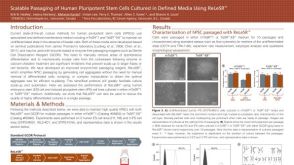 科学海报Scalable Passaging of Human Pluripotent Stem Cells Cultured in Defined Media Using ReLeSR™
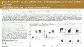 科学海报Stroma-Free, Serum-Free Expansion and Differentiation of Hematopoietic Stem and Progenitor Cells to the T Cell Lineage
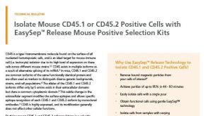 技术公告Isolate Mouse CD45.1 or CD45.2 Positive Cells with EasySep™ Release Mouse Positive Selection Kits
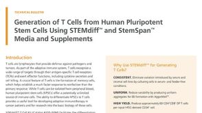 技术公告Generation of T Cells from Human Pluripotent Stem Cells Using STEMdiff™ and StemSpan™ Media and Supplements
 沪公网安备31010102008431号