Serrero G and Lepak NM (APR 1997)
Biochemical and biophysical research communications 233 1 200--2
Prostaglandin F2alpha receptor (FP receptor) agonists are potent adipose differentiation inhibitors for primary culture of adipocyte precursors in defined medium.
Prostaglandin F2alpha inhibits adipose differentiation of primary culture of adipocyte precursors and of the adipogenic cell line 1246 in defined medium. In the present paper,we investigated the effect of FP receptor agonists cloprostenol and fluprostenol on the differentiation of newborn rat adipocyte precursors in primary culture. The results show that cloprostenol and fluprostenol are very potent inhibitors of adipose differentiation. Dose response studies indicate that both agonists are more potent than PGF2alpha in inhibiting adipocyte precursors differentiation. 50% inhibition of adipose differentiation was observed at a concentration of 3 x 10(-12) M for cloprostenol and 3 to 10 x 10(-11) M for fluprostenol respectively whereas the PGF2alpha concentration required to elicit the same effect was 10(-8) M. In contrast compounds structurally related to PGE2 such as 17-phenyl trinor PGE2 had no effect on adipose differentiation except when added at a 10,000-fold higher concentration.
View Publication
Gibbons JJ et al. (DEC 2009)
Seminars in oncology 36 Suppl 3 S3--S17
Mammalian target of rapamycin: discovery of rapamycin reveals a signaling pathway important for normal and cancer cell growth.
Since the discovery of rapamycin,considerable progress has been made in unraveling the details of the mammalian target of rapamycin (mTOR) signaling network,including the upstream mechanisms that modulate mTOR signaling functions,and the roles of mTOR in the regulation of mRNA translation and other cell growth-related responses. mTOR is found in two different complexes within the cell,mTORC1 and mTORC2,but only mTORC1 is sensitive to inhibition by rapamycin. mTORC1 is a master controller of protein synthesis,integrating signals from growth factors within the context of the energy and nutritional conditions of the cell. Activated mTORC1 regulates protein synthesis by directly phosphorylating 4E-binding protein 1 (4E-BP1) and p70S6K (S6K),translation initiation factors that are important to cap-dependent mRNA translation,which increases the level of many proteins that are needed for cell cycle progression,proliferation,angiogenesis,and survival pathways. In normal physiology,the roles of mTOR in both glucose and lipid catabolism underscore the importance of the mTOR pathway in the production of metabolic energy in quantities sufficient to fuel cell growth and mitotic cell division. Several oncogenes and tumor-suppressor genes that activate mTORC1,often through the phosphatidylinositol 3-kinase (PI3K)/AKT pathway,are frequently dysregulated in cancer. Novel analogs of rapamycin (temsirolimus,everolimus,and deforolimus),which have improved pharmaceutical properties,were designed for oncology indications. Clinical trials of these analogs have already validated the importance of mTOR inhibition as a novel treatment strategy for several malignancies. Inhibition of mTOR now represents an attractive anti-tumor target,either alone or in combination with strategies to target other pathways that may overcome resistance. The far-reaching downstream consequences of mTOR inhibition make defining the critical molecular effector mechanisms that mediate the anti-tumor response and associated biomarkers that predict responsiveness to mTOR inhibitors a challenge and priority for the field.
View Publication
产品类型:
产品号#:
73362
73364
100-1050
产品名:
雷帕霉素
雷帕霉素
雷帕霉素
Yang YM et al. (JUN 2013)
Cell stem cell 12 6 713--26
A small molecule screen in stem-cell-derived motor neurons identifies a kinase inhibitor as a candidate therapeutic for ALS.
Amyotrophic lateral sclerosis (ALS) is a rapidly progressing neurodegenerative disease,characterized by motor neuron (MN) death,for which there are no truly effective treatments. Here,we describe a new small molecule survival screen carried out using MNs from both wild-type and mutant SOD1 mouse embryonic stem cells. Among the hits we found,kenpaullone had a particularly impressive ability to prolong the healthy survival of both types of MNs that can be attributed to its dual inhibition of GSK-3 and HGK kinases. Furthermore,kenpaullone also strongly improved the survival of human MNs derived from ALS-patient-induced pluripotent stem cells and was more active than either of two compounds,olesoxime and dexpramipexole,that recently failed in ALS clinical trials. Our studies demonstrate the value of a stem cell approach to drug discovery and point to a new paradigm for identification and preclinical testing of future ALS therapeutics.
View Publication
产品类型:
产品号#:
72782
产品名:
Kenpaullone
(Aug 2024)
STAR Protocols 5 3
Protocol for generation and engineering of thyroid cell lineages using CRISPR-Cas9 editing to recapitulate thyroid cancer histotype progression
SummaryThyroid carcinoma represents the first malignancy among the endocrine organs. Investigating the cellular hierarchy and the mechanisms underlying the initiation of thyroid carcinoma is crucial in thyroid cancer research. Here,we present a protocol for deriving thyroid cell lineage from human embryonic stem cells. We also describe steps for engineering thyroid progenitor cells utilizing CRISPR-Cas9 technology,which can be used to perform in vivo studies,thus facilitating the development of representative thyroid tumorigenesis models.For complete details on the use and execution of this protocol,please refer to Veschi et al.1 Graphical abstract Highlights•Differentiation protocol for thyroid cell lineages from human embryonic stem cells•CRISPR-Cas9-mediated cellular engineering for common thyroid cancer genetic alteration•Orthotopic injection of thyroid progenitors to recapitulate thyroid cancer progression Publisher’s note: Undertaking any experimental protocol requires adherence to local institutional guidelines for laboratory safety and ethics. Thyroid carcinoma represents the first malignancy among the endocrine organs. Investigating the cellular hierarchy and the mechanisms underlying the initiation of thyroid carcinoma is crucial in thyroid cancer research. Here,we present a protocol for deriving thyroid cell lineage from human embryonic stem cells. We also describe steps for engineering thyroid progenitor cells utilizing CRISPR-Cas9 technology,which can be used to perform in vivo studies,thus facilitating the development of representative thyroid tumorigenesis models.
View Publication
产品类型:
产品号#:
05110
85850
85857
产品名:
STEMdiff™定型内胚层检测试剂盒
mTeSR™1
mTeSR™1
(Nov 2024)
Molecular Therapy. Methods & Clinical Development 32 4
Generation and maintenance of kidney and kidney cancer organoids from patient-derived material for drug development and precision oncology
Despite significant advancements in targeted- and immunotherapies,millions of patients with cancer still succumb to the disease each year. In renal cell carcinoma,up to 25% of metastatic patients do not respond to first-line therapies. This reality underscores the urgent need for innovative or repurposed therapies to effectively treat these patients. Patient-derived organoids represent a promising model for evaluating treatment efficacy and toxicity,offering a potential breakthrough in personalized medicine. However,utilizing organoid models for drug screening presents several challenges. Our protocol aims to address these obstacles by outlining a practical approach to successfully isolate and cultivate patient-derived renal cell carcinoma and kidney organoids for treatment screening purposes. Graphical abstract Patient-derived organoids represent a promising model for evaluating treatment efficacy and toxicity,offering a potential breakthrough in personalized medicine. Nowak-Sliwinska and colleagues present a detailed protocol for obtaining kidney and kidney cancer organoids for drug development and precision oncology.
View Publication
产品类型:
产品号#:
17899
产品名:
EasySep™ 死细胞去除 (Annexin V) 试剂盒
Booth L et al. (AUG 2015)
Journal of cellular physiology 230 8 1982--98
OSU-03012 and Viagra Treatment Inhibits the Activity of Multiple Chaperone Proteins and Disrupts the Blood-Brain Barrier: Implications for Anti-Cancer Therapies.
We examined the interaction between OSU-03012 (also called AR-12) with phosphodiesterase 5 (PDE5) inhibitors to determine the role of the chaperone glucose-regulated protein (GRP78)/BiP/HSPA5 in the cellular response. Sildenafil (Viagra) interacted in a greater than additive fashion with OSU-03012 to kill stem-like GBM cells. Treatment of cells with OSU-03012/sildenafil: abolished the expression of multiple oncogenic growth factor receptors and plasma membrane drug efflux pumps and caused a rapid degradation of GRP78 and other HSP70 and HSP90 family chaperone proteins. Decreased expression of plasma membrane receptors and drug efflux pumps was dependent upon enhanced PERK-eIF2α-ATF4-CHOP signaling and was blocked by GRP78 over-expression. In vivo OSU-03012/sildenafil was more efficacious than treatment with celecoxib and sildenafil at killing tumor cells without damaging normal tissues and in parallel reduced expression of ABCB1 and ABCG2 in the normal brain. The combination of OSU-03012/sildenafil synergized with low concentrations of sorafenib to kill tumor cells,and with lapatinib to kill ERBB1 over-expressing tumor cells. In multiplex assays on plasma and human tumor tissue from an OSU-03012/sildenafil treated mouse,we noted a profound reduction in uPA signaling and identified FGF and JAK1/2 as response biomarkers for potentially suppressing the killing response. Inhibition of FGFR signaling and to a lesser extent JAK1/2 signaling profoundly enhanced OSU-03012/sildenafil lethality.
View Publication
产品类型:
产品号#:
05750
05751
产品名:
NeuroCult™ NS-A 基础培养基(人)
NeuroCult™ NS-A 扩增试剂盒(人)
Marchetti S et al. (MAY 2002)
Journal of cell science 115 Pt 10 2075--85
Endothelial cells genetically selected from differentiating mouse embryonic stem cells incorporate at sites of neovascularization in vivo.
Large scale purification of endothelial cells is of great interest as it could improve tissue transplantation,reperfusion of ischemic tissues and treatment of pathologies in which an endothelial cell dysfunction exists. In this study,we describe a novel genetic approach that selects for endothelial cells from differentiating embryonic stem (ES) cells. Our strategy is based on the establishment of ES-cell clones that carry an integrated puromycin resistance gene under the control of a vascular endothelium-specific promoter,tie-1. Using EGFP as a reporter gene,we first confirmed the endothelial specificity of the tie-1 promoter in the embryoid body model and in cells differentiated in 2D cultures. Subsequently,tie-1-EGFP ES cells were used as recipients for the tie-1-driven puror transgene. The resulting stable clones were expanded and differentiated for seven days in the presence of VEGF before puromycin selection. As expected,puromycin-resistant cells were positive for EGFP and also expressed several endothelial markers,including CD31,CD34,VEGFR-1,VEGFR-2,Tie-1,VE-cadherin and ICAM-2. Release from the puromycin selection resulted in the appearance of alpha-smooth muscle actin-positive cells. Such cells became more numerous when the population was cultured on laminin-1 or in the presence of TGF-beta1,two known inducers of smooth muscle cell differentiation. The hypothesis that endothelial cells or their progenitors may differentiate towards a smooth muscle cell phenotype was further supported by the presence of cells expressing both CD31 and alpha-smooth muscle actin markers. Finally,we show that purified endothelial cells can incorporate into the neovasculature of transplanted tumors in nude mice. Taken together,these results suggest that application of endothelial lineage selection to differentiating ES cells may become a useful approach for future pro-angiogenic and endothelial cell replacement therapies.
View Publication
IGF-1 enhances cell proliferation and survival during early differentiation of mesenchymal stem cells to neural progenitor-like cells
BACKGROUND There has been increasing interest recently in the plasticity of mesenchymal stem cells (MSCs) and their potential to differentiate into neural lineages. To unravel the roles and effects of different growth factors in the differentiation of MSCs into neural lineages,we have differentiated MSCs into neural lineages using different combinations of growth factors. Based on previous studies of the roles of insulin-like growth factor 1 (IGF-1) in neural stem cell isolation in the laboratory,we hypothesized that IGF-1 can enhance proliferation and reduce apoptosis in neural progenitor-like cells (NPCs) during differentiation of MSCs into NCPs.We induced MSCs differentiation under four different combinations of growth factors: (A) EGF%+%bFGF,(B) EGF%+%bFGF%+%IGF-1,(C) EGF%+%bFGF%+%LIF,(D) EGF%+%bFGF%+%BDNF,and (E) without growth factors,as a negative control. The neurospheres formed were characterized by immunofluorescence staining against nestin,and the expression was measured by flow cytometry. Cell proliferation and apoptosis were also studied by MTS and Annexin V assay,respectively,at three different time intervals (24 hr,3 days,and 5 days). The neurospheres formed in the four groups were then terminally differentiated into neuron and glial cells. RESULTS The four derived NPCs showed a significantly higher expression of nestin than was shown by the negative control. Among the groups treated with growth factors,NPCs treated with IGF-1 showed the highest expression of nestin. Furthermore,NPCs derived using IGF-1 exhibited the highest cell proliferation and cell survival among the treated groups. The NPCs derived from IGF-1 treatment also resulted in a better yield after the terminal differentiation into neurons and glial cells than that of the other treated groups. CONCLUSIONS Our results suggested that IGF-1 has a crucial role in the differentiation of MSCs into neuronal lineage by enhancing the proliferation and reducing the apoptosis in the NPCs. This information will be beneficial in the long run for improving both cell-based and cell-free therapy for neurodegenerative diseases.
View Publication
产品类型:
产品号#:
05771
产品名:
Lee SJ et al. (DEC 2014)
Stem Cells and Development 23 23 2831--2840
Adult Stem Cells from the Hyaluronic Acid-Rich Node and Duct System Differentiate into Neuronal Cells and Repair Brain Injury
The existence of a hyaluronic acid-rich node and duct system (HAR-NDS) within the lymphatic and blood vessels was demonstrated previously. The HAR-NDS was enriched with small (3.0-5.0 μm in diameter),adult stem cells with properties similar to those of the very small embryonic-like stem cells (VSELs). Sca-1(+)Lin(-)CD45(-) cells were enriched approximately 100-fold in the intravascular HAR-NDS compared with the bone marrow. We named these adult stem cells node and duct stem cells (NDSCs)." NDSCs formed colonies on C2C12 feeder layers were positive for fetal alkaline phosphatase and could be subcultured on the feeder layers. NDSCs were Oct4(+)Nanog(+)SSEA-1(+)Sox2(+) while VSELs were Oct4(+)Nanog(+)SSEA-1(+)Sox2(-). NDSCs had higher sphere-forming efficiency and proliferative potential than VSELs and they were found to differentiate into neuronal cells in vitro. Injection of NDSCs into mice partially repaired ischemic brain damage. Thus we report the discovery of potential adult stem cells that may be involved in tissue regeneration. The intravascular HAR-NDS may serve as a route that delivers these stem cells to their target tissues.
View Publication
产品类型:
产品号#:
05700
产品名:
NeuroCult™ 基础培养基(小鼠和大鼠)
Hu K et al. (APR 2011)
Blood 117 14 e109--19
Efficient generation of transgene-free induced pluripotent stem cells from normal and neoplastic bone marrow and cord blood mononuclear cells.
Reprogramming blood cells to induced pluripotent stem cells (iPSCs) provides a novel tool for modeling blood diseases in vitro. However,the well-known limitations of current reprogramming technologies include low efficiency,slow kinetics,and transgene integration and residual expression. In the present study,we have demonstrated that iPSCs free of transgene and vector sequences could be generated from human BM and CB mononuclear cells using non-integrating episomal vectors. The reprogramming described here is up to 100 times more efficient,occurs 1-3 weeks faster compared with the reprogramming of fibroblasts,and does not require isolation of progenitors or multiple rounds of transfection. Blood-derived iPSC lines lacked rearrangements of IGH and TCR,indicating that their origin is non-B- or non-T-lymphoid cells. When cocultured on OP9,blood-derived iPSCs could be differentiated back to the blood cells,albeit with lower efficiency compared to fibroblast-derived iPSCs. We also generated transgene-free iPSCs from the BM of a patient with chronic myeloid leukemia (CML). CML iPSCs showed a unique complex chromosomal translocation identified in marrow sample while displaying typical embryonic stem cell phenotype and pluripotent differentiation potential. This approach provides an opportunity to explore banked normal and diseased CB and BM samples without the limitations associated with virus-based methods.
View Publication

 EasySep™小鼠TIL(CD45)正选试剂盒


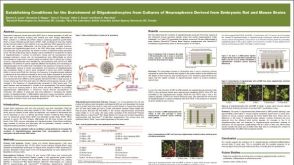 科学海报Establishing Conditions for the Enrichment of Oligodendrocytes from Cultures of Neurospheres Derived from Embryonic Rat and Mouse Brains
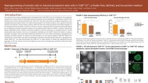 科学海报Reprogramming of Somatic Cells to Induced Pluripotent Stem Cells in TeSR™-E7™, a Feeder-Free, Defined, and Low-Protein Medium
 沪公网安备31010102008431号