Wattanapanitch M et al. (SEP 2014)
PloS one 9 9 e106952
Dual small-molecule targeting of SMAD signaling stimulates human induced pluripotent stem cells toward neural lineages.
Incurable neurological disorders such as Parkinson's disease (PD),Huntington's disease (HD),and Alzheimer's disease (AD) are very common and can be life-threatening because of their progressive disease symptoms with limited treatment options. To provide an alternative renewable cell source for cell-based transplantation and as study models for neurological diseases,we generated induced pluripotent stem cells (iPSCs) from human dermal fibroblasts (HDFs) and then differentiated them into neural progenitor cells (NPCs) and mature neurons by dual SMAD signaling inhibitors. Reprogramming efficiency was improved by supplementing the histone deacethylase inhibitor,valproic acid (VPA),and inhibitor of p160-Rho associated coiled-coil kinase (ROCK),Y-27632,after retroviral transduction. We obtained a number of iPS colonies that shared similar characteristics with human embryonic stem cells in terms of their morphology,cell surface antigens,pluripotency-associated gene and protein expressions as well as their in vitro and in vivo differentiation potentials. After treatment with Noggin and SB431542,inhibitors of the SMAD signaling pathway,HDF-iPSCs demonstrated rapid and efficient differentiation into neural lineages. Six days after neural induction,neuroepithelial cells (NEPCs) were observed in the adherent monolayer culture,which had the ability to differentiate further into NPCs and neurons,as characterized by their morphology and the expression of neuron-specific transcripts and proteins. We propose that our study may be applied to generate neurological disease patient-specific iPSCs allowing better understanding of disease pathogenesis and drug sensitivity assays.
View Publication
产品类型:
产品号#:
05850
05857
05870
05875
07923
85850
85857
85870
85875
产品名:
Dispase (1 U/mL)
mTeSR™1
mTeSR™1
Lindgren AG et al. (JAN 2015)
Cell regeneration (London,England) 4 1 1
ETV2 expression increases the efficiency of primitive endothelial cell derivation from human embryonic stem cells.
BACKGROUND: Endothelial cells line the luminal surface of blood vessels and form a barrier between the blood and other tissues of the body. Ets variant 2 (ETV2) is transiently expressed in both zebrafish and mice and is necessary and sufficient for vascular endothelial cell specification. Overexpression of this gene in early zebrafish and mouse embryos results in ectopic appearance of endothelial cells. Ectopic expression of ETV2 in later development results in only a subset of cells responding to the signal.backslashnbackslashnFINDINGS: We have examined the expression pattern of ETV2 in differentiating human embryonic stem cells (ESCs) to determine when the peak of ETV2 expression occurs. We show that overexpression of ETV2 in differentiating human ESC is able to increase the number of endothelial cells generated when administered during or after the endogenous peak of gene expression.backslashnbackslashnCONCLUSIONS: Addition of exogenous ETV2 to human ESCs significantly increased the number of cells expressing angioblast genes without arterial or venous specification. This may be a viable solution to generate in vitro endothelial cells for use in research and in the clinic.
View Publication
产品类型:
产品号#:
05850
05857
05870
05875
85850
85857
85870
85875
产品名:
mTeSR™1
mTeSR™1
Hu W et al. (AUG 2015)
Cell stem cell 17 2 204--12
Direct Conversion of Normal and Alzheimer's Disease Human Fibroblasts into Neuronal Cells by Small Molecules.
Neuronal conversion from human fibroblasts can be induced by lineage-specific transcription factors; however,the introduction of ectopic genes limits the therapeutic applications of such induced neurons (iNs). Here,we report that human fibroblasts can be directly converted into neuronal cells by a chemical cocktail of seven small molecules,bypassing a neural progenitor stage. These human chemical-induced neuronal cells (hciNs) resembled hiPSC-derived neurons and human iNs (hiNs) with respect to morphology,gene expression profiles,and electrophysiological properties. This approach was further applied to generate hciNs from familial Alzheimer's disease patients. Taken together,our transgene-free and chemical-only approach for direct reprogramming of human fibroblasts into neurons provides an alternative strategy for modeling neurological diseases and for regenerative medicine.
View Publication
产品类型:
产品号#:
72052
72054
72112
72114
72292
72302
72304
72307
72308
72392
72394
72462
72642
73792
73794
100-1042
100-0249
100-1044
产品名:
CHIR99021
CHIR99021
Forskolin
Forskolin
丙戊酸(钠盐)
Y-27632(二盐酸盐)
Y-27632(二盐酸盐)
Y-27632(二盐酸盐)
Y-27632(二盐酸盐)
RepSox(盐酸盐)
RepSox(盐酸盐)
Gö6983
SP600125
RepSox
RepSox
CHIR99021
Forskolin
Y-27632(二盐酸盐)
Kang YK et al. (MAR 2016)
Blood research 51 1 31--6
Humanizing NOD/SCID/IL-2Rγnull (NSG) mice using busulfan and retro-orbital injection of umbilical cord blood-derived CD34(+) cells.
BACKGROUND Humanized mouse models are still under development,and various protocols exist to improve human cell engraftment and function. METHODS Fourteen NOD/SCID/IL-2Rγnull (NSG) mice (4‒5 wk old) were conditioned with busulfan and injected with human umbilical cord blood (hUCB)-derived CD34(+) hematopoietic stem cells (HSC) via retro-orbital sinuses. The bone marrow (BM),spleen,and peripheral blood (PB) were analyzed 8 and 12 weeks after HSC transplantation. RESULTS Most of the NSG mice tolerated the regimen well. The percentage of hCD45(+) and CD19(+) cells rose significantly in a time-dependent manner. The median percentage of hCD45(+)cells in the BM was 55.5% at week 8,and 67.2% at week 12. The median percentage of hCD45(+) cells in the spleen at weeks 8 and 12 was 42% and 51%,respectively. The median percentage of hCD19(+) cells in BM at weeks 8 and 12 was 21.5% and 39%,respectively (P=0.04). Similarly,the median percentage of hCD19(+) cells in the spleen at weeks 8 and 12 was 10% and 24%,respectively (P=0.04). The percentage of hCD19(+) B cells in PB was 23% at week 12. At week 8,hCD3(+) T cells were barely detectable,while hCD7(+) was detected in the BM and spleen. The percentage of hCD3(+) T cells was 2‒3% at week 12 in the BM,spleen,and PB of humanized NSG mice. CONCLUSION We adopted a simplified protocol for establishing humanized NSG mice. We observed a higher engraftment rate of human CD45(+) cells than earlier studies without any significant toxicity. And human CD45(+) cell engraftment at week 8 was comparable to that of week 12.
View Publication
产品类型:
产品号#:
15026
15066
产品名:
RosetteSep™人造血祖细胞富集抗体混合物
RosetteSep™人造血祖细胞富集抗体混合物
Vauchez K et al. (NOV 2009)
Molecular therapy : the journal of the American Society of Gene Therapy 17 11 1948--58
Aldehyde dehydrogenase activity identifies a population of human skeletal muscle cells with high myogenic capacities.
Aldehyde dehydrogenase 1A1 (ALDH) activity is one hallmark of human bone marrow (BM),umbilical cord blood (UCB),and peripheral blood (PB) primitive progenitors presenting high reconstitution capacities in vivo. In this study,we have identified ALDH(+) cells within human skeletal muscles,and have analyzed their phenotypical and functional characteristics. Immunohistofluorescence analysis of human muscle tissue sections revealed rare endomysial cells. Flow cytometry analysis using the fluorescent substrate of ALDH,Aldefluor,identified brightly stained (ALDH(br)) cells with low side scatter (SSC(lo)),in enzymatically dissociated muscle biopsies,thereafter abbreviated as SMALD(+) (for skeletal muscle ALDH(+)) cells. Phenotypical analysis discriminated two sub-populations according to CD34 expression: SMALD(+)/CD34(-) and SMALD(+)/CD34(+) cells. These sub-populations did not initially express endothelial (CD31),hematopoietic (CD45),and myogenic (CD56) markers. Upon sorting,however,whereas SMALD(+)/CD34(+) cells developed in vitro as a heterogeneous population of CD56(-) cells able to differentiate in adipoblasts,the SMALD(+)/CD34(-) fraction developed in vitro as a highly enriched population of CD56(+) myoblasts able to form myotubes. Moreover,only the SMALD(+)/CD34(-) population maintained a strong myogenic potential in vivo upon intramuscular transplantation. Our results suggest that ALDH activity is a novel marker for a population of new human skeletal muscle progenitors presenting a potential for cell biology and cell therapy.
View Publication
产品类型:
产品号#:
01700
01705
01701
01702
产品名:
ALDEFLUOR™ 试剂盒
ALDEFLUOR™ DEAB试剂, 1.5 mM, 1 mL
ALDEFLUOR™检测缓冲液
Jiang X et al. (SEP 2010)
Blood 116 12 2112--21
Properties of CD34+ CML stem/progenitor cells that correlate with different clinical responses to imatinib mesylate.
Imatinib mesylate (IM) induces clinical remissions in chronic-phase chronic myeloid leukemia (CML) patients but IM resistance remains a problem. We recently identified several features of CML CD34(+) stem/progenitor cells expected to confer resistance to BCR-ABL-targeted therapeutics. From a study of 25 initially chronic-phase patients,we now demonstrate that some,but not all,of these parameters correlate with subsequent clinical response to IM therapy. CD34(+) cells from the 14 IM nonresponders demonstrated greater resistance to IM than the 11 IM responders in colony-forming cell assays in vitro (P textless .001) and direct sequencing of cloned transcripts from CD34(+) cells further revealed a higher incidence of BCR-ABL kinase domain mutations in the IM nonresponders (10%-40% vs 0%-20% in IM responders,P textless .003). In contrast,CD34(+) cells from IM nonresponders and IM responders were not distinguished by differences in BCR-ABL or transporter gene expression. Interestingly,one BCR-ABL mutation (V304D),predicted to destabilize the interaction between p210(BCR-ABL) and IM,was detectable in 14 of 20 patients. T315I mutant CD34(+) cells found before IM treatment in 2 of 20 patients examined were preferentially amplified after IM treatment. Thus,2 properties of pretreatment CML stem/progenitor cells correlate with subsequent response to IM therapy. Prospective assessment of these properties may allow improved patient management.
View Publication
Recombinant erythroid Kruppel-like factor fused to GATA1 up-regulates delta- and gamma-globin expression in erythroid cells.
The β-hemoglobinopathies sickle cell disease and β-thalassemia are among the most common human genetic disorders worldwide. Hemoglobin A2 (HbA2,α₂δ₂) and fetal hemoglobin (HbF,α₂γ₂) both inhibit the polymerization of hemoglobin S,which results in erythrocyte sickling. Expression of erythroid Kruppel-like factor (EKLF) and GATA1 is critical for transitioning hemoglobin from HbF to hemoglobin A (HbA,α₂β₂) and HbA2. The lower levels of δ-globin expression compared with β-globin expression seen in adulthood are likely due to the absence of an EKLF-binding motif in the δ-globin proximal promoter. In an effort to up-regulate δ-globin to increase HbA2 expression,we created a series of EKLF-GATA1 fusion constructs composed of the transactivation domain of EKLF and the DNA-binding domain of GATA1,and then tested their effects on hemoglobin expression. EKLF-GATA1 fusion proteins activated δ-,γ-,and β-globin promoters in K562 cells,and significantly up-regulated δ- and γ-globin RNA transcript and protein expression in K562 and/or CD34(+) cells. The binding of EKLF-GATA1 fusion proteins at the GATA1 consensus site in the δ-globin promoter was confirmed by chromatin immunoprecipitation assay. Our studies demonstrate that EKLF-GATA1 fusion proteins can enhance δ-globin expression through interaction with the δ-globin promoter,and may represent a new genetic therapeutic approach to β-hemoglobinopathies.
View Publication
产品类型:
产品号#:
02690
09600
09650
产品名:
StemSpan™ CC100
StemSpan™ SFEM
StemSpan™ SFEM
Dutta D et al. (APR 2011)
Stem cells 29 4 618--28
Self-renewal versus lineage commitment of embryonic stem cells: protein kinase C signaling shifts the balance.
The intricate molecular mechanisms that regulate ESC pluripotency are incompletely understood. Prior research indicated that activation of the Janus kinase-signal transducer and activator of transcription (STAT3) pathway or inhibition of extracellular signal-regulated kinase/glycogen synthase kinase 3 (ERK/GSK3) signaling maintains mouse ESC (mESC) pluripotency. Here,we demonstrate that inhibition of protein kinase C (PKC) isoforms maintains mESC pluripotency without the activation of STAT3 or inhibition of ERK/GSK3 signaling pathways. Our analyses revealed that the atypical PKC isoform,PKCζ plays an important role in inducing lineage commitment in mESCs through a PKCζ-nuclear factor kappa-light-chain-enhancer of activated B cells signaling axis. Furthermore,inhibition of PKC isoforms permits derivation of germline-competent ESCs from mouse blastocysts and also facilitates reprogramming of mouse embryonic fibroblasts toward induced pluripotent stem cells. Our results indicate that PKC signaling is critical to balancing ESC self-renewal and lineage commitment.
View Publication
产品类型:
产品号#:
72462
产品名:
Gö6983
Peng Y et al. (NOV 2012)
Journal of Tissue Engineering and Regenerative Medicine 6 10 e74----86
Human fibroblast matrices bio-assembled under macromolecular crowding support stable propagation of human embryonic stem cells.
Stable pluripotent feeder-free propagation of human embryonic stem cells (hESCs) prior to their therapeutic applications remains a major challenge. Matrigel™ (BD Singapore) is a murine sarcoma-derived extracellular matrix (ECM) widely used as a cell-free support combined with conditioned or chemically defined media; however,inherent xenogenic and immunological threats invalidate it for clinical applications. Using human fibrogenic cells to generate ECM is promising but currently suffers from inefficient and time-consuming deposition in vitro. We recently showed that macromolecular crowding (MMC) accelerated ECM deposition substantially in vitro. In the current study,we used dextran sulfate 500 kDa as a macromolecular crowder to induce WI-38 fetal human lung fibroblasts at 0.5% serum condition to deposit human ECM in three days. After decellularization,the generated ECMs allowed stable propagation of H9 hESCs over 20 passages in chemically-defined medium (mTEsR1) with an overall improved outcome compared to Matrigel in terms of population doubling while retaining teratoma formation and differentiation capacity. Of significance,only ECMs generated by MMC allowed the successful propagation of hESCs. ECMs were highly complex and in contrast to Matrigel,contained no vitronectin but did contain collagen XII,ig-h3 and novel for hESC-supporting human matrices,substantial amounts of transglutaminase 2. Genome-wide analysis of promoter DNA methylation states revealed high overall similarity between human ECM- and Matrigel-cultured hESCs; however,distinct differences were observed with 49 genes associated with a variety of cellular functions. Thus,human ECMs deposited by MMC by selected fibroblast lines are a suitable human microenvironment for stable hESC propagation and clinically translational settings.
View Publication
产品类型:
产品号#:
05850
05857
05870
05875
07923
85850
85857
85870
85875
产品名:
Dispase (1 U/mL)
mTeSR™1
mTeSR™1
Sharma A et al. (JUN 2013)
Journal of Biological Chemistry 288 25 18439--18447
The role of SIRT6 protein in aging and reprogramming of human induced pluripotent stem cells
Aging is known to be the single most important risk factor for multiple diseases. Sirtuin 6,or SIRT6,has recently been identified as a critical regulator of transcription,genome stability,telomere integrity,DNA repair,and metabolic homeostasis. A knockout mouse model of SIRT6 has displayed dramatic phenotypes of accelerated aging. In keeping with its role in aging,we demonstrated that human dermal fibroblasts (HDFs) from older human subjects were more resistant to reprogramming by classic Yamanaka factors than those from younger human subjects,but the addition of SIRT6 during reprogramming improved such efficiency in older HDFs substantially. Despite the importance of SIRT6,little is known about the molecular mechanism of its regulation. We show,for the first,time posttranscriptional regulation of SIRT6 by miR-766 and inverse correlation in the expression of this microRNA in HDFs from different age groups. Our results suggest that SIRT6 regulates miR-766 transcription via a feedback regulatory loop,which has implications for the modulation of SIRT6 expression in reprogramming of aging cells.
View Publication
产品类型:
产品号#:
05850
05857
05870
05875
85850
85857
85870
85875
产品名:
mTeSR™1
mTeSR™1
Bao X et al. ( 2016)
Methods in molecular biology (Clifton,N.J.) 1481 183--196
Directed Endothelial Progenitor Differentiation from Human Pluripotent Stem Cells Via Wnt Activation Under Defined Conditions.
Efficient derivation of endothelial cells and their progenitors from human pluripotent stem cells (hPSCs) can facilitate studies of human vascular development,disease modeling,drug discovery,and cell-based therapy. Here we provide a detailed protocol for directing hPSCs to functional endothelial cells and their progenitors in a completely defined,growth factor- and serum-free system by temporal modulation of Wnt/$$-catenin signaling via small molecules. We demonstrate a 10-day,two-stage process that recapitulates endothelial cell development,in which hPSCs first differentiate to endothelial progenitors that then generate functional endothelial cells and smooth muscle cells. Methods to characterize endothelial cell identity and function are also described.
View Publication

 EasySep™小鼠TIL(CD45)正选试剂盒


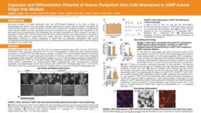 科学海报Expansion and Differentiation Potential of Human Pluripotent Stem Cells Maintained in cGMP Animal Origin-Free Medium
 沪公网安备31010102008431号