Guan BX et al. (MAY 2014)
IEEE/ACM transactions on computational biology and bioinformatics / IEEE,ACM 11 3 604--611
Bio-Driven Cell Region Detection in Human Embryonic Stem Cell Assay.
This paper proposes a bio-driven algorithm that detects cell regions automatically in the human embryonic stem cell (hESC) images obtained using a phase contrast microscope. The algorithm uses both statistical intensity distributions of foreground/hESCs and background/substrate as well as cell property for cell region detection. The intensity distributions of foreground/hESCs and background/substrate are modeled as a mixture of two Gaussians. The cell property is translated into local spatial information. The algorithm is optimized by parameters of the modeled distributions and cell regions evolve with the local cell property. The paper validates the method with various videos acquired using different microscope objectives. In comparison with the state-of-the-art methods,the proposed method is able to detect the entire cell region instead of fragmented cell regions. It also yields high marks on measures such as Jacard similarity,Dice coefficient,sensitivity and specificity. Automated detection by the proposed method has the potential to enable fast quantifiable analysis of hESCs using large data sets which are needed to understand dynamic cell behaviors.
View Publication
产品类型:
产品号#:
05850
05857
05870
05875
85850
85857
85870
85875
产品名:
mTeSR™1
mTeSR™1
Xia N et al. (FEB 2016)
Scientific Reports 6 20270
Transcriptional comparison of human induced and primary midbrain dopaminergic neurons
Generation of induced dopaminergic (iDA) neurons may provide a significant step forward towards cell replacement therapy for Parkinson's disease (PD). To study and compare transcriptional programs of induced cells versus primary DA neurons is a preliminary step towards characterizing human iDA neurons. We have optimized a protocol to efficiently generate iDA neurons from human pluripotent stem cells (hPSCs). We then sequenced the transcriptomes of iDA neurons derived from 6 different hPSC lines and compared them to that of primary midbrain (mDA) neurons. We identified a small subset of genes with altered expression in derived iDA neurons from patients with Parkinson's Disease (PD). We also observed that iDA neurons differ significantly from primary mDA neurons in global gene expression,especially in genes related to neuron maturation level. Results suggest iDA neurons from patient iPSCs could be useful for basic and translational studies,including in vitro modeling of PD. However,further refinement of methods of induction and maturation of neurons may better recapitulate full development of mDA neurons from hPSCs.
View Publication
M. L. Price et al. (Apr 2025)
Journal of Molecular Endocrinology 74 4
Identification of anti-resorptive GPCRs by high-content imaging in human osteoclasts
Osteoporosis diagnoses are increasing in the ageing population,and although some treatments exist,these have several disadvantages,highlighting the need to identify new drug targets. G protein-coupled receptors (GPCRs) are transmembrane proteins whose surface expression and extracellular activation make them desirable drug targets. Our previous studies have identified 144 GPCR genes to be expressed in primary human osteoclasts,which could provide novel drug targets. The development of high-throughput assays to assess osteoclast activity would improve the efficiency at which we could assess the effect of GPCR activation on human bone cells and could be utilised for future compound screening. Here,we assessed the utility of a high-content imaging (HCI) assay that measured cytoplasmic-to-nuclear translocation of the nuclear factor of activated T cells-1 (NFATc1),a transcription factor that is essential for osteoclast differentiation,and resorptive activity. We first demonstrated that the HCI assay detected changes in NFATc1 nuclear translocation in human primary osteoclasts using GIPR as a positive control,and then developed an automated analysis platform to assess NFATc1 in nuclei in an efficient and unbiased manner. We assessed six GPCRs simultaneously and identified four receptors (FFAR2,FFAR4,FPR1 and GPR35) that reduced osteoclast activity. Bone resorption assays and measurements of TRAP activity verified that activation of these GPCRs reduced osteoclast activity,and that receptor-specific antagonists prevented these effects. These studies demonstrate that HCI of NFATc1 can accurately assess osteoclast activity in human cells,reducing observer bias and increasing efficiency of target detection for future osteoclast-targeted osteoporosis therapies.
View Publication
产品类型:
产品号#:
15028
15068
产品名:
RosetteSep™人单核细胞富集抗体混合物
RosetteSep™人单核细胞富集抗体混合物
E. Bulaeva et al. (jun 2020)
Blood
MYC-induced human acute myeloid leukemia requires a continuing IL3/GM-CSF co-stimulus.
Hematopoietic clones with leukemogenic mutations arise in healthy people as they age,but progression to acute myeloid leukemia (AML) is rare. Recent evidence suggests that the microenvironment may play an important role in modulating human AML population dynamics. To investigate this concept further,we examined the combined and separate effects of an oncogene (c-MYC) and exposure to IL3,GM-CSF and SCF on the experimental genesis of a human AML in xenografted immunodeficient mice. Initial experiments showed that normal human CD34+ blood cells transduced with a lentiviral MYC vector and then transplanted into immunodeficient mice produced a hierarchically organized,rapidly fatal and serially transplantable blast population,phenotypically and transcriptionally similar to human AML cells,but only in mice producing IL3,GM-CSF and SCF transgenically,or in regular mice in which the cells were exposed to IL3 or GM-CSF delivered using a co-transduction strategy. In their absence,the MYC+ human cells produced a normal repertoire of lymphoid and myeloid progeny in transplanted mice for many months but,upon transfer to secondary mice producing the human cytokines,the MYC+ cells rapidly generated AML. Indistinguishable diseases were also obtained efficiently from both primitive (CD34+CD38-) and late (GMPs) cells. These findings underscore the critical role that these cytokines can play in activating a malignant state in normally differentiating human hematopoietic cells in which MYC expression has been deregulated. They also introduce a robust experimental model of human leukemogenesis to further elucidate key mechanisms involved and test strategies to suppress them.
View Publication
L. L. A. van Dijk et al. (Apr 2026)
Virus Research 368 1
Innate airway responses shape permissiveness to human respiratory syncytial virus
The interaction between human respiratory syncytial virus (HRSV) and the innate immune system has been demonstrated both in vitro and in vivo. Disruption of interferon (IFN) signaling pathways increases susceptibility and permissiveness to HRSV infection,whereas pretreatment of cells with IFN confers (partial) resistance. This suggests that HRSV disease severity is likely influenced by a pre-existing antiviral state of the respiratory epithelium,driven by baseline or primed expression of type I and type III IFNs. Here,we investigated whether prior exposure to respiratory bacteria or viruses alters in vitro susceptibility and permissiveness to HRSV infection by shaping an antiviral state using both immortalized cell lines and airway organoid models. In A549 cells,pre-exposure to S. aureus had the most significant impact by reducing HRSV infection and inducing robust interferon responses. However,this effect was not reproduced in airway organoids. Conversely,sequential virus infection experiments in airway organoids revealed that prior infection with human parainfluenza virus type 3 (HPIV‑3) reduced the spread of subsequent HRSV infection. In addition to interferon signaling this proved to be associated with epithelial damage mediated by HPIV-3 infection. Collectively,these findings show that HRSV susceptibility and permissiveness are influenced by the up- or downregulation of specific anti- and pro-viral factors induced by prior bacterial or viral exposure,together with the maintenance or disruption of epithelial integrity. Understanding these interactions could be crucial when identifying specific risk groups for severe HRSV-associated disease and the development of targeted HRSV interventions.
View Publication
产品类型:
产品号#:
05001
05021
05022
100-0620
产品名:
PneumaCult™-ALI 培养基
PneumaCult™-ALI 培养基含12 mm Transwell®插件
PneumaCult™-ALI 培养基含6.5 mm Transwell®插件
PneumaCult™ Apical-Out气道类器官培养基
Wang X et al. (DEC 2010)
Blood 116 26 5972--82
Sequential treatment of CD34+ cells from patients with primary myelofibrosis with chromatin-modifying agents eliminate JAK2V617F-positive NOD/SCID marrow repopulating cells.
Because primary myelofibrosis (PMF) originates at the level of the pluripotent hematopoietic stem cell (HSC),we examined the effects of various therapeutic agents on the in vitro and in vivo behavior of PMF CD34(+) cells. Treatment of PMF CD34(+) cells with chromatin-modifying agents (CMAs) but not hydroxyurea,Janus kinase 2 (JAK2) inhibitors,or low doses of interferon-α led to the generation of greater numbers of CD34(+) chemokine (C-X-C motif) receptor (CXCR)4(+) cells,which were capable of migrating in response to chemokine (C-X-C motif) ligand (CXCL)12 and resulted in a reduction in the proportion of hematopoietic progenitor cells (HPCs) that were JAK2V617F(+). Furthermore,sequential treatment of PMF CD34(+) cells but not normal CD34(+) cells with decitabine (5-aza-2'-deoxycytidine [5azaD]),followed by suberoylanilide hydroxamic acid (SAHA; 5azaD/SAHA),or trichostatin A (5azaD/TSA) resulted in a higher degree of apoptosis. Two to 6 months after the transplantation of CMAs treated JAK2V617F(+) PMF CD34(+) cells into nonobese diabetic/severe combined immunodeficient (SCID)/IL-2Rγ(null) mice,the percentage of JAK2V617F/JAK2(total) in human CD45(+) marrow cells was dramatically reduced. These findings suggest that both PMF HPCs,short-term and long-term SCID repopulating cells (SRCs),are JAK2V617F(+) and that JAK2V617F(+) HPCs and SRCs can be eliminated by sequential treatment with CMAs. Sequential treatment with CMAs,therefore,represents a possible effective means of treating PMF at the level of the malignant SRC.
View Publication
产品类型:
产品号#:
04230
产品名:
MethoCult™ H4230
Kang H et al. (DEC 2015)
Molecular therapy. Nucleic acids 4 October e268
CCR5 Disruption in Induced Pluripotent Stem Cells Using CRISPR/Cas9 Provides Selective Resistance of Immune Cells to CCR5-tropic HIV-1 Virus.
The chemokine (C-C motif) receptor 5 (CCR5) serves as an HIV-1 co-receptor and is essential for cell infection with CCR5-tropic viruses. Loss of functional receptor protects against HIV infection. Here,we report the successful targeting of CCR5 in GFP-marked human induced pluripotent stem cells (iPSCs) using CRISPR/Cas9 with single and dual guide RNAs (gRNAs). Following CRISPER/Cas9-mediated gene editing using a single gRNA,12.5% of cell colonies demonstrated CCR5 editing,of which 22.2% showed biallelic editing as determined by a Surveyor nuclease assay and direct sequencing. The use of dual gRNAs significantly increased the efficacy of CCR5 editing to 27% with a biallelic gene alteration frequency of 41%. To ensure the homogeneity of gene editing within cells,we used single cell sorting to establish clonal iPSC lines. Single cell-derived iPSC lines with homozygous CCR5 mutations displayed the typical characteristics of pluripotent stem cells and differentiated efficiently into hematopoietic cells,including macrophages. Although macrophages from both wild-type and CCR5-edited iPSCs supported CXCR4-tropic virus replication,macrophages from CCR5-edited iPSCs were uniquely resistant to CCR5-tropic virus challenge. This study demonstrates the feasibility of applying iPSC technology for the study of the role of CCR5 in HIV infection in vitro,and generation of HIV-resistant cells for potential therapeutic applications.
View Publication
产品类型:
产品号#:
05850
05857
05870
05875
84435
84445
85850
85857
85870
85875
产品名:
mTeSR™1
mTeSR™1
Tan WL et al. (JAN 2017)
Cardiovascular Research 113 3 298--309
A landscape of circular RNA expression in the human heart
AIMS: Circular RNA (circRNA) is a newly validated class of single-stranded RNA,ubiquitously expressed in mammalian tissues and possessing key functions including acting as microRNA sponges and as transcriptional regulators by binding to RNA-binding proteins. While independent studies confirm the expression of circRNA in various tissue types,genome-wide circRNA expression in the heart has yet to be described in detail. METHODS AND RESULTS: We performed deep RNA-sequencing on ribosomal-depleted RNA isolated from 12 human hearts,25 mouse hearts and across a 28-day differentiation time-course of human embryonic stem cell-derived cardiomyocytes. Using purpose-designed bioinformatics tools,we uncovered a total of 15 318 and 3017 cardiac circRNA within human and mouse,respectively. Their abundance generally correlates with the abundance of their cognate linear RNA,but selected circRNAs exist at disproportionately higher abundance. Top highly expressed circRNA corresponded to key cardiac genes including Titin (TTN),RYR2,and DMD. The most abundant cardiac-expressed circRNA is a cytoplasmic localized single-exon circSLC8A1-1. The longest human transcript TTN alone generates up to 415 different exonic circRNA isoforms,the majority (83%) of which originates from the I-band domain. Finally,we confirmed the expression of selected cardiac circRNA by RT-PCR,Sanger sequencing and single molecule RNA-fluorescence in situ hybridization. CONCLUSIONS: Our data provide a detailed circRNA expression landscape in hearts. There is a high-abundance of specific cardiac-expressed circRNA. These findings open up a new avenue for future investigation into this emerging class of RNA.
View Publication


 EasySep™小鼠TIL(CD45)正选试剂盒





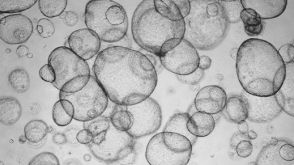 实验方案Detection of CYP3A4 Activity in Human Hepatic Organoids by LC-MS

 沪公网安备31010102008431号