E. Menares et al. (sep 2019)
Nature communications 10 1 4401
Tissue-resident memory CD8+ T cells amplify anti-tumor immunity by triggering antigen spreading through dendritic cells.
Tissue-resident memory CD8+ T (Trm) cells mediate potent local innate and adaptive immune responses and play a central role against solid tumors. However,whether Trm cells cross-talk with dendritic cells (DCs) to support anti-tumor immunity remains unclear. Here we show that antigen-specific activation of skin Trm cells leads to maturation and migration to draining lymph nodes of cross-presenting dermal DCs. Tumor rejection mediated by Trm cells triggers the spread of cytotoxic CD8+ T cell responses against tumor-derived neo- and self-antigens via dermal DCs. These responses suppress the growth of intradermal tumors and disseminated melanoma lacking the Trm cell-targeted epitope. Moreover,analysis of RNA sequencing data from human melanoma tumors reveals that enrichment of a Trm cell gene signature associates with DC activation and improved survival. This work unveils the ability of Trm cells to amplify the breath of cytotoxic CD8+ T cell responses through DCs,thereby strengthening anti-tumor immunity.
View Publication
产品类型:
产品号#:
09605
09655
产品名:
StemSpan™ SFEM II
StemSpan™ SFEM II
(Apr 2025)
NPJ Vaccines 10
Emulsion adjuvant-induced uric acid release modulates optimal immunogenicity by targeting dendritic cells and B cells
Squalene-based emulsion (SE) adjuvants like MF59 and AS03 are used in protein subunit vaccines against influenza virus (e.g.,Fluad,Pandemrix,Arepanrix) and SARS-CoV-2 (e.g.,Covifenz,SKYCovione). We demonstrate the critical role of uric acid (UA),a damage-associated molecular pattern (DAMP),in triggering immunogenicity by SE adjuvants. In mice,SE adjuvants elevated DAMP levels in draining lymph nodes. Strikingly,inhibition of UA synthesis reduced vaccine-induced innate immunity,subsequently impairing optimal antibody and T cell responses. In vivo treatment with UA crystals elicited partial adjuvant effects. In vitro stimulation with UA crystals augmented the activation of dendritic cells (DCs) and B cells and altered multiple pathways in these cells,including inflammation and antigen presentation in DCs and cell proliferation in B cells. In an influenza vaccine model,UA contributed to protection against influenza viral infection. These results demonstrate the importance of DAMPs,specifically the versatile role of UA in the immunogenicity of SE adjuvants,by regulating DCs and B cells.
View Publication
Lister R et al. (NOV 2009)
Nature 462 7271 315--22
Human DNA methylomes at base resolution show widespread epigenomic differences.
DNA cytosine methylation is a central epigenetic modification that has essential roles in cellular processes including genome regulation,development and disease. Here we present the first genome-wide,single-base-resolution maps of methylated cytosines in a mammalian genome,from both human embryonic stem cells and fetal fibroblasts,along with comparative analysis of messenger RNA and small RNA components of the transcriptome,several histone modifications,and sites of DNA-protein interaction for several key regulatory factors. Widespread differences were identified in the composition and patterning of cytosine methylation between the two genomes. Nearly one-quarter of all methylation identified in embryonic stem cells was in a non-CG context,suggesting that embryonic stem cells may use different methylation mechanisms to affect gene regulation. Methylation in non-CG contexts showed enrichment in gene bodies and depletion in protein binding sites and enhancers. Non-CG methylation disappeared upon induced differentiation of the embryonic stem cells,and was restored in induced pluripotent stem cells. We identified hundreds of differentially methylated regions proximal to genes involved in pluripotency and differentiation,and widespread reduced methylation levels in fibroblasts associated with lower transcriptional activity. These reference epigenomes provide a foundation for future studies exploring this key epigenetic modification in human disease and development.
View Publication
产品类型:
产品号#:
05850
05857
05870
05875
85850
85857
85870
85875
产品名:
mTeSR™1
mTeSR™1
Lam RS et al. ( 2017)
PloS one 12 1 e0169506
Functional Maturation of Human Stem Cell-Derived Neurons in Long-Term Cultures.
Differentiated neurons can be rapidly acquired,within days,by inducing stem cells to express neurogenic transcription factors. We developed a protocol to maintain long-term cultures of human neurons,called iNGNs,which are obtained by inducing Neurogenin-1 and Neurogenin-2 expression in induced pluripotent stem cells. We followed the functional development of iNGNs over months and they showed many hallmark properties for neuronal maturation,including robust electrical and synaptic activity. Using iNGNs expressing a variant of channelrhodopsin-2,called CatCh,we could control iNGN activity with blue light stimulation. In combination with optogenetic tools,iNGNs offer opportunities for studies that require precise spatial and temporal resolution. iNGNs developed spontaneous network activity,and these networks had excitatory glutamatergic synapses,which we characterized with single-cell synaptic recordings. AMPA glutamatergic receptor activity was especially dominant in postsynaptic recordings,whereas NMDA glutamatergic receptor activity was absent from postsynaptic recordings but present in extrasynaptic recordings. Our results on long-term cultures of iNGNs could help in future studies elucidating mechanisms of human synaptogenesis and neurotransmission,along with the ability to scale-up the size of the cultures.
View Publication
产品类型:
产品号#:
05854
05855
85850
85857
85870
85875
产品名:
mFreSR™
mFreSR™
mTeSR™1
mTeSR™1
Tsikritsis D et al. (MAY 2016)
Cytometry. Part A : the journal of the International Society for Analytical Cytology 1--23
Label-free biomarkers of human embryonic stem cell differentiation to hepatocytes.
Three different label-free,minimally invasive,live single cell analysis techniques were used to characterize embryonic stem cells,and the hepatocytes into which they were differentiated. Atomic Force Microscopy measures the cell's mechanical properties,Raman spectroscopy measures its chemical properties,and dielectrophoresis measures the membrane's capacitance. We were able to assign cell type of individual cells with accuracies of 96.5% (Atomic Force Microscopy),92.5 % (Raman spectroscopy),and *** % (Dielectrophoresis). These techniques,used either independently or in combination,offer label-free methods to study individual living cells. Although they can be applied to any phenotypical or environmental change,these techniques have most potential in human cell therapies where the use of biomarkers is best avoided. If all three properties are independent,then a combined accuracy of *** % can be achieved in cell characterization. We suggest how these methods could be combined into one microfluidic chip for cell sorting,and how they can be applied to cell culture.
View Publication
产品类型:
产品号#:
05850
05857
05870
05875
85850
85857
85870
85875
产品名:
mTeSR™1
mTeSR™1
Bajpai VK et al. (JAN 2017)
Stem cells (Dayton,Ohio)
Reprogramming Postnatal Human Epidermal Keratinocytes Toward Functional Neural Crest Fates.
During development,neural crest (NC) cells are induced by signaling events at the neural plate border of all vertebrate embryos. Initially arising within the central nervous system,NC cells subsequently undergo an epithelial to mesenchymal transition to migrate into the periphery,where they differentiate into diverse cell types. Here we provide evidence that postnatal human epidermal keratinocytes (KC),in response to fibroblast growth factor 2 and insulin like growth factor 1 signals,can be reprogrammed toward a NC fate. Genome-wide transcriptome analyses show that keratinocyte-derived NC cells are similar to those derived from human embryonic stem cells. Moreover,they give rise in vitro and in vivo to NC derivatives such as peripheral neurons,melanocytes,Schwann cells and mesenchymal cells (osteocytes,chondrocytes,adipocytes,and smooth muscle cells). By demonstrating that human keratin-14+ KC can form NC cells,even from clones of single cells,our results have important implications in stem cell biology and regenerative medicine. Stem Cells 2017.
View Publication
产品类型:
产品号#:
70022
70071
产品名:
Quadrato G et al. (MAY 2017)
Nature 545 7652 48--53
Cell diversity and network dynamics in photosensitive human brain organoids.
In vitro models of the developing brain such as three-dimensional brain organoids offer an unprecedented opportunity to study aspects of human brain development and disease. However,the cells generated within organoids and the extent to which they recapitulate the regional complexity,cellular diversity and circuit functionality of the brain remain undefined. Here we analyse gene expression in over 80,000 individual cells isolated from 31 human brain organoids. We find that organoids can generate a broad diversity of cells,which are related to endogenous classes,including cells from the cerebral cortex and the retina. Organoids could be developed over extended periods (more than 9 months),allowing for the establishment of relatively mature features,including the formation of dendritic spines and spontaneously active neuronal networks. Finally,neuronal activity within organoids could be controlled using light stimulation of photosensitive cells,which may offer a way to probe the functionality of human neuronal circuits using physiological sensory stimuli.
View Publication
产品类型:
产品号#:
05850
05857
05870
05875
85850
85857
85870
85875
产品名:
mTeSR™1
mTeSR™1
Liebmann JE et al. ( 1993)
British journal of cancer 68 6 1104--1109
Cytotoxic studies of paclitaxel (Taxol) in human tumour cell lines.
The cytotoxicity of paclitaxel against eight human tumour cell lines has been studied with in vitro clonogenic assays. The fraction of surviving cells fell sharply after exposure for 24 h to paclitaxel concentrations ranging from 2 to 20 nM; the paclitaxel IC50 was found to range between 2.5 and 7.5 nM. Increasing the paclitaxel concentration above 50 nM,however,resulted in no additional cytotoxicity after a 24 h drug exposure. Cells incubated in very high concentrations of paclitaxel (10,000 nM) had an increase in survival compared with cells treated with lower concentrations of the drug. Prolonging the time of exposure of cells to paclitaxel from 24 to 72 h increased cytotoxicity from 5 to 200 fold in different cell lines. Exponentially growing cells were more sensitive to paclitaxel than were cells in the plateau phase of growth. Cremophor EL,the diluent in which the clinical preparation of paclitaxel is formulated,antagonised paclitaxel at concentrations of 0.135% (v/v). These data suggest that paclitaxel will be most effective clinically when there is prolonged exposure of tumour to the drug. Further,it appears that modest concentrations (i.e.,50 nM) should be as effective as higher concentrations of paclitaxel. Finally,we have noted that Cremophor EL is a biologically active diluent and,at high concentrations (0.135% v/v),can antagonise paclitaxel cytotoxicity.
View Publication
产品类型:
产品号#:
73312
73314
产品名:
紫杉醇
紫杉醇
McIntyre BAS et al. (JUL 2015)
Innate immunity 21 5 504--511
Innate immune response of human pluripotent stem cell-derived airway epithelium.
The acquisition of innate immune response is requisite to having bona fide differentiation of airway epithelium. Procedures developed to differentiate lung airway from human pluripotent stem cells (hPSCs) have demonstrated anecdotal evidence for innate immune response,but an in-depth exploration of response levels is lacking. Herein,using an established method of airway epithelial generation from hPSCs,we show that hPSC-derived epithelial cells are able to up-regulate expression of TNF$\$,IL8 and IL1$\$ response to challenge with bacterial endotoxin LPS,but lack response from genes associated with innate immune response in other cell types. Further,stimulation of cells with TNF-$\$ in auto-induction of TNF$\$,as well as cytokine responses of IL8 and IL1$\$ The demonstration of innate immune induction in hPSC-derived airway epithelia gives further strength to the functionality of in vitro protocols aimed at generating differentiated airway cells that can potentially be used in a translational setting. Finally,we propose that innate immune challenge of airway epithelium from human pluripotent stem cell sources be used as a robust validation of functional in vitro differentiation.
View Publication

 EasySep™小鼠TIL(CD45)正选试剂盒


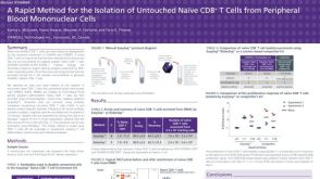 科学海报Cell Isolation to Obtain Untouched Naïve CD8 T Cells from Peripheral Blood Mononuclear Cells (PBMCs)


 沪公网安备31010102008431号