
 EasySep™小鼠TIL(CD45)正选试剂盒


搜索结果: 'methocult media formulations for human hematopoietic cells serum containing'
-

产品类型:
产品号#:
100-0560
产品名:
L -丁硫氨酸-(S,R)-亚砜亚胺
-
产品类型:
产品号#:
100-0548
100-0549
产品名:
3,3',5-三碘- l -甲状腺原氨酸(钠盐水合物)
3,3',5-三碘- l -甲状腺原氨酸(钠盐水合物)
-
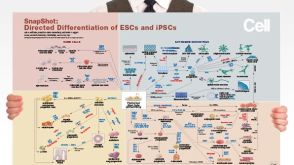 挂图Directed Differentiation of Pluripotent Stem Cells Strategies for differentiating ES and iPS cells into ectoderm, endoderm or mesoderm
 -
产品类型:
产品号#:
05850
05857
05870
05875
07903
85850
85857
85870
85875
产品名:
0.1% 明胶水溶液
mTeSR™1
mTeSR™1
-

-
产品类型:
产品号#:
05850
05857
05870
05875
85850
85857
85870
85875
产品名:
mTeSR™1
mTeSR™1
-
产品类型:
产品号#:
03800
03801
03802
03803
03804
03805
03806
产品名:
ClonaCell™-HY杂交瘤试剂盒
ClonaCell™-HY培养基A
ClonaCell™-HY 培养基 B
ClonaCell™-HY 培养基 C
ClonaCell™-HY 培养基 D
ClonaCell™-HY 培养基 E
ClonaCell™-HY PEG
-
产品类型:
产品号#:
21000
20119
20155
18081
18081RF
产品名:
RoboSep™- S
RoboSep™ 吸头组件抛光剂
RoboSep™分选管套装(9个塑料管)
-
产品类型:
产品号#:
05620
产品名:
MammoCult™ 人源培养基套装
-
产品类型:
产品号#:
73012
73014
产品名:
布雷非德菌素A
布雷非德菌素A
-
产品类型:
产品号#:
21000
20119
20155
19752
19752RF
产品名:
RoboSep™- S
RoboSep™ 吸头组件抛光剂
RoboSep™分选管套装(9个塑料管)


 沪公网安备31010102008431号