
 EasySep™小鼠TIL(CD45)正选试剂盒


搜索结果: 'methocult media formulations for human hematopoietic cells serum containing'
-

产品类型:
产品号#:
73432
73434
产品名:
SU11274
SU11274, 5 mg
-
产品类型:
产品号#:
73262
73264
产品名:
(S) -MG132
(S) -MG132
-
产品类型:
产品号#:
05711
100-1281
产品名:
NeuroCult™ SM1 神经添加物
NeuroCult™ SM1 神经添加物
-
产品类型:
产品号#:
05700
05750
05751
产品名:
NeuroCult™ 基础培养基(小鼠和大鼠)
NeuroCult™ NS-A 基础培养基(人)
NeuroCult™ NS-A 扩增试剂盒(人)
-
产品类型:
产品号#:
19254
19254RF
19854
19854RF
产品名:
EasySep™人Naïve B细胞富集试剂盒
RoboSep™ 人Naïve B细胞富集试剂盒含滤芯吸头
EasySep™小鼠B细胞分选试剂盒
RoboSep™ 小鼠B细胞分选试剂盒
-
产品类型:
产品号#:
01700
01705
01701
01702
14056
14066
28600
19056
19056RF
19756
19756RF
产品名:
ALDEFLUOR™ 试剂盒
ALDEFLUOR™ DEAB试剂, 1.5 mM, 1 mL
ALDEFLUOR™检测缓冲液
L-Calc™有限稀释软件
-
产品类型:
产品号#:
15021
15061
产品名:
RosetteSep™人T细胞富集抗体混合物
RosetteSep™人T细胞富集抗体混合物
-
产品类型:
产品号#:
05850
05857
05870
05875
85850
85857
85870
85875
产品名:
mTeSR™1
mTeSR™1
-
产品类型:
产品号#:
05001
05021
05022
产品名:
PneumaCult™-ALI 培养基
PneumaCult™-ALI 培养基含12 mm Transwell®插件
PneumaCult™-ALI 培养基含6.5 mm Transwell®插件
-
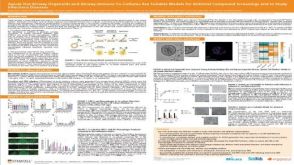 科学海报Apical-Out Airway Organoids and Airway-Immune Co-Cultures Are Suitable Models for Antiviral Compound Screenings and to Study Infectious Diseases
产品类型:
Conference:
ATS 2023
产品号#:
产品名:
-
产品类型:
产品号#:
100-0784
100-0956
10971
10981
10991
产品名:
ImmunoCult™ 人CD3/CD28 T细胞激活剂
ImmunoCult™ XF培养基
ImmunoCult™ 人CD3/CD28 T细胞激活剂
ImmunoCult™ XF 人T细胞扩增培养基,500 mL
ImmunoCult™ 人CD3/CD28 T细胞激活剂
-
产品类型:
产品号#:
15028
15068
产品名:
RosetteSep™人单核细胞富集抗体混合物
RosetteSep™人单核细胞富集抗体混合物

 沪公网安备31010102008431号