Rac1 is essential for intraembryonic hematopoiesis and for the initial seeding of fetal liver with definitive hematopoietic progenitor cells.
Definitive hematopoietic stem and progenitor cells (HSCs/Ps) originating from the yolk sac and/or para-aorta-splanchno-pleura/aorta-gonad-mesonephros are hypothesized to colonize the fetal liver,but mechanisms involved are poorly defined. The Rac subfamily of Rho GTPases has been shown to play essential roles in HSC/P localization to the bone marrow following transplantation. Here,we study the role of Rac1 in HSC/P migration during ontogeny and seeding of fetal liver. Using a triple-transgenic approach,we have deleted Rac1 in HSCs/Ps during very early embryonic development. Without Rac1,there was a decrease in circulating HSCs/Ps in the blood of embryonic day (E) 10.5 embryos,while yolk sac definitive hematopoiesis was quantitatively normal. Intraembryonic hematopoiesis was significantly impaired in Rac1-deficient embryos,culminating with absence of intra-aortic clusters and fetal liver hematopoiesis. At E10.5,Rac1-deficient HSCs/Ps displayed decreased transwell migration and impaired inter-action with the microenvironment in migration-dependent assays. These data suggest that Rac1 plays an important role in HSC/P migration during embryonic development and is essential for the emergence of intraembryonic hematopoiesis.
View Publication
Xaymardan M et al. (AUG 2009)
Stem cells (Dayton,Ohio) 27 8 1911--20
c-Kit function is necessary for in vitro myogenic differentiation of bone marrow hematopoietic cells.
In recent years,the differentiation of bone marrow cells (BMCs) into myocytes has been extensively investigated,but the findings remain inconclusive. The purpose of this study was to determine the conditions necessary to induce myogenic differentiation in short-term cultures of adult BMCs,and to identify the BMC subpopulation responsible for this phenomenon. We report that high-density cultures of murine hematopoietic BMCs gave rise to spontaneous beating cell clusters in the presence of vascular endothelial and fibroblast growth factors. These clusters originated from c-kit(pos) cells. The formation of the clusters could be completely blocked by adding a c-kit/tyrosine kinase inhibitor,Gleevec (imatinib mesylate; Novartis International,Basel,Switzerland,http://www.novartis.com),to the culture. Cluster formation was also blunted in BMCs from c-kit-deficient (Kit(W)/Kit(W-v)) mice. Clustered cells expressed cardiomyocyte-specific transcription factor genes Gata-4 and Nkx2.5,sarcomeric proteins beta-MHC and MLC-2v,and ANF and connexin-43. Immunostaining revealed alpha-sarcomeric actinin expression in more than 90% of clustered cells. Under electron microscopy,the clustered cells exhibited a sarcomeric myofiber arrangement and z-bands. This study defines the microenvironment required to achieve a reproducible in vitro model of beating,myogenic cell clusters. This model could be used to examine the mechanisms responsible for the postnatal myogenic differentiation of BMCs. Our results identify c-kit(pos) bone marrow hematopoietic cells as the source of the myogenic clusters.
View Publication
Leung CG et al. (JUL 2007)
The Journal of experimental medicine 204 7 1603--11
Requirements for survivin in terminal differentiation of erythroid cells and maintenance of hematopoietic stem and progenitor cells.
Survivin,which is the smallest member of the inhibitor of apoptosis protein (IAP) family,is a chromosomal passenger protein that mediates the spindle assembly checkpoint and cytokinesis,and also functions as an inhibitor of apoptosis. Frequently overexpressed in human cancers and not expressed in most adult tissues,survivin has been proposed as an attractive target for anticancer therapies and,in some cases,has even been touted as a cancer-specific gene. Survivin is,however,expressed in proliferating adult cells,including human hematopoietic stem cells,T-lymphocytes,and erythroid cells throughout their maturation. Therefore,it is unclear how survivin-targeted anticancer therapies would impact steady-state blood development. To address this question,we used a conditional gene-targeting strategy and abolished survivin expression from the hematopoietic compartment of mice. We show that inducible deletion of survivin leads to ablation of the bone marrow,with widespread loss of hematopoietic progenitors and rapid mortality. Surprisingly,heterozygous deletion of survivin causes defects in erythropoiesis in a subset of the animals,with a dramatic reduction in enucleated erythrocytes and the presence of immature megaloblastic erythroblasts. Our studies demonstrate that survivin is essential for steady-state hematopoiesis and survival of the adult,and further,that a high level of survivin expression is critical for proper erythroid differentiation.
View Publication
产品类型:
产品号#:
19756
19756RF
产品名:
J. Qiu et al. (dec 2022)
STAR protocols 3 4 101828
Protocol to identify and analyze mouse and human quiescent hematopoietic stem cells using flow cytometry combined with confocal imaging.
Mitochondrial membrane potential (MMP) segregates functionally distinct subsets within highly purified hematopoietic stem cells (HSCs). Here,we detail a protocol for FACS isolation of MMP sub-fractions of phenotypically defined mouse and human HSCs. These steps are followed by high-/super-resolution immunofluorescence microscopy of HSCs' lysosomes. While the protocol describes the isolation of quiescent HSCs,which are the most potent subsets,it could also be applied to other HSC subsets. This protocol overcomes some experimental challenges associated with low HSC numbers. For complete details on the use and execution of this protocol,please refer to Liang et al. (2020) and Qiu et al. (2021).
View Publication
产品类型:
产品号#:
09600
18000
19856
09650
19856RF
产品名:
StemSpan™ SFEM
EasySep™磁极
EasySep™小鼠造血祖细胞分选试剂盒
StemSpan™ SFEM
RoboSep™ 小鼠造血祖细胞分选试剂盒
Zhang CC et al. (FEB 2006)
Proceedings of the National Academy of Sciences of the United States of America 103 7 2184--9
Prion protein is expressed on long-term repopulating hematopoietic stem cells and is important for their self-renewal.
Although the wild-type prion protein (PrP) is abundant and widely expressed in various types of tissues and cells,its physiological function(s) remain unknown,and PrP knockout mice do not exhibit overt and undisputed phenotypes. Here we showed that PrP is expressed on the surface of several bone marrow cell populations successively enriched in long-term (LT) hematopoietic stem cells (HSCs) using flow cytometry analysis. Affinity purification of the PrP-positive and -negative fractions from these populations,followed by competitive bone marrow reconstitution assays,shows that all LT HSCs express PrP. HSCs from PrP-null bone marrow exhibited impaired self-renewal in serial transplantation of lethally irradiated mouse recipients both in the presence and absence of competitors. When treated with a cell cycle-specific myelotoxic agent,the animals reconstituted with PrP-null HSCs exhibit increased sensitivity to hematopoietic cell depletion. Ectopic expression of PrP in PrP-null bone marrow cells by retroviral infection rescued the defective hematopoietic engraftment during serial transplantation. Therefore,PrP is a marker for HSCs and supports their self-renewal.
View Publication
产品类型:
产品号#:
03630
03434
03444
09600
09650
28600
产品名:
MethoCult™ M3630
MethoCult™ GF M3434
MethoCult™ GF M3434
StemSpan™ SFEM
StemSpan™ SFEM
L-Calc™有限稀释软件
Phondeechareon T et al. (OCT 2016)
Annals of hematology 95 10 1617--1625
Generation of induced pluripotent stem cells as a potential source of hematopoietic stem cells for transplant in PNH patients.
Paroxysmal nocturnal hemoglobinuria (PNH) is an acquired hemolytic anemia caused by lack of CD55 and CD59 on blood cell membrane leading to increased sensitivity of blood cells to complement. Hematopoietic stem cell transplantation (HSCT) is the only curative therapy for PNH,however,lack of HLA-matched donors and post-transplant complications are major concerns. Induced pluripotent stem cells (iPSCs) derived from patients are an attractive source for generating autologous HSCs to avoid adverse effects resulting from allogeneic HSCT. The disease involves only HSCs and their progeny; therefore,other tissues are not affected by the mutation and may be used to produce disease-free autologous HSCs. This study aimed to derive PNH patient-specific iPSCs from human dermal fibroblasts (HDFs),characterize and differentiate to hematopoietic cells using a feeder-free protocol. Analysis of CD55 and CD59 expression was performed before and after reprogramming,and hematopoietic differentiation. Patients' dermal fibroblasts expressed CD55 and CD59 at normal levels and the normal expression remained after reprogramming. The iPSCs derived from PNH patients had typical pluripotent properties and differentiation capacities with normal karyotype. After hematopoietic differentiation,the differentiated cells expressed early hematopoietic markers (CD34 and CD43) with normal CD59 expression. The iPSCs derived from HDFs of PNH patients have normal levels of CD55 and CD59 expression and hold promise as a potential source of HSCs for autologous transplantation to cure PNH patients.
View Publication
产品类型:
产品号#:
05850
05857
05870
05875
07923
07920
04435
04445
85850
85857
85870
85875
07922
产品名:
Dispase (1 U/mL)
ACCUTASE™
MethoCult™ H4435 Enriched
MethoCult™ H4435 Enriched
mTeSR™1
mTeSR™1
ACCUTASE™
S. S. De Ravin et al. (APR 2016)
Nature biotechnology 34 4 424--9
Targeted gene addition in human CD34(+) hematopoietic cells for correction of X-linked chronic granulomatous disease.
Gene therapy with genetically modified human CD34(+) hematopoietic stem and progenitor cells (HSPCs) may be safer using targeted integration (TI) of transgenes into a genomic 'safe harbor' site rather than random viral integration. We demonstrate that temporally optimized delivery of zinc finger nuclease mRNA via electroporation and adeno-associated virus (AAV) 6 delivery of donor constructs in human HSPCs approaches clinically relevant levels of TI into the AAVS1 safe harbor locus. Up to 58{\%} Venus(+) HSPCs with 6-16{\%} human cell marking were observed following engraftment into mice. In HSPCs from patients with X-linked chronic granulomatous disease (X-CGD),caused by mutations in the gp91phox subunit of the NADPH oxidase,TI of a gp91phox transgene into AAVS1 resulted in ∼15{\%} gp91phox expression and increased NADPH oxidase activity in ex vivo-derived neutrophils. In mice transplanted with corrected HSPCs,4-11{\%} of human cells in the bone marrow expressed gp91phox. This method for TI into AAVS1 may be broadly applicable to correction of other monogenic diseases.
View Publication
J. Yen et al. (NOV 2018)
Scientific reports 8 1 16304
TRIAMF: A New Method for Delivery of Cas9 Ribonucleoprotein Complex to Human Hematopoietic Stem Cells.
CRISPR/Cas9 mediated gene editing of patient-derived hematopoietic stem and progenitor cells (HSPCs) ex vivo followed by autologous transplantation of the edited HSPCs back to the patient can provide a potential cure for monogenic blood disorders such as $\beta$-hemoglobinopathies. One challenge for this strategy is efficient delivery of the ribonucleoprotein (RNP) complex,consisting of purified Cas9 protein and guide RNA,into HSPCs. Because $\beta$-hemoglobinopathies are most prevalent in developing countries,it is desirable to have a reliable,efficient,easy-to-use and cost effective delivery method. With this goal in mind,we developed TRansmembrane Internalization Assisted by Membrane Filtration (TRIAMF),a new method to quickly and effectively deliver RNPs into HSPCs by passing a RNP and cell mixture through a filter membrane. We achieved robust gene editing in HSPCs using TRIAMF and demonstrated that the multilineage colony forming capacities and the competence for engraftment in immunocompromised mice of HSPCs were preserved post TRIAMF treatment. TRIAMF is a custom designed system using inexpensive components and has the capacity to process HSPCs at clinical scale.
View Publication

 EasySep™小鼠TIL(CD45)正选试剂盒


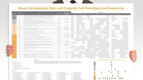 挂图Mouse Hematopoietic Stem and Progenitor Cell Phenotyping Overview of mouse HSPC subset surface markers and frequencies发布日期: 08/10/2018
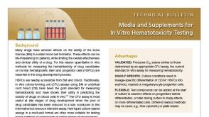 技术公告Media and Supplements for In Vitro Hematotoxicity Testing

 沪公网安备31010102008431号