Lee J et al. ( 2012)
Angewandte Chemie (International ed. in English) 51 50 12509--12513
A novel small molecule facilitates the reprogramming of human somatic cells into a pluripotent state and supports the maintenance of an undifferentiated state of human pluripotent stem cells.
Booster of pluripotency: RSC133,a new synthetic derivative of indoleacrylic acid/indolepropionic acid,exhibits dual activity by inhibiting histone deacetylase and DNA methyltransferase. Furthermore it potently improves the reprogramming of human somatic cells into a pluripotent state and aids the growth and maintenance of human pluripotent stem cells (hPSCs).
View Publication
产品类型:
产品号#:
73392
产品名:
RSC-133
Warren L et al. (NOV 2010)
Cell stem cell 7 5 618--630
Highly efficient reprogramming to pluripotency and directed differentiation of human cells with synthetic modified mRNA
Clinical application of induced pluripotent stem cells (iPSCs) is limited by the low efficiency of iPSC derivation and the fact that most protocols modify the genome to effect cellular reprogramming. Moreover,safe and effective means of directing the fate of patient-specific iPSCs toward clinically useful cell types are lacking. Here we describe a simple,nonintegrating strategy for reprogramming cell fate based on administration of synthetic mRNA modified to overcome innate antiviral responses. We show that this approach can reprogram multiple human cell types to pluripotency with efficiencies that greatly surpass established protocols. We further show that the same technology can be used to efficiently direct the differentiation of RNA-induced pluripotent stem cells (RiPSCs) into terminally differentiated myogenic cells. This technology represents a safe,efficient strategy for somatic cell reprogramming and directing cell fate that has broad applicability for basic research,disease modeling,and regenerative medicine. ?? 2010 Elsevier Inc.
View Publication
产品类型:
产品号#:
04434
04444
05850
05857
05870
05875
07913
27100
27150
85850
85857
85870
85875
产品名:
MethoCult™ H4434 Classic
MethoCult™ H4434 Classic
Dispase(5 U/mL)
35 mm培养皿
35 mm培养皿
mTeSR™1
mTeSR™1
Abuljadayel IS (JAN 2003)
Current medical research and opinion 19 5 355--75
Induction of stem cell-like plasticity in mononuclear cells derived from unmobilised adult human peripheral blood.
Undifferentiated pluripotent stem cells with flexible developmental potentials are not normally found in peripheral blood. However,such cells have recently been reported to reside in the bone marrow. Herein are reported methods of inducing pluripotency in cells derived from unmobilised adult human peripheral blood. In response to the inclusion of purified CR3/43 monoclonal antibody (mAb) to well-established culture conditions,mononuclear cells (MNC) obtained from a single blood donor are converted into pluripotent haematopoietic,neuronal and cardiomyogenic progenitor stem cells or undifferentiated stem cells. The haematopoietic stem cells are CD34+,clonogenic and have been shown to repopulate non-obese diabetic/severe combined immunodeficient (NOD/SCID) mice. The neuronal precursors transcribe the primitive stem cell markers OCT-4 and nestin,and on maturation,differentially stain positive for neuronal,glial or oligodendrocyte-specific antigens. The cardiomyogenic progenitor stem cells form large bodies of asynchronously beating cells and differentiate into mature cardiomyocytes which transcribe GATA-4. The undifferentiated stem cells do not express haematopoietic-associated markers,are negative for major histocompatibility complex (MHC) class I and II antigens,transcribe high levels of OCT-4 and form embryoid body (EB)-like structures. This induction of stem cell-like plasticity in MNC may have proceeded by a process of retrodifferentiation but,in any case,could have profound clinical and pharmacological implications. Finally,the flexibility and the speed by which a variety of stem cell classes can be generated ex vivo from donor blood could potentially transfer this novel process into a less invasive automated clinical procedure.
View Publication
产品类型:
产品号#:
04434
04444
产品名:
MethoCult™ H4434 Classic
MethoCult™ H4434 Classic
J. Shao et al. (FEB 2017)
Scientific reports 7 42363
Experimental Study of the Biological Properties of Human Embryonic Stem Cell-Derived Retinal Progenitor Cells.
Retinal degenerative diseases are among the leading causes of blindness worldwide,and cell replacement is considered as a promising therapeutic. However,the resources of seed cells are scarce. To further explore this type of therapy,we adopted a culture system that could harvest a substantial quantity of retinal progenitor cells (RPCs) from human embryonic stem cells (hESCs) within a relatively short period of time. Furthermore,we transplanted these RPCs into the subretinal spaces of Royal College of Surgeons (RCS) rats. We quantified the thickness of the treated rats' outer nuclear layers (ONLs) and explored the visual function via electroretinography (ERG). It was found that the differentiated cells expressed RPC markers and photoreceptor progenitor markers. The transplanted RPCs survived for at least 12 weeks,resulting in beneficial effects on the morphology of the host retina,and led to a significant improvement in the visual function of the treated animals. These therapeutic effects suggest that the hESCs-derived RPCs could delay degeneration of the retina and partially restore visual function.
View Publication
Burkholderia pseudomallei-loaded cells act as a Trojan horse to invade the brain during endotoxemia.
Neurologic melioidosis occurs in both human and animals; however,the mechanism by which the pathogen Burkholderia pseudomallei invades the central nervous system (CNS) remains unclear. B. pseudomallei-loaded Ly6C cells have been suggested as a putative portal; however,during melioidosis,lipopolysaccharide (LPS) can drive disruption of the blood-brain barrier (BBB). This study aims to test whether the Trojan horse-like mechanism occurs during endotoxemia. The expression levels of cerebral cytokines,chemokines and cell adhesion molecules; the activation of astrocytes,microglia and endothelial cells; and the increased vascular permeability and brain-infiltrating leukocytes were evaluated using B. pseudomallei,B. thailandensis,B. cenocepacia and B. multivorans LPS-induced brains. Accordingly,different degrees of BBB damage in those brains with endotoxemia were established. The B. multivorans LPS-induced brain exhibited the highest levels of disruptive BBB according to the above mediators/indicators. Into these distinct groups of endotoxemic mice,B. pseudomallei-loaded Ly6C cells or free B. pseudomallei were adoptively transferred at equal bacterial concentrations (103 CFU). The bacterial load and number of cases of meningeal neutrophil infiltration in the brains of animals treated with B. pseudomallei-loaded Ly6C cells were higher than those in brains induced by free B. pseudomallei in any of the endotoxemic groups. In particular,these results were reproducible in B. multivorans LPS-induced brains. We suggest that B. pseudomallei-loaded cells can act as a Trojan horse and are more effective than free B. pseudomallei in invading the CNS under septic or endotoxemic conditions even when there is a high degree of BBB disruption.
View Publication
产品类型:
产品号#:
产品名:
R. M. Eichenberger et al. ( 2018)
Frontiers in immunology 9 850
Hookworm Secreted Extracellular Vesicles Interact With Host Cells and Prevent Inducible Colitis in Mice.
Gastrointestinal (GI) parasites,hookworms in particular,have evolved to cause minimal harm to their hosts,allowing them to establish chronic infections. This is mediated by creating an immunoregulatory environment. Indeed,hookworms are such potent suppressors of inflammation that they have been used in clinical trials to treat inflammatory bowel diseases (IBD) and celiac disease. Since the recent description of helminths (worms) secreting extracellular vesicles (EVs),exosome-like EVs from different helminths have been characterized and their salient roles in parasite-host interactions have been highlighted. Here,we analyze EVs from the rodent parasite Nippostrongylus brasiliensis,which has been used as a model for human hookworm infection. N. brasiliensis EVs (Nb-EVs) are actively internalized by mouse gut organoids,indicating a role in driving parasitism. We used proteomics and RNA-Seq to profile the molecular composition of Nb-EVs. We identified 81 proteins,including proteins frequently present in exosomes (like tetraspanin,enolase,14-3-3 protein,and heat shock proteins),and 27 sperm-coating protein-like extracellular proteins. RNA-Seq analysis revealed 52 miRNA species,many of which putatively map to mouse genes involved in regulation of inflammation. To determine whether GI nematode EVs had immunomodulatory properties,we assessed their potential to suppress GI inflammation in a mouse model of inducible chemical colitis. EVs from N. brasiliensis but not those from the whipworm Trichuris muris or control vesicles from grapes protected against colitic inflammation in the gut of mice that received a single intraperitoneal injection of EVs. Key cytokines associated with colitic pathology (IL-6,IL-1$\beta$,IFN$\gamma$,and IL-17a) were significantly suppressed in colon tissues from EV-treated mice. By contrast,high levels of the anti-inflammatory cytokine IL-10 were detected in Nb-EV-treated mice. Proteins and miRNAs contained within helminth EVs hold great potential application in development of drugs to treat helminth infections as well as chronic non-infectious diseases resulting from a dysregulated immune system,such as IBD.
View Publication
产品类型:
产品号#:
05504
06005
产品名:
MesenCult™ 成骨刺激试剂盒(小鼠)
IntestiCult™ 类器官生长培养基 (小鼠)
M. Epeldegui et al. (jun 2019)
Scientific reports 9 1 9371
Elevated numbers of PD-L1 expressing B cells are associated with the development of AIDS-NHL.
The risk for non-Hodgkin lymphoma (NHL) is markedly increased in persons living with human immunodeficiency virus (HIV) infection,and remains elevated in those on anti-retroviral therapy (cART). Both the loss of immunoregulation of Epstein-Barr virus (EBV) infected cells,as well as chronic B-cell activation,are believed to contribute to the genesis of AIDS-related NHL (AIDS-NHL). However,the mechanisms that lead to AIDS-NHL have not been completely defined. A subset of B cells that is characterized by the secretion of IL10,as well as the expression of the programmed cell death ligand-1 (PD-L1/CD274),was recently described. These PD-L1+ B cells can exert regulatory function,including the dampening of T-cell activation,by interacting with the program cell death protein (PD1) on target cells. The role of PD-L1+ B cells in the development of AIDS-NHL has not been explored. We assessed B cell PD-L1 expression on B cells preceding AIDS-NHL diagnosis in a nested case-control study of HIV+ subjects who went on to develop AIDS-NHL,as well as HIV+ subjects who did not,using multi-color flow cytometry. Archival frozen viable PBMC were obtained from the UCLA Multicenter AIDS Cohort Study (MACS). It was seen that the number of CD19+CD24++CD38++and CD19+PD-L1+cells was significantly elevated in cases 1-4 years prior to AIDS-NHL diagnosis,compared to controls,raising the possibility that these cells may play a role in the etiology of AIDS-NHL. Interestingly,most PD-L1+ expression on CD19+ cells was seen on CD19+CD24++CD38++ cells. In addition,we showed that HIV can directly induce PD-L1 expression on B cells through interaction of virion-associated CD40L with CD40 on B cells.
View Publication
Expression of breast cancer resistance protein in blast cells from patients with acute leukemia.
Breast cancer resistance protein (BCRP) is a novel member of the adenosine triphosphate-binding cassette superfamily of transport proteins. Transfection and enforced expression of BCRP in drug-sensitive cells confer resistance to mitoxantrone,doxorubicin,daunorubicin,and topotecan. We studied blast cells from 21 acute leukemia patients (20 acute myeloid leukemia,1 acute lymphocytic leukemia) for the expression of BCRP mRNA using a quantitative reverse-transcription polymerase chain reaction assay. BCRP mRNA expression varied more than 1000-fold among the samples tested,with low or barely detectable expression in half of the samples. Seven samples (33%) had relatively high expression of BCRP mRNA. High expression of BCRP did not correlate strongly with high expression of P-glycoprotein,suggesting that BCRP may cause resistance to certain antileukemic drugs in P-glycoprotein-negative cases. High expression of BCRP mRNA is sufficiently frequent in AML to warrant more extensive investigations to determine the relation of disease subtype and treatment outcome to BCRP expression and function.
View Publication
产品类型:
产品号#:
产品名:
Schlecht G et al. (SEP 2004)
Blood 104 6 1808--15
Murine plasmacytoid dendritic cells induce effector/memory CD8+ T-cell responses in vivo after viral stimulation.
Like their human counterparts,mouse plasmacytoid dendritic cells (pDCs) play a central role in innate immunity against viral infections,but their capacity to prime T cells in vivo remains unknown. We show here that virus-activated pDCs differentiate into antigen-presenting cells able to induce effector/memory CD8(+) T-cell responses in vivo against both epitopic peptides and endogenous antigen,whereas pDCs activated by synthetic oligodeoxynucleotides containing unmethylated cytosine-guanine motifs (CpG) acquire only the ability to recall antigen-experienced T-cell responses. We also show that immature pDCs are unable to induce effector or regulatory CD8(+) T-cell responses. Thus,murine pDCs take part in both innate and adaptive immune responses by directly priming naive CD8(+) T cells during viral infection.
View Publication
产品类型:
产品号#:
09600
09650
产品名:
StemSpan™ SFEM
StemSpan™ SFEM
Blanco J et al. (DEC 2004)
The Journal of biological chemistry 279 49 51305--14
High level of coreceptor-independent HIV transfer induced by contacts between primary CD4 T cells.
Cell-to-cell virus transmission is one of the most efficient mechanisms of human immunodeficiency virus (HIV) spread,requires CD4 and coreceptor expression in target cells,and may also lead to syncytium formation and cell death. Here,we show that in addition to this classical coreceptor-mediated transmission,the contact between HIV-producing cells and primary CD4 T cells lacking the appropriate coreceptor induced the uptake of HIV particles by target cells in the absence of membrane fusion or productive HIV replication. HIV uptake by CD4 T cells required cellular contacts mediated by the binding of gp120 to CD4 and intact actin cytoskeleton. HIV antigens taken up by CD4 T cells were rapidly endocytosed to trypsin-resistant compartments inducing a partial disappearance of CD4 molecules from the cell surface. Once the cellular contact was stopped,captured HIV were released as infectious particles. Electron microscopy revealed that HIV particles attached to the surface of target cells and accumulated in large (0.5-1.0 microm) intracellular vesicles containing 1-14 virions,without any evidence for massive clathrin-mediated HIV endocytosis. The capture of HIV particles into trypsin-resistant compartments required the availability of the gp120 binding site of CD4 but was independent of the intracytoplasmic tail of CD4. In conclusion,we describe a novel mechanism of HIV transmission,activated by the contact of infected and uninfected primary CD4 T cells,by which HIV could exploit CD4 T cells lacking the appropriate coreceptor as an itinerant virus reservoir.
View Publication
产品类型:
产品号#:
15022
15062
产品名:
RosetteSep™人CD4+ T细胞富集抗体混合物
RosetteSep™人CD4+ T细胞富集抗体混合物
Ben-Kasus T et al. (JUL 2005)
Biochemical pharmacology 70 1 121--33
Metabolic activation of zebularine, a novel DNA methylation inhibitor, in human bladder carcinoma cells.
Zebularine (2(1H)-pyrimidinone riboside,Zeb),a synthetic analogue of cytidine that is a potent inhibitor of cytidine deaminase,has been recently identified as a general inhibitor of DNA methylation. This inhibition of DNA methyltransferase (DNMT) is hypothesized to be mechanism-based and result from formation of a covalent complex between the enzyme and zebularine-substituted DNA. Metabolic activation of Zeb thus requires that it be phosphorylated and incorporated into DNA. We have quantitatively assessed the phosphorylation and DNA incorporation of Zeb in T24 cells using 2-[(14)C]-Zeb in conjunction with gradient anion-exchange HPLC and selected enzymatic and spectroscopic analyses. The corresponding 5'-mono-,di- and triphosphates of Zeb were readily formed in a dose- and time-dependent manner. Two additional Zeb-containing metabolites were tentatively identified as diphosphocholine (Zeb-DP-Chol) and diphosphoethanolamine adducts. Intracellular concentrations of Zeb-TP and Zeb-DP-Chol were similar and greatly exceeded those of other metabolites. DNA incorporation occurred but was surpassed by that of RNA by at least seven-fold. Equivalent levels and similar intracellular metabolic patterns were also observed in the Molt-4 (human T-lymphoblasts) and MC38 (murine colon carcinoma) cell lines. For male BALB/c nu/nu mice implanted s.c. with the EJ6 variant of T24 bladder carcinoma and treated i.p. with 500mg/kg 2-[(14)C]-Zeb,the in vivo phosphorylation pattern of Zeb in tumor tissue examined 24h after drug administration was similar to that observed in vitro. The complex metabolism of Zeb and its limited DNA incorporation suggest that these are the reasons why it is less potent than either 5-azacytidine or 5-aza-2'-deoxycytidine and requires higher doses for equivalent inhibition of DNMT.
View Publication

 EasySep™小鼠TIL(CD45)正选试剂盒


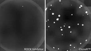 新闻STEMCELL Technologies to Launch CloneR™ to Facilitate Genome Editing of Human Pluripotent Stem Cells
 沪公网安备31010102008431号