Kumagai T et al. (JUN 2003)
Journal of the National Cancer Institute 95 12 896--905
Vitamin D2 analog 19-nor-1,25-dihydroxyvitamin D2: antitumor activity against leukemia, myeloma, and colon cancer cells.
BACKGROUND: 1,25-Dihydroxyvitamin D(3) inhibits growth of several types of human cancer cells in vitro,but its therapeutic use is hampered because it causes hypercalcemia. 19-nor-1,25-Dihydroxyvitamin D(2) (paricalcitol) is a noncalcemic vitamin D analog that is approved by the Food and Drug Administration for the treatment of secondary hyperparathyroidism. We investigated the antitumor activity and mechanism of action of paricalcitol in vitro and in vivo. METHODS: Effects of paricalcitol on proliferation,the cell cycle,differentiation,and apoptosis were examined in cancer cell lines. Effects on tumor growth were examined with colon cancer cell xenografts in nude mice (five in the experimental group and five in the control group). The interaction of paricalcitol with the vitamin D receptor (VDR) in mononuclear spleen cells and myeloid stem cells from wild-type and VDR knockout mice was examined. All statistical tests were two-sided. RESULTS: Paricalcitol inhibited the proliferation of myeloid leukemia cell lines HL-60,NB-4,and THP-1 cells at an effective dose that inhibited growth 50% (ED(50)) of 2.4-5.8 x 10(-9) M by inducing cell cycle arrest and differentiation. Paricalcitol inhibited the proliferation of NCI-H929 myeloma cells at an ED(50) of 2.0 x 10(-10) M by inducing cell cycle arrest and apoptosis. Paricalcitol also inhibited the proliferation of colon cancer cell lines HT-29 (ED(50) = 1.7 x 10(-8) M) and SW837 (ED(50) = 3.2 x 10(-8) M). HT-29 colon cancer xenografts in paricalcitol-treated nude mice were smaller (1044 mm(3) and 1752 mm(3),difference = 708 mm(3),95% confidence interval = 311 to 1104 mm(3); P =.03) and weighed less (1487 mg and 4162 mg,difference = 2675 mg,95% confidence interval = 2103 to 3248 mg; Ptextless.001) than those in vehicle-treated mice. Paricalcitol induced committed myeloid hematopoietic stem cells from wild-type but not from VDR knockout mice to differentiate as macrophages. CONCLUSION: Paricalcitol has anticancer activity against myeloid leukemia,myeloma,and colon cancer cells that may be mediated through the VDR. Because it has been approved by the Food and Drug Administration,clinical trials of this agent in certain cancers are reasonable.
View Publication
产品类型:
产品号#:
03234
产品名:
MethoCult™ M3234
Cohen-Haguenauer O et al. (FEB 2006)
Proceedings of the National Academy of Sciences of the United States of America 103 7 2340--5
In vivo repopulation ability of genetically corrected bone marrow cells from Fanconi anemia patients.
Fanconi anemia (FA) is a rare inherited genomic instability syndrome representing one of the best examples of hematopoietic stem cell deficiency. Although FA might be an excellent candidate for bone marrow (BM) genetic correction ex vivo,knockout animal models are not sufficient to guide preclinical steps,and gene therapy attempts have proven disappointing so far. Contributing to these poor results is a characteristic and dramatic early BM-cells die-off when placed in culture. We show here that human primary FA BM cell survival can be ameliorated by using specific culture conditions that limit oxidative stress. When coupled with retrovirus-mediated transfer of the main complementation group FANCA-cDNA,we could achieve long-term reconstitution of the stem cell compartment both in vitro and in vivo. Gene-corrected BM cultures grew for textgreater120 days,and after cultured cell transplantation into NOD/SCID mice,clonogenic human cells carrying the FANCA transgene could be detected 6 months after transduction. By comparison,untransduced cells died in culture by 15 days. Of necessity for ethical reasons,experiments were conducted on a very limited number of primary BM cells. By using low cytokine regimen and conditions matching regulatory requirements,a contingent of gene-corrected cells slowly emerges with an unmet potential for in vivo engraftment. Future therapeutic applications of stem cells might be expanding from these data. In addition,we provide a model of gene-corrected human primary cell growth that carries the potential to better delineate the combined role of both DNA damage and oxidative stress in the pathogenesis of FA.
View Publication
Song DH et al. (AUG 2000)
Journal of Biological Chemistry 275 31 23790--97
Endogenous protein kinase CK2 participates in Wnt signaling in mammary epithelial cells
Protein kinase CK2 (formerly casein kinase II) is a serine/threonine kinase overexpressed in many human tumors,transformed cell lines,and rapidly proliferating tissues. Recent data have shown that many cancers involve inappropriate reactivation of Wnt signaling through ectopic expression of Wnts themselves,as has been seen in a number of human breast cancers,or through mutation of intermediates in the Wnt pathway,such as adenomatous polyposis coli or beta-catenin,as described in colon and other cancers. Wnts are secreted factors that are important in embryonic development,but overexpression of certain Wnts,such as Wnt-1,leads to proliferation and transformation of cells. We report that upon stable transfection of Wnt-1 into the mouse mammary epithelial cell line C57MG,morphological changes and increased proliferation are accompanied by increased levels of CK2,as well as of beta-catenin. CK2 and beta-catenin co-precipitate with the Dvl proteins,which are Wnt signaling intermediates. A major phosphoprotein of the size of beta-catenin appears in in vitro kinase reactions performed on the Dvl immunoprecipitates. In vitro translated beta-catenin,Dvl-2,and Dvl-3 are phosphorylated by CK2. The selective CK2 inhibitor apigenin blocks proliferation of Wnt-1-transfected cells,abrogates phosphorylation of beta-catenin,and reduces beta-catenin and Dvl protein levels. These results demonstrate that endogenous CK2 is a positive regulator of Wnt signaling and growth of mammary epithelial cells.
View Publication
产品类型:
产品号#:
03800
03801
03802
03803
03804
03805
03806
产品名:
ClonaCell™-HY杂交瘤试剂盒
ClonaCell™-HY培养基A
ClonaCell™-HY 培养基 B
ClonaCell™-HY 培养基 C
ClonaCell™-HY 培养基 D
ClonaCell™-HY 培养基 E
ClonaCell™-HY PEG
Lang J et al. (SEP 2016)
Stem cell reports 7 3 341--354
Modeling Dengue Virus-Hepatic Cell Interactions Using Human Pluripotent Stem Cell-Derived Hepatocyte-like Cells.
The development of dengue antivirals and vaccine has been hampered by the incomplete understanding of molecular mechanisms of dengue virus (DENV) infection and pathology,partly due to the limited suitable cell culture or animal models that can capture the comprehensive cellular changes induced by DENV. In this study,we differentiated human pluripotent stem cells (hPSCs) into hepatocytes,one of the target cells of DENV,to investigate various aspects of DENV-hepatocyte interaction. hPSC-derived hepatocyte-like cells (HLCs) supported persistent and productive DENV infection. The activation of interferon pathways by DENV protected bystander cells from infection and protected the infected cells from massive apoptosis. Furthermore,DENV infection activated the NF-$$B pathway,which led to production of proinflammatory cytokines and downregulated many liver-specific genes such as albumin and coagulation factor V. Our study demonstrates the utility of hPSC-derived hepatocytes as an in vitro model for DENV infection and reveals important aspects of DENV-host interactions.
View Publication
产品类型:
产品号#:
05850
05857
05870
05875
85850
85857
85870
85875
产品名:
mTeSR™1
mTeSR™1
Asai A et al. (MAR 2017)
Development (Cambridge,England) 144 6 1056--1064
Paracrine signals regulate human liver organoid maturation from induced pluripotent stem cells.
A self-organizing organoid model provides a new approach to study the mechanism of human liver organogenesis. Previous animal models documented that simultaneous paracrine signaling and cell-to-cell surface contact regulate hepatocyte differentiation. To dissect the relative contributions of the paracrine effects,we first established a liver organoid using human induced pluripotent stem cells (iPSCs),mesenchymal stem cells (MSCs) and human umbilical vein endothelial cells (HUVECs) as previously reported. Time-lapse imaging showed that hepatic-specified endoderm iPSCs (HE-iPSCs) self-assembled into three-dimensional organoids,resulting in hepatic gene induction. Progressive differentiation was demonstrated by hepatic protein production after in vivo organoid transplantation. To assess the paracrine contributions,we employed a Transwell system in which HE-iPSCs were separately co-cultured with MSCs and/or HUVECs. Although the three-dimensional structure did not form,their soluble factors induced a hepatocyte-like phenotype in HE-iPSCs,resulting in the expression of bile salt export pump. In conclusion,the mesoderm-derived paracrine signals promote hepatocyte maturation in liver organoids,but organoid self-organization requires cell-to-cell surface contact. Our in vitro model demonstrates a novel approach to identify developmental paracrine signals regulating the differentiation of human hepatocytes.
View Publication
产品类型:
产品号#:
05850
05857
05870
05875
85850
85857
85870
85875
产品名:
mTeSR™1
mTeSR™1
Ohtsuka T et al. (JAN 2006)
Molecular and cellular neurosciences 31 1 109--22
Visualization of embryonic neural stem cells using Hes promoters in transgenic mice.
In the central nervous system,neural stem cells proliferate in the ventricular zone (VZ) and sequentially give rise to both neurons and glial cells in a temporally and spatially regulated manner,suggesting that stem cells may differ from one another in different brain regions and at different developmental stages. For the purpose of marking and purifying neural stem cells to ascertain whether such differences exist,we generated transgenic mice using promoters from Hes genes (pHes1 or pHes5) to drive expression of destabilized enhanced green fluorescent protein. In the developing brains of these transgenic mice,GFP expression was restricted to undifferentiated cells in the VZ,which could asymmetrically produce a Numb-positive neuronal daughter and a GFP-positive progenitor cell in clonal culture,indicating that they retain the capacity to self-renew. Our results suggest that pHes-EGFP transgenic mice can be used to explore similarities and differences among neural stem cells during development.
View Publication
产品类型:
产品号#:
05700
05701
05702
产品名:
NeuroCult™ 基础培养基(小鼠和大鼠)
NeuroCult™ 扩增添加物(小鼠和大鼠)
NeuroCult™扩增试剂盒(小鼠和大鼠)
Takahashi K et al. (NOV 2007)
Cell 131 5 861--72
Induction of pluripotent stem cells from adult human fibroblasts by defined factors.
Successful reprogramming of differentiated human somatic cells into a pluripotent state would allow creation of patient- and disease-specific stem cells. We previously reported generation of induced pluripotent stem (iPS) cells,capable of germline transmission,from mouse somatic cells by transduction of four defined transcription factors. Here,we demonstrate the generation of iPS cells from adult human dermal fibroblasts with the same four factors: Oct3/4,Sox2,Klf4,and c-Myc. Human iPS cells were similar to human embryonic stem (ES) cells in morphology,proliferation,surface antigens,gene expression,epigenetic status of pluripotent cell-specific genes,and telomerase activity. Furthermore,these cells could differentiate into cell types of the three germ layers in vitro and in teratomas. These findings demonstrate that iPS cells can be generated from adult human fibroblasts.
View Publication
产品类型:
产品号#:
05860
05880
05850
05857
05870
05875
72602
85850
85857
85870
85875
产品名:
OAC1
mTeSR™1
mTeSR™1
Trilck et al. ( 2013)
Orphanet journal of rare diseases 8 144
Niemann-Pick type C1 patient-specific induced pluripotent stem cells display disease specific hallmarks.
BACKGROUND: Niemann-Pick type C1 disease (NPC1) is a rare progressive neurodegenerative disorder caused by mutations in the NPC1 gene. In this lysosomal storage disorder the intracellular transport and sequestration of several lipids like cholesterol is severely impaired,resulting in an accumulation of lipids in late endosomes and lysosomes. The neurological manifestation of the disease is caused by dysfunction and cell death in the central nervous system. Several animal models were used to analyze the impaired pathways. However,the underlying pathogenic mechanisms are still not completely understood and the genetic variability in humans cannot be reflected in these models. Therefore,a human model using patient-specific induced pluripotent stem cells provides a promising approach. METHODS: We reprogrammed human fibroblasts from a NPC1 patient and a healthy control by retroviral transduction with Oct4,Klf4,Sox2 and c-Myc. The obtained human induced pluripotent stem cells (hiPSCs) were characterized by immunocytochemical analyses. Neural progenitor cells were generated and patch clamp recordings were performed for a functional analysis of derived neuronal cells. Filipin stainings and the Amplex Red assay were used to demonstrate and quantify cholesterol accumulation. RESULTS: The hiPSCs expressed different stem cell markers,e.g. Nanog,Tra-1-81 and SSEA4. Using the embryoid body assay,the cells were differentiated in cells of all three germ layers and induced teratoma in immunodeficient mice,demonstrating their pluripotency. In addition,neural progenitor cells were derived and differentiated into functional neuronal cells. Patch clamp recordings revealed voltage dependent channels,spontaneous action potentials and postsynaptic currents. The accumulation of cholesterol in different tissues is the main hallmark of NPC1. In this study we found an accumulation of cholesterol in fibroblasts of a NPC1 patient,derived hiPSCs,and neural progenitor cells,but not in cells derived from fibroblasts of a healthy individual. These findings were quantified by the Amplex Red assay,demonstrating a significantly elevated cholesterol level in cells derived from fibroblasts of a NPC1 patient. CONCLUSIONS: We generated a neuronal model based on induced pluripotent stem cells derived from patient fibroblasts,providing a human in vitro model to study the pathogenic mechanisms of NPC1 disease.
View Publication
产品类型:
产品号#:
05850
05857
05870
05875
07923
85850
85857
85870
85875
产品名:
Dispase (1 U/mL)
mTeSR™1
mTeSR™1
Sousa-Ferreira L et al. ( 2014)
PloS one 9 3 e88917
Fluoxetine induces proliferation and inhibits differentiation of hypothalamic neuroprogenitor cells in vitro.
A significant number of children undergo maternal exposure to antidepressants and they often present low birth weight. Therefore,it is important to understand how selective serotonin reuptake inhibitors (SSRIs) affect the development of the hypothalamus,the key center for metabolism regulation. In this study we investigated the proliferative actions of fluoxetine in fetal hypothalamic neuroprogenitor cells and demonstrate that fluoxetine induces the proliferation of these cells,as shown by increased neurospheres size and number of proliferative cells (Ki-67+ cells). Moreover,fluoxetine inhibits the differentiation of hypothalamic neuroprogenitor cells,as demonstrated by decreased number of mature neurons (Neu-N+ cells) and increased number of undifferentiated cells (SOX-2+ cells). Additionally,fluoxetine-induced proliferation and maintenance of hypothalamic neuroprogenitor cells leads to changes in the mRNA levels of appetite regulator neuropeptides,including Neuropeptide Y (NPY) and Cocaine-and-Amphetamine-Regulated-Transcript (CART). This study provides the first evidence that SSRIs affect the development of hypothalamic neuroprogenitor cells in vitro with consequent alterations on appetite neuropeptides.
View Publication
产品类型:
产品号#:
73142
73144
产品名:
Medina EA et al. (OCT 2014)
Leukemia 28 10 2080--9
PKA/AMPK signaling in relation to adiponectin's antiproliferative effect on multiple myeloma cells.
Obesity increases the risk of developing multiple myeloma (MM). Adiponectin is a cytokine produced by adipocytes,but paradoxically decreased in obesity,that has been implicated in MM progression. Herein,we evaluated how prolonged exposure to adiponectin affected the survival of MM cells as well as putative signaling mechanisms. Adiponectin activates protein kinase A (PKA),which leads to decreased AKT activity and increased AMP-activated protein kinase (AMPK) activation. AMPK,in turn,induces cell cycle arrest and apoptosis. Adiponectin-induced apoptosis may be mediated,at least in part,by the PKA/AMPK-dependent decline in the expression of the enzyme acetyl-CoA-carboxylase (ACC),which is essential to lipogenesis. Supplementation with palmitic acid,the preliminary end product of fatty acid synthesis,rescues MM cells from adiponectin-induced apoptosis. Furthermore,5-(tetradecyloxy)-2-furancarboxylic acid (TOFA),an ACC inhibitor,exhibited potent antiproliferative effects on MM cells that could also be inhibited by fatty acid supplementation. Thus,adiponectin's ability to reduce survival of MM cells appears to be mediated through its ability to suppress lipogenesis. Our findings suggest that PKA/AMPK pathway activators,or inhibitors of ACC,may be useful adjuvants to treat MM. Moreover,the antimyeloma effect of adiponectin supports the concept that hypoadiponectinemia,as occurs in obesity,promotes MM tumor progression.
View Publication

 EasySep™小鼠TIL(CD45)正选试剂盒


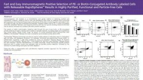 科学海报Positive Selection of PE- or Biotin-Conjugated Antibody Labeled Cells with Releasable Rapidspheres™
 沪公网安备31010102008431号