Axlund SD et al. (FEB 2013)
Hormones & cancer 4 1 36--49
Progesterone-inducible cytokeratin 5-positive cells in luminal breast cancer exhibit progenitor properties.
Progestins play a deleterious role in the onset of breast cancer,yet their influence on existing breast cancer and tumor progression is not well understood. In luminal estrogen receptor (ER)- and progesterone receptor (PR)-positive breast cancer,progestins induce a fraction of cells to express cytokeratin 5 (CK5),a marker of basal epithelial and progenitor cells in the normal breast. CK5(+) cells lose expression of ER and PR and are relatively quiescent,increasing their resistance to endocrine and chemotherapy compared to intratumoral CK5(-)ER(+)PR(+) cells. Characterization of live CK5(+) cells has been hampered by a lack of means for their direct isolation. Here,we describe optical (GFP) and bioluminescent (luciferase) reporter models to quantitate and isolate CK5(+) cells in luminal breast cancer cell lines utilizing the human KRT5 gene promoter and a viral vector approach. Using this system,we confirmed that the induction of GFP(+)/CK5(+) cells is specific to progestins,is dependent on PR,can be blocked by antiprogestins,and does not occur with other steroid hormones. Progestin-induced,fluorescence-activated cell sorting-isolated CK5(+) cells had lower ER and PR mRNA,were slower cycling,and were relatively more invasive and sphere forming than their CK5(-) counterparts in vitro. Repeated progestin treatment and selection of GFP(+) cells enriched for a persistent population of CK5(+) cells,suggesting that this transition can be semi-permanent. These data support that in PR(+) breast cancers,progestins induce a subpopulation of CK5(+)ER(-)PR(-) cells with enhanced progenitor properties and have implications for treatment resistance and recurrence in luminal breast cancer.
View Publication
产品类型:
产品号#:
05620
产品名:
MammoCult™ 人源培养基套装
Zhang Y et al. (JUN 2013)
Neuron 78 5 785--798
Rapid single-step induction of functional neurons from human pluripotent stem cells
Available methods for differentiating human embryonic stem cells (ESCs) and induced pluripotent cells (iPSCs) into neurons are often cumbersome,slow,and variable. Alternatively,human fibroblasts can be directly converted into induced neuronal (iN) cells. However,with present techniques conversion is inefficient,synapse formation is limited,and only small amounts of neurons can be generated. Here,we show that human ESCs and iPSCs can be converted into functional iN cells with nearly 100% yield and purity in less than 2weeks by forced expression of a single transcription factor. The resulting ES-iN or iPS-iN cells exhibit quantitatively reproducible properties independent of the cell line of origin,form mature pre- and postsynaptic specializations,and integrate into existing synaptic networks when transplanted into mouse brain. As illustrated by selected examples,our approach enables large-scale studies of human neurons for questions such as analyses of human diseases,examination of human-specific genes,and drug screening
View Publication
产品类型:
产品号#:
05850
05857
05870
05875
85850
85857
85870
85875
产品名:
mTeSR™1
mTeSR™1
Wagner JP et al. (AUG 2014)
Journal of pediatric surgery 49 8 1319--24; discussion 1324--5
INTRODUCTION Hirschsprung's disease is characterized by a developmental arrest of neural crest cell migration,causing distal aganglionosis. Transplanted cells derived from the neural crest may regenerate enteric ganglia in this condition. We investigated the potential of skin-derived precursor cells (SKPs) to engraft and to differentiate into enteric ganglia in aganglionic rat intestine in vivo. METHODS Adult Lewis rat jejunal segments were separated from intestinal continuity and treated with benzalkonium chloride to induce aganglionosis. Ganglia were identified via immunohistochemical stains for S100 and β-III tubulin (TUJ1). SKPs were procured from neonatal Lewis rats expressing enhanced green fluorescent protein (GFP) and cultured in neuroglial-selective media. SKP cell line expansion was quantified,and immunophenotypes were assessed by immunocytochemistry. Aganglionic segments underwent SKP transplantation 21-79days after benzalkonium chloride treatment. The presence of GFP+cells,mature neurons,and mature glia was evaluated at posttransplant days 1,6,and 9. RESULTS Benzalkonium chloride-induced aganglionosis persisted for at least 85days. Prior to differentiation,SKPs expressed S100,denoting neural crest lineage,and nestin,a marker of neuronal precursors. Differentiated SKPs in vitro expressed GFAP,a marker of glial differentiation,as well as TUJ1 and several enteric neurotransmitters. After transplantation,GFP+structures resembling ganglia were identified between longitudinal and circular smooth muscle layers. CONCLUSION SKPs are capable of engraftment,migration,and differentiation within aganglionic rodent intestine in vivo. Differentiated SKPs generate structures that resemble enteric ganglia. Our observations suggest that SKPs represent a potential gangliogenic therapeutic agent for Hirschsprung's disease.
View Publication
产品类型:
产品号#:
05771
产品名:
Ghezzi S et al. (APR 2017)
Antiviral research 140 13--17
Heparin prevents Zika virus induced-cytopathic effects in human neural progenitor cells.
The recent Zika virus (ZIKV) outbreak,which mainly affected Brazil and neighbouring states,demonstrated the paucity of information concerning the epidemiology of several flaviruses,but also highlighted the lack of available agents with which to treat such emerging diseases. Here,we show that heparin,a widely used anticoagulant,while exerting a modest inhibitory effect on Zika Virus replication,fully prevents virus-induced cell death of human neural progenitor cells (NPCs).
View Publication
产品类型:
产品号#:
85850
85857
85870
85875
产品名:
mTeSR™1
mTeSR™1
T. J. Bussian et al. (SEP 2018)
Nature
Clearance of senescent glial cells prevents tau-dependent pathology and cognitive decline.
Cellular senescence,which is characterized by an irreversible cell-cycle arrest1 accompanied by a distinctive secretory phenotype2,can be induced through various intracellular and extracellular factors. Senescent cells that express the cell cycle inhibitory protein p16INK4A have been found to actively drive naturally occurring age-related tissue deterioration3,4 and contribute to several diseases associated with ageing,including atherosclerosis5 and osteoarthritis6. Various markers of senescence have been observed in patients with neurodegenerative diseases7-9; however,a role for senescent cells in the aetiology of these pathologies is unknown. Here we show a causal link between the accumulation of senescent cells and cognition-associated neuronal loss. We found that the MAPTP301SPS19 mouse model of tau-dependent neurodegenerative disease10 accumulates p16INK4A-positive senescent astrocytes and microglia. Clearance of these cells as they arise using INK-ATTAC transgenic mice prevents gliosis,hyperphosphorylation of both soluble and insoluble tau leading to neurofibrillary tangle deposition,and degeneration of cortical and hippocampal neurons,thus preserving cognitive function. Pharmacological intervention with a first-generation senolytic modulates tau aggregation. Collectively,these results show that senescent cells have a role in the initiation and progression of tau-mediated disease,and suggest that targeting senescent cells may provide a therapeutic avenue for the treatment of these pathologies.
View Publication
产品类型:
产品号#:
18970
18970RF
产品名:
EasySep™小鼠CD11b正选试剂盒II
RoboSep™ 小鼠CD11b正选试剂盒II
C. A. Egelston et al. (OCT 2018)
Nature communications 9 1 4297
Human breast tumor-infiltrating CD8+ T cells retain polyfunctionality despite PD-1 expression.
Functional CD8+ T cells in human tumors play a clear role in clinical prognosis and response to immunotherapeutic interventions. PD-1 expression in T cells involved in chronic infections and tumors such as melanoma often correlates with a state of T-cell exhaustion. Here we interrogate CD8+ tumor-infiltrating lymphocytes (TILs) from human breast and melanoma tumors to explore their functional state. Despite expression of exhaustion hallmarks,such as PD-1 expression,human breast tumor CD8+ TILs retain robust capacity for production of effector cytokines and degranulation capacity. In contrast,melanoma CD8+ TILs display dramatic reduction of cytokine production and degranulation capacity. We show that CD8+ TILs from human breast tumors can potently kill cancer cells via bi-specific antibodies. Our data demonstrate that CD8+ TILs in human breast tumors retain polyfunctionality,despite PD-1 expression,and suggest that they may be harnessed for effective immunotherapies.
View Publication
产品类型:
产品号#:
17853
17853RF
19159
19159RF
100-0699
产品名:
EasySep™人CD8正选试剂盒 II
RoboSep™ 人CD8正选试剂盒 II
EasySep™人记忆CD8+ T细胞富集试剂盒
RoboSep™ 人记忆CD8+ T细胞富集试剂盒
EasySep™人CD8阳性选择试剂盒II
D. Gerace et al. ( 2019)
Methods in molecular biology (Clifton,N.J.) 2029 197--214
Lentiviral vectors are the method of choice for stable gene modification of a variety of cell types. However,the efficiency with which they transduce target cells varies significantly,in particular their typically poor capacity to transduce primary stem cells. Here we describe the isolation and enrichment of murine bone-marrow mesenchymal stem cells (MSCs) via fluorescence-activated cell sorting (FACS); the cloning,production,and concentration of high-titer second generation lentiviral vectors via combined tangential flow filtration (TFF) and ultracentrifugation; and the subsequent high-efficiency gene modification of MSCs into insulin-producing cells via overexpression of the furin-cleavable human insulin (INS-FUR) gene.
View Publication
A. Borek-Dorosz et al. (nov 2022)
Journal of advanced research 41 191--203
Raman-based spectrophenotyping of the most important cells of the immune system.
INTRODUCTION Human peripheral blood mononuclear cells (PBMCs) are a heterogeneous population of cells that includes T and B lymphocytes. The total number of lymphocytes and their percentage in the blood can be a marker for the diagnosis of several human diseases. Currently,cytometric methods are widely used to distinguish subtypes of leukocytes and quantify their number. These techniques use cell immunophenotyping,which is limited by the number of fluorochrome-labeled antibodies that can be applied simultaneously. OBJECTIVE B and T lymphocytes were isolated from peripheral blood obtained from healthy human donors. METHODS The immunomagnetic negative selection was used for the enrichment of B and T cells fractions,and their purity was assessed by flow cytometry. Isolated cells were fixed with 0.5% glutaraldehyde and measured using confocal Raman imaging. K-means cluster analysis,principal component analysis and partial least squares discriminant methods were applied for the identification of spectroscopic markers to distinguish B and T cells. HPLC was the reference method for identifying carotene in T cells. RESULTS Reliable discrimination between T and B lymphocytes based on their spectral profile has been demonstrated using label-free Raman imaging and chemometric analysis. The presence of carotene in T lymphocytes (in addition to the previously reported in plasma) was confirmed and for the first time unequivocally identified as $\beta$-carotene. In addition,the molecular features of the lymphocytes nuclei were found to support the discriminant analysis. It has been shown that although the presence of carotenoids in T cells depends on individual donor variability,the reliable differentiation between lymphocytes is possible based on Raman spectra collected from individual cells. CONCLUSIONS This proves the potential of Raman spectroscopy in clinical diagnostics to automatically differentiate between cells that are an important component of our immune system.
View Publication
hESC- and hiPSC-derived Schwann cells are molecularly comparable and functionally equivalent
Establishing robust models of human myelinating Schwann cells is critical for studying peripheral nerve injury and disease. Stem cell differentiation has emerged as a key human cell model and disease motivating development of Schwann cell differentiation protocols. Human embryonic stem cells (hESCs) are considered the ideal pluripotent cell but ethical concerns regarding their use have propelled the popularity of human induced pluripotent stem cells (hiPSCs). Given that the equivalence of hESCs and hiPSCs remains controversial,we sought to compare the molecular and functional equivalence of hESC- and hiPSC-derived Schwann cells generated with our previously reported protocol. We identified only modest transcriptome differences by RNA sequencing and insignificant proteome differences by antibody array. Additionally,both cell types comparably improved nerve regeneration and function in a chronic denervation and regeneration animal model. Our findings demonstrate that Schwann cells derived from hESCs and hiPSCs with our protocol are molecularly comparable and functionally equivalent. Subject areas: Neuroscience,Cell biology,Stem cells research,Transcriptomics
View Publication
产品类型:
产品号#:
100-0483
100-0484
产品名:
Hausser Scientificᵀᴹ 明线血球计数板
ReLeSR™
J. Slamecka et al. (Sep 2024)
iScience 27 10
Highly efficient generation of self-renewing trophoblast from human pluripotent stem cells
Human pluripotent stem cells (hPSCs) represent a powerful model system to study early developmental processes. However,lineage specification into trophectoderm (TE) and trophoblast (TB) differentiation remains poorly understood,and access to well-characterized placental cells for biomedical research is limited,largely depending on fetal tissues or cancer cell lines. Here,we developed novel strategies enabling highly efficient TE specification that generates cytotrophoblast (CTB) and multinucleated syncytiotrophoblast (STB),followed by the establishment of trophoblast stem cells (TSCs) capable of differentiating into extravillous trophoblast (EVT) and STB after long-term expansion. We confirmed stepwise and controlled induction of lineage- and cell-type-specific genes consistent with developmental biology principles and benchmarked typical features of placental cells using morphological,biochemical,genomics,epigenomics,and single-cell analyses. Charting a well-defined roadmap from hPSCs to distinct placental phenotypes provides invaluable opportunities for studying early human development,infertility,and pregnancy-associated diseases. Subject areas: Natural sciences,Biological sciences,Cell biology,Stem cells research
View Publication

 EasySep™小鼠TIL(CD45)正选试剂盒


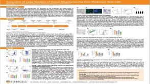 科学海报Generation of Large Numbers of Human Megakaryocytes from Pluripotent Stem Cells
 沪公网安备31010102008431号