Lane ME et al. ( 2001)
Cancer research 61 16 6170--6177
A novel cdk2-selective inhibitor, SU9516, induces apoptosis in colon carcinoma cells.
Recent studies have indicated that the development of cyclin-dependent kinase (cdk)2 inhibitors that deregulate E2F are a plausible pharmacological strategy for novel antineoplastic agents. We show here that 3-[1-(3H-Imidazol-4-yl)-meth-(Z)-ylidene]-5-methoxy-1,3-dihydro-indol-2-one (SU9516),a novel 3-substituted indolinone compound,binds to and selectively inhibits the activity of cdk2. This inhibition results in a time-dependent decrease (4-64%) in the phosphorylation of the retinoblastoma protein pRb,an increase in caspase-3 activation (5-84%),and alterations in cell cycle resulting in either a G(0)-G(1) or a G(2)-M block. We also report here cell line differences in the cdk-dependent phosphorylation of pRb. These findings demonstrate that SU9516 is a selective cdk2 inhibitor and support the theory that compounds that inhibit cdk2 are viable resources in the development of new antineoplastic agents.
View Publication
产品类型:
产品号#:
73452
产品名:
SU9516
Wu X et al. (DEC 2002)
Journal of the American Chemical Society 124 49 14520--1
A small molecule with osteogenesis-inducing activity in multipotent mesenchymal progenitor cells.
Purmorphamine,which is a 2,6,9-trisubstituted purine compound,was discovered through cell-based high-throughput screening from a heterocycle combinatorial library. It differentiates multipotent mesenchymal progenitor cells into an osteoblast lineage. It will serve as a unique chemical tool to study the molecular mechanisms of osteogenesis of stem cells and bone development.
View Publication
产品类型:
产品号#:
72202
72204
100-1049
产品名:
Purmorphamine
Purmorphamine
Purmorphamine
Laird DJ et al. (DEC 2005)
Cell 123 7 1351--60
Stem cells are units of natural selection in a colonial ascidian.
Stem cells are highly conserved biological units of development and regeneration. Here we formally demonstrate that stem cell lineages are also legitimate units of natural selection. In a colonial ascidian,Botryllus schlosseri,vascular fusion between genetically distinct individuals results in cellular parasitism of somatic tissues,gametes,or both. We show that genetic hierarchies of somatic and gametic parasitism following fusion can be replicated by transplanting cells between colonies. We prospectively isolate a population of multipotent,self-renewing stem cells that retain their competitive phenotype upon transplantation. Their single-cell contribution to either somatic or germline fates,but not to both,is consistent with separate lineages of somatic and germline stem cells or pluripotent stem cells that differentiate according to the niche in which they land. Since fusion is restricted to individuals that share a fusion/histocompatibility allele,these data suggest that histocompatibility genes in Botryllus evolved to protect the body from parasitic stem cells usurping asexual or sexual inheritance.
View Publication
产品类型:
产品号#:
01700
01705
01701
01702
产品名:
ALDEFLUOR™ 试剂盒
ALDEFLUOR™ DEAB试剂, 1.5 mM, 1 mL
ALDEFLUOR™检测缓冲液
Fulcher JA et al. (JUL 2006)
Journal of immunology (Baltimore,Md. : 1950) 177 1 216--26
Galectin-1-matured human monocyte-derived dendritic cells have enhanced migration through extracellular matrix.
Dendritic cells (DCs) are potent mediators of the immune response,and can be activated by exogenous pathogen components. Galectin-1 is a member of the conserved beta-galactoside-binding lectin family that binds galactoside residues on cell surface glycoconjugates. Galectin-1 is known to play a role in immune regulation via action on multiple immune cells. However,its effects on human DCs are unknown. In this study,we show that galectin-1 induces a phenotypic and functional maturation in human monocyte-derived DCs (MDDCs) similar to but distinct from the activity of the exogenous pathogen stimuli,LPS. Immature human MDDCs exposed to galectin-1 up-regulated cell surface markers characteristic of DC maturation (CD40,CD83,CD86,and HLA-DR),secreted high levels of IL-6 and TNF-alpha,stimulated T cell proliferation,and showed reduced endocytic capacity,similar to LPS-matured MDDCs. However,unlike LPS-matured DCs,galectin-1-treated MDDCs did not produce the Th1-polarizing cytokine IL-12. Microarray analysis revealed that in addition to modulating many of the same DC maturation genes as LPS,galectin-1 also uniquely up-regulated a significant subset of genes related to cell migration through the extracellular matrix (ECM). Indeed,compared with LPS,galectin-1-treated human MDDCs exhibited significantly better chemotactic migration through Matrigel,an in vitro ECM model. Our findings show that galectin-1 is a novel endogenous activator of human MDDCs that up-regulates a significant subset of genes distinct from those regulated by a model exogenous stimulus (LPS). One unique effect of galectin-1 is to increase DC migration through the ECM,suggesting that galectin-1 may be an important component in initiating an immune response.
View Publication
产品类型:
产品号#:
15022
15062
15028
15068
产品名:
RosetteSep™人CD4+ T细胞富集抗体混合物
RosetteSep™人CD4+ T细胞富集抗体混合物
RosetteSep™人单核细胞富集抗体混合物
RosetteSep™人单核细胞富集抗体混合物
Benvenuti S et al. (OCT 2007)
Journal of endocrinological investigation 30 9 RC26--30
Rosiglitazone stimulates adipogenesis and decreases osteoblastogenesis in human mesenchymal stem cells.
Thiazolidinediones (TZD) are widely prescribed for the treatment of Type 2 diabetes. Increased loss of bone mass and a higher incidence of fractures have been associated with the use of this class of drugs in post-menopausal women. In vitro studies performed in rodent cell models indicated that rosiglitazone (RGZ),one of the TZD,inhibited osteoblastogenesis and induced adipogenesis in bone marrow progenitor cells. The objective of the present study was to determine for the first time the RGZ-dependent shift from osteoblastogenesis toward adipogenesis using a human cell model. To this purpose,bone marrow-derived mesenchymal stem cells were characterized and induced to differentiate along osteogenic and adipogenic lineages. We found that the exposure to RGZ potentiated adipogenic differentiation and shifted the differentiation toward an osteogenic phenotype into an adipogenic phenotype,as assessed by the appearance of lipid droplets. Accordingly,RGZ markedly increased the expression of the typical marker of adipogenesis fatty-acid binding protein 4,whereas it reduced the expression of Runx2,a marker of osteoblastogenesis. This is the first demonstration that RGZ counteracts osteoblastogenesis and induces a preferential differentiation into adipocytes in human mesenchymal stem cells.
View Publication
产品类型:
产品号#:
72622
72624
产品名:
罗格列酮(Rosiglitazone)
罗格列酮(Rosiglitazone)
Xing Q et al. (AUG 2014)
Digestive and liver disease : official journal of the Italian Society of Gastroenterology and the Italian Association for the Study of the Liver 46 8 731--737
Hepatectomised patient sera promote hepatocyte differentiation of human-induced pluripotent stem cells.
Background: Human induced pluripotent stem cells,which can be differentiated into hepatocyte-like cells,could provide a source for liver regeneration and bio-artificial liver devices. However,the functionality of hepatocyte-like cells is significantly lower than that of primary hepatocytes. Aims: To investigate whether serum from patients undergoing hepatectomy might promote differentiation from human induced pluripotent stem cells to hepatocyte-like cells. Methods: Serum from patients undergoing hepatectomy (acquired pre-hepatectomy and 3. hours,1 day and 3 days post-hepatectomy) was used to replace foetal bovine serum when differentiating human induced pluripotent stem cells into hepatocyte-like cells. Properties of hepatocyte-like cells were assessed and compared with cells cultured in foetal bovine serum. Results: The differentiation efficiency and functionality of hepatocyte-like cells cultured in human serum 3. hours and 1 day post-hepatectomy were superior to those cultured in foetal bovine serum and human serum pre-hepatectomy. Human serum 3 days post-hepatectomy had an equal effect to that of human serum pre-hepatectomy. Some cytochrome P450 isozyme transcript levels of hepatocyte-like cells cultured in human serum were higher than those cultured in foetal bovine serum. Conclusion: Human serum,particularly that acquired relatively soon after hepatectomy,can enhance the differentiation efficiency and functionality of hepatocyte-like cells derived from human induced pluripotent stem cells. textcopyright 2014 Editrice Gastroenterologica Italiana S.r.l.
View Publication
Molecular beacon-enabled purification of living cells by targeting cell type-specific mRNAs.
Molecular beacons (MBs) are dual-labeled oligonucleotides that fluoresce only in the presence of complementary mRNA. The use of MBs to target specific mRNAs allows sorting of specific cells from a mixed cell population. In contrast to existing approaches that are limited by available surface markers or selectable metabolic characteristics,the MB-based method enables the isolation of a wide variety of cells. For example,the ability to purify specific cell types derived from pluripotent stem cells (PSCs) is important for basic research and therapeutics. In addition to providing a general protocol for MB design,validation and nucleofection into cells,we describe how to isolate a specific cell population from differentiating PSCs. By using this protocol,we have successfully isolated cardiomyocytes differentiated from mouse or human PSCs (hPSCs) with ∼ 97% purity,as confirmed by electrophysiology and immunocytochemistry. After designing MBs,their ordering and validation requires 2 weeks,and the isolation process requires 3 h.
View Publication
产品类型:
产品号#:
05850
05857
05870
05875
85850
85857
85870
85875
产品名:
mTeSR™1
mTeSR™1
Byrne SM et al. (FEB 2015)
Nucleic Acids Research 43 3 e21
Multi-kilobase homozygous targeted gene replacement in human induced pluripotent stem cells.
Sequence-specific nucleases such as TALEN and the CRISPR/Cas9 system have so far been used to disrupt,correct or insert transgenes at precise locations in mammalian genomes. We demonstrate efficient 'knock-in' targeted replacement of multi-kilobase genes in human induced pluripotent stem cells (iPSC). Using a model system replacing endogenous human genes with their mouse counterpart,we performed a comprehensive study of targeting vector design parameters for homologous recombination. A 2.7 kilobase (kb) homozygous gene replacement was achieved in up to 11% of iPSC without selection. The optimal homology arm length was around 2 kb,with homology length being especially critical on the arm not adjacent to the cut site. Homologous sequence inside the cut sites was detrimental to targeting efficiency,consistent with a synthesis-dependent strand annealing (SDSA) mechanism. Using two nuclease sites,we observed a high degree of gene excisions and inversions,which sometimes occurred more frequently than indel mutations. While homozygous deletions of 86 kb were achieved with up to 8% frequency,deletion frequencies were not solely a function of nuclease activity and deletion size. Our results analyzing the optimal parameters for targeting vector design will inform future gene targeting efforts involving multi-kilobase gene segments,particularly in human iPSC.
View Publication
产品类型:
产品号#:
05850
05857
05870
05875
85850
85857
85870
85875
产品名:
mTeSR™1
mTeSR™1
Martinez RA et al. (MAY 2015)
Nucleic acids research 43 10 e65
Genome engineering of isogenic human ES cells to model autism disorders
Isogenic pluripotent stem cells are critical tools for studying human neurological diseases by allowing one to study the effects of a mutation in a fixed genetic background. Of particular interest are the spectrum of autism disorders,some of which are monogenic such as Timothy syndrome (TS); others are multigenic such as the microdeletion and microduplication syndromes of the 16p11.2 chromosomal locus. Here,we report engineered human embryonic stem cell (hESC) lines for modeling these two disorders using locus-specific endonucleases to increase the efficiency of homology-directed repair (HDR). We developed a system to: (1) computationally identify unique transcription activator-like effector nuclease (TALEN) binding sites in the genome using a new software program,TALENSeek,(2) assemble the TALEN genes by combining golden gate cloning with modified constructs from the FLASH protocol,and (3) test the TALEN pairs in an amplification-based HDR assay that is more sensitive than the typical non-homologous end joining assay. We applied these methods to identify,construct,and test TALENs that were used with HDR donors in hESCs to generate an isogenic TS cell line in a scarless manner and to model the 16p11.2 copy number disorder without modifying genomic loci with high sequence similarity.
View Publication
产品类型:
产品号#:
05850
05857
05870
05875
85850
85857
85870
85875
产品名:
mTeSR™1
mTeSR™1
Paik KJ et al. (MAR 2014)
Plastic and reconstructive surgery 133 3 Suppl 174
Abstract 158: Identification of BMP-Responsive Long Noncoding RNAs in Pluripotent Cells.
Kempf H et al. (SEP 2015)
Nature protocols 10 9 1345--1361
Cardiac differentiation of human pluripotent stem cells in scalable suspension culture.
Cardiomyocytes (CMs) generated from human pluripotent stem cells (hPSCs) are a potential cell source for regenerative therapies,drug discovery and disease modeling. All these applications require a routine supply of relatively large quantities of in vitro-generated CMs. This protocol describes a suspension culture-based strategy for the generation of hPSC-CMs as cell-only aggregates,which facilitates process development and scale-up. Aggregates are formed for 4 d in hPSC culture medium followed by 10 d of directed differentiation by applying chemical Wnt pathway modulators. The protocol is applicable to static multiwell formats supporting fast adaptation to specific hPSC line requirements. We also demonstrate how to apply the protocol using stirred tank bioreactors at a 100-ml scale,providing a well-controlled upscaling platform for CM production. In bioreactors,the generation of 40-50 million CMs per differentiation batch at textgreater80% purity without further lineage enrichment can been achieved within 24 d.
View Publication


 EasySep™小鼠TIL(CD45)正选试剂盒




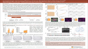 科学海报Increased Single-Cell Cloning Efficiency of Human Pluripotent Stem Cells Using CloneR™

 沪公网安备31010102008431号