
 EasySep™小鼠TIL(CD45)正选试剂盒


搜索结果: 'methocult media formulations for human hematopoietic cells serum containing'
-

产品类型:
产品号#:
05850
05857
05870
05875
05790
05792
05793
85850
85857
85870
85875
05794
05795
产品名:
BrainPhys™神经元培养基
BrainPhys™神经元培养基和SM1试剂盒
BrainPhys™ 神经元培养基N2-A和SM1试剂盒
mTeSR™1
mTeSR™1
BrainPhys™原代神经元试剂盒
BrainPhys™ hPSC 神经元试剂盒
-
产品类型:
产品号#:
05750
05751
产品名:
NeuroCult™ NS-A 基础培养基(人)
NeuroCult™ NS-A 扩增试剂盒(人)
-
产品类型:
产品号#:
100-0276
100-1130
产品名:
mTeSR™ Plus
mTeSR™ Plus
-
产品类型:
产品号#:
07801
07811
07851
07861
10971
10991
15021
15061
18060
18061
产品名:
Lymphoprep™
Lymphoprep™
ImmunoCult™ 人CD3/CD28 T细胞激活剂
ImmunoCult™ 人CD3/CD28 T细胞激活剂
RosetteSep™人T细胞富集抗体混合物
RosetteSep™人T细胞富集抗体混合物
Lymphoprep™
Lymphoprep™
-
产品类型:
产品号#:
100-0536
100-0537
产品名:
Ac-DEVD-CHO (Trifluoroacetate Salt)
Ac-DEVD-CHO (Trifluoroacetate Salt)
-
产品类型:
产品号#:
100-0538
100-0539
产品名:
SANT-1
SANT-1
-
产品类型:
产品号#:
05850
05857
05870
05875
85850
85857
85870
85875
产品名:
mTeSR™1
mTeSR™1
-
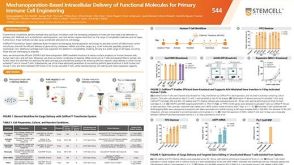 科学海报Mechanoporation-Based Intracellular Delivery of Functional Molecules for Primary Immune Cell Engineering
产品类型:
Conference:
AAI 2026
产品号#:
产品名:
-
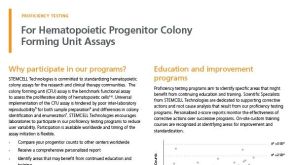 产品手册Registration Form - Proficiency Testing Programs (For Countries Serviced by a Distributor)
产品类型:
品牌:
MethoCult
产品号#:
00602
00603
00608
00609
产品名:
-
产品类型:
产品号#:
05850
05857
05870
05875
85850
85857
85870
85875
产品名:
mTeSR™1
mTeSR™1


 沪公网安备31010102008431号