
 EasySep™小鼠TIL(CD45)正选试剂盒


搜索结果: 'methocult media formulations for human hematopoietic cells serum containing'
-

产品类型:
产品号#:
100-0956
10981
产品名:
ImmunoCult™ XF培养基
ImmunoCult™ XF 人T细胞扩增培养基,500 mL
-
产品类型:
产品号#:
100-0956
产品名:
ImmunoCult™ XF培养基
-
产品类型:
产品号#:
100-0276
100-1130
产品名:
mTeSR™ Plus
mTeSR™ Plus
-
产品类型:
产品号#:
100-0276
100-1130
产品名:
mTeSR™ Plus
mTeSR™ Plus
-
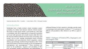 研究综述Endothelial Progenitor Cells and Endothelial Cells
产品类型:
细胞类型:
内皮细胞
产品号#:
产品名:
发布日期: 06/01/2015 -
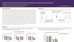 科学海报Fast and Easy Enrichment of Cell Subsets for HLA Analysis
产品类型:
Conference:
EFI 2012
产品号#:
15470
15450
15420
15460
15425
15465
15430
15415
85415
85420
85450
85460
产品名:
SepMate™-15 (IVD)
SepMate™-15 (IVD)
SepMate™-50 (IVD)
SepMate™-50 (IVD)
-
产品类型:
产品号#:
100-0276
100-1130
产品名:
mTeSR™ Plus
mTeSR™ Plus
-
产品类型:
产品号#:
18958
18958RF
产品名:
EasySep™小鼠CD90.1正选试剂盒
RoboSep™ 小鼠CD90.1正选试剂盒
-
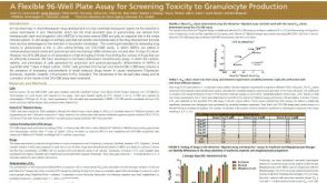 科学海报A Flexible 96-Well Plate Assay for Screening Toxicity to Granulocyte Production
产品类型:
Conference:
SOT 2015
产品号#:
09704
产品名:
HemaTox™髓系试剂盒

 沪公网安备31010102008431号