B cells are not drivers of stromal cell activation during acute CNS infection
BackgroundCNS stromal cells,especially fibroblasts and endothelial cells,support leukocyte accumulation through upregulation of adhesion molecules and lymphoid chemokines. While chronically activated fibroblast networks can drive pathogenic immune cell aggregates known as tertiary lymphoid structures (TLS),early stromal cell activation during CNS infection can support anti-viral T cells. However,the cell types and factors driving early stromal cell activation is poorly explored.AimsA neurotropic murine coronavirus (mCoV) infection model was used to better characterize signals that promote fibroblast networks supporting accumulation of antiviral lymphocytes. Based on the early appearance of IgD+ B cells with unknown functions during several CNS infections,we probed their potential to activate stromal cells through lymphotoxin β (LTβ),a molecule critical in maintaining fibroblast-networks in lymphoid tissues as well as promoting TLS in autoimmunity and cancers.ResultsKinetic analysis of stromal cell activation in olfactory bulbs and brains revealed that upregulation of adhesion molecules and lymphoid chemokines Ccl19,Ccl21 and Cxcl13 closely tracked viral replication. Immunohistochemistry revealed that upregulation of the fibroblast marker podoplanin (PDPN) at meningeal and perivascular sites mirrored kinetics of RNA expression. Moreover,both B cells and T cells colocalized to areas of PDPN reactivity,supporting a potential role in regulating stromal cell activation. However,specific depletion of LTβ from B cells using Mb1-creERT2 x Ltβfl/fl mice had no effect on T or B cell recruitment or viral replication. B cell depletion by anti-CD20 antibody also had no adverse effects. Surprisingly,LTβR agonism reduced viral control and parenchymal T cell localization despite increasing stromal cell lymphoid chemokines and PDPN. Additional assessment of direct stromal cell activation by the viral RNA mimic poly I:C showed induction of Pdpn and Ccl19 preceding Ltb.ConclusionsNeither B cell-derived LTβ or B cells are primary drivers of stromal cell activation networks in the CNS following mCoV infection. Although supplementary agonist mediated LTβR engagement confirmed a role for LTβ in enhancing PDPN and lymphoid chemokine expression,it impeded T cell migration to the CNS parenchyma and viral control. Our data overall indicate that stromal cells can integrate LTβR signals to tune their activation,but that LTβ is not necessarily essential and can even dysregulate protective antiviral T cell functions.Supplementary InformationThe online version contains supplementary material available at 10.1186/s12974-025-03491-7.
View Publication
Gene expression analysis in osteoblastic differentiation from peripheral blood mesenchymal stem cells.
MSCs are known to have an extensive proliferative potential and ability to differentiate in various cell types. Osteoblastic differentiation from mesenchymal progenitor cells is an important step of bone formation,though the pattern of gene expression during differentiation is not yet well understood. Here,to investigate the possibility to obtain a model for in vitro bone differentiation using mesenchymal stem cells (hMSCs) from human subjects non-invasively,we developed a method to obtain hMSCs-like cells from peripheral blood by a two step method that included an enrichment of mononuclear cells followed by depletion of unwanted cells. Using these cells,we analyzed the expression of transcription factor genes (runt-related transcription factor 2 (RUNX2) and osterix (SP7)) and bone related genes (osteopontin (SPP1),osteonectin (SPARC) and collagen,type I,alpha 1 (COLIA1)) during osteoblastic differentiation. Our results demonstrated that hMSCs can be obtained from peripheral blood and that they are able to generate CFU-F and to differentiate in osteoblast and adipocyte; in this study,we also identified a possible gene expression timing during osteoblastic differentiation that provided a powerful tool to study bone physiology.
View Publication
产品类型:
产品号#:
15128
15168
产品名:
RosetteSep™人间充质干细胞富集抗体混合物
RosetteSep™人间充质干细胞富集抗体混合物
Meng G et al. (APR 2011)
Stem cells and development 20 4 583--91
Rapid isolation of undifferentiated human pluripotent stem cells from extremely differentiated colonies
Conventionally,researchers remove spontaneously differentiated areas in human pluripotent stem cell (hPSC) colonies by using a finely drawn glass pipette or a commercially available syringe needle. However,when extreme differentiation occurs,it is inefficient to purify the remaining undifferentiated cells,as these undifferentiated areas are too small to be isolated completely with the mechanical method. Antibodies can be utilized to purify the rare undifferentiated cells; however,this type of purification cannot be used in xeno-free culture systems. To avoid the loss of valuable hPSCs,we developed a novel method to isolate undifferentiated hPSCs from extremely differentiated colonies that could be easily adapted to xeno-free culture conditions. This protocol involves dissecting away differentiated areas,dissociating the remaining colony into clumps,seeding small clumps into new dishes,and picking undifferentiated colonies for expansion. Using this method,we routinely achieve completely undifferentiated colonies in one passage without the use of antibody-based purification.
View Publication
产品类型:
产品号#:
05850
05857
05870
05875
07923
85850
85857
85870
85875
产品名:
Dispase (1 U/mL)
mTeSR™1
mTeSR™1
Lumelsky N et al. (MAY 2001)
Science (New York,N.Y.) 292 5520 1389--94
Differentiation of embryonic stem cells to insulin-secreting structures similar to pancreatic islets.
Although the source of embryonic stem (ES) cells presents ethical concerns,their use may lead to many clinical benefits if differentiated cell types can be derived from them and used to assemble functional organs. In pancreas,insulin is produced and secreted by specialized structures,islets of Langerhans. Diabetes,which affects 16 million people in the United States,results from abnormal function of pancreatic islets. We have generated cells expressing insulin and other pancreatic endocrine hormones from mouse ES cells. The cells self-assemble to form three-dimensional clusters similar in topology to normal pancreatic islets where pancreatic cell types are in close association with neurons. Glucose triggers insulin release from these cell clusters by mechanisms similar to those employed in vivo. When injected into diabetic mice,the insulin-producing cells undergo rapid vascularization and maintain a clustered,islet-like organization.
View Publication
产品类型:
产品号#:
06902
06952
00321
00322
00323
00324
00325
产品名:
Abdelwahab SF et al. (DEC 2003)
Proceedings of the National Academy of Sciences of the United States of America 100 25 15006--10
HIV-1-suppressive factors are secreted by CD4+ T cells during primary immune responses.
CD4+ T cells are required for immunity against many viral infections,including HIV-1 where a positive correlation has been observed between strong recall responses and low HIV-1 viral loads. Some HIV-1-specific CD4+ T cells are preferentially infected with HIV-1,whereas others escape infection by unknown mechanisms. One possibility is that some CD4+ T cells are protected from infection by the secretion of soluble HIV-suppressive factors,although it is not known whether these factors are produced during primary antigen-specific responses. Here,we show that soluble suppressive factors are produced against CXCR4 and CCR5 isolates of HIV-1 during the primary immune response of human CD4+ T cells. This activity requires antigenic stimulation of naïve CD4+ T cells. One anti-CXCR4 factor is macrophage-derived chemokine (chemokine ligand 22,CCL22),and anti-CCR5 factors include macrophage inflammatory protein-1 alpha (CCL3),macrophage inflammatory protein-1 beta (CCL4),and RANTES (regulated upon activation of normal T cells expressed and secreted) (CCL5). Intracellular staining confirms that CD3+CD4+ T cells are the source of the prototype HIV-1-inhibiting chemokines CCL22 and CCL4. These results show that CD4+ T cells secrete an evolving HIV-1-suppressive activity during the primary immune response and that this activity is comprised primarily of CC chemokines. The data also suggest that production of such factors should be considered in the design of vaccines against HIV-1 and as a mechanism whereby the host can control infections with this virus.
View Publication
产品类型:
产品号#:
09500
09600
09650
19155
19155RF
产品名:
BIT 9500血清替代物
StemSpan™ SFEM
StemSpan™ SFEM
Fang H et al. (APR 2005)
Journal of immunology (Baltimore,Md. : 1950) 174 8 4966--71
Anthrax lethal toxin blocks MAPK kinase-dependent IL-2 production in CD4+ T cells.
Anthrax lethal toxin (LT) is a critical virulence factor that cleaves and inactivates MAPK kinases (MAPKKs) in host cells and has been proposed as a therapeutic target in the treatment of human anthrax infections. Despite the potential use of anti-toxin agents in humans,the standard activity assays for anthrax LT are currently based on cytotoxic actions of anthrax LT that are cell-,strain-,and species-specific,which have not been demonstrated to occur in human cells. We now report that T cell proliferation and IL-2 production inversely correlate with anthrax LT levels in human cell assays. The model CD4+ T cell tumor line,Jurkat,is a susceptible target for the specific protease action of anthrax LT. Anthrax LT cleaves and inactivates MAPKKs in Jurkat cells,whereas not affecting proximal or parallel TCR signal transduction pathways. Moreover,anthrax LT specifically inhibits PMA/ionomycin- and anti-CD3-induced IL-2 production in Jurkat cells. An inhibitor of the protease activity of anthrax LT completely restores IL-2 production by anthrax LT-treated Jurkat cells. Anthrax LT acts on primary CD4+ T cells as well,cleaving MAPKKs and leading to a 95% reduction in anti-CD3-induced proliferation and IL-2 production. These findings not only will be useful in the development of new human cell-based bioassays for the activity of anthrax LT,but they also suggest new mechanisms that facilitate immune evasion by Bacillus anthracis. Specifically,anthrax LT inhibits IL-2 production and proliferative responses in CD4+ T cells,thereby blocking functions that are pivotal in the regulation of immune responses.
View Publication
产品类型:
产品号#:
15022
15062
产品名:
RosetteSep™人CD4+ T细胞富集抗体混合物
RosetteSep™人CD4+ T细胞富集抗体混合物
Piccirillo SGM et al. (DEC 2006)
Nature 444 7120 761--5
Bone morphogenetic proteins inhibit the tumorigenic potential of human brain tumour-initiating cells.
Transformed,oncogenic precursors,possessing both defining neural-stem-cell properties and the ability to initiate intracerebral tumours,have been identified in human brain cancers. Here we report that bone morphogenetic proteins (BMPs),amongst which BMP4 elicits the strongest effect,trigger a significant reduction in the stem-like,tumour-initiating precursors of human glioblastomas (GBMs). Transient in vitro exposure to BMP4 abolishes the capacity of transplanted GBM cells to establish intracerebral GBMs. Most importantly,in vivo delivery of BMP4 effectively blocks the tumour growth and associated mortality that occur in 100% of mice after intracerebral grafting of human GBM cells. We demonstrate that BMPs activate their cognate receptors (BMPRs) and trigger the Smad signalling cascade in cells isolated from human glioblastomas (GBMs). This is followed by a reduction in proliferation,and increased expression of markers of neural differentiation,with no effect on cell viability. The concomitant reduction in clonogenic ability,in the size of the CD133+ population and in the growth kinetics of GBM cells indicates that BMP4 reduces the tumour-initiating cell pool of GBMs. These findings show that the BMP-BMPR signalling system--which controls the activity of normal brain stem cells--may also act as a key inhibitory regulator of tumour-initiating,stem-like cells from GBMs and the results also identify BMP4 as a novel,non-cytotoxic therapeutic effector,which may be used to prevent growth and recurrence of GBMs in humans.
View Publication
产品类型:
产品号#:
05751
产品名:
NeuroCult™ NS-A 扩增试剂盒(人)
Qin H et al. (FEB 2007)
The Journal of biological chemistry 282 8 5842--52
Regulation of apoptosis and differentiation by p53 in human embryonic stem cells.
The essentially infinite expansion potential and pluripotency of human embryonic stem cells (hESCs) makes them attractive for cell-based therapeutics. In contrast to mouse embryonic stem cells (mESCs),hESCs normally undergo high rates of spontaneous apoptosis and differentiation,making them difficult to maintain in culture. Here we demonstrate that p53 protein accumulates in apoptotic hESCs induced by agents that damage DNA. However,despite the accumulation of p53,it nevertheless fails to activate the transcription of its target genes. This inability of p53 to activate its target genes has not been observed in other cell types,including mESCs. We further demonstrate that p53 induces apoptosis of hESCs through a mitochondrial pathway. Reducing p53 expression in hESCs in turn reduces both DNA damage-induced apoptosis as well as spontaneous apoptosis. Reducing p53 expression also reduces spontaneous differentiation and slows the differentiation rate of hESCs. Our studies reveal the important roles of p53 as a critical mediator of human embryonic stem cells survival and differentiation.
View Publication
产品类型:
产品号#:
72062
72064
72802
72804
产品名:
环状 Pifithrin-α(Cyclic Pifithrin-Alpha)
环状 Pifithrin-α (Hydrobromide)
Pifithrin-mu
Liu Y et al. (APR 2014)
British journal of cancer 110 8 2063--2071
Lack of correlation of stem cell markers in breast cancer stem cells.
BACKGROUND Various markers are used to identify the unique sub-population of breast cancer cells with stem cell properties. Whether these markers are expressed in all breast cancers,identify the same population of cells,or equate to therapeutic response is controversial. METHODS We investigated the expression of multiple cancer stem cell markers in human breast cancer samples and cell lines in vitro and in vivo,comparing across and within samples and relating expression with growth and therapeutic response to doxorubicin,docetaxol and radiotherapy. RESULTS CD24,CD44,ALDH and SOX2 expression,the ability to form mammospheres and side-population cells are variably present in human cancers and cell lines. Each marker identifies a unique rather than common population of cancer cells. In vivo,cells expressing these markers are not specifically localized to the presumptive stem cell niche at the tumour/stroma interface. Repeated therapy does not consistently enrich cells expressing these markers,although ER-negative cells accumulate. CONCLUSIONS Commonly employed methods identify different cancer cell sub-populations with no consistent therapeutic implications,rather than a single population of cells. The relationships of breast cancer stem cells to clinical parameters will require identification of specific markers or panels for the individual cancer.
View Publication
产品类型:
产品号#:
01700
01705
01702
产品名:
ALDEFLUOR™ 试剂盒
ALDEFLUOR™ DEAB试剂, 1.5 mM, 1 mL
ALDEFLUOR™检测缓冲液
Lu Q et al. (DEC 2014)
PLoS ONE 9 12 e114949
Negligible immunogenicity of induced pluripotent stem cells derived from human skin fibroblasts
Human induced pluripotent stem cells (hiPSCs) have potential applications in cell replacement therapy and regenerative medicine. However,limited information is available regarding the immunologic features of iPSCs. In this study,expression of MHC and T cell co-stimulatory molecules in hiPSCs,and the effects on activation,proliferation and cytokine production in allogeneic human peripheral blood mononuclear cells were examined. We found that no-integrate hiPSCs had no MHC-II and T cell co-stimulatory molecules expressions but had moderate level of MHC-I and HLA-G expressions. In contrast to human skin fibroblasts (HSFs) which significantly induced allogeneic T cell activation and proliferation,hiPSCs failed to induce allogeneic CD45+ lymphocyte and CD8+ T cell activation and proliferation but could induce a low level of allogeneic CD4+ T cell proliferation. Unlike HSFs which induced allogeneic lymphocytes to produce high levels of IFN-γ,TNF-α and IL-17,hiPSCs only induced allogeneic lymphocytes to produce IL-2 and IL-10,and promote IL-10-secreting regulatory T cell (Treg) generation. Our study suggests that the integration-free hiPSCs had low or negligible immunogenicity,which may result from their induction of IL-10-secreting Treg.
View Publication
产品类型:
产品号#:
05850
05857
05870
05875
85850
85857
85870
85875
产品名:
mTeSR™1
mTeSR™1
Halder D et al. ( 2015)
Molecular bioSystems
Synthetic small molecules that induce neuronal differentiation in neuroblastoma and fibroblast cells.
An investigation was conducted to demonstrate that neurodazine (Nz) and neurodazole (Nzl),two imidazole-based small molecules,promote neuronal differentiation in both neuroblastoma and fibroblast cells. The results show that differentiated cells generated by treatment with Nz and Nzl express neuron-specific markers. The ability of Nz and Nzl to induce neurogenesis of neuroblastoma and fibroblast cells was found to be comparable to those of the known neurogenic factors,retinoic acid and trichostatin A. In addition,the cells differentiated by Nz and Nzl are observed to express different isoforms of glutamate receptors. The results of signaling pathway studies reveal that two substances enhance neurogenesis in neuroblastoma cells by activating Wnt and Shh signaling pathways and neurogenesis in fibroblast cells by mainly activating the Wnt signaling pathway. Observations made in the present study suggest that Nz and Nzl will serve as chemical tools to generate specific populations of neuronal cells from readily available and simply manageable cells.
View Publication


 EasySep™小鼠TIL(CD45)正选试剂盒





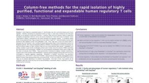 科学海报Rapid Isolation of Highly Purified, Functional and Expandable Human Regulatory T Cells
 沪公网安备31010102008431号