Tamaki T et al. (MAY 2002)
The Journal of cell biology 157 4 571--7
Identification of myogenic-endothelial progenitor cells in the interstitial spaces of skeletal muscle.
Putative myogenic and endothelial (myo-endothelial) cell progenitors were identified in the interstitial spaces of murine skeletal muscle by immunohistochemistry and immunoelectron microscopy using CD34 antigen. Enzymatically isolated cells were characterized by fluorescence-activated cell sorting on the basis of cell surface antigen expression,and were sorted as a CD34+ and CD45- fraction. Cells in this fraction were approximately 94% positive for Sca-1,and mostly negative (textless3% positive) for CD14,31,49,144,c-kit,and FLK-1. The CD34+/45- cells formed colonies in clonal cell cultures and colony-forming units displayed the potential to differentiate into adipocytes,endothelial,and myogenic cells. The CD34+/45- cells fully differentiated into vascular endothelial cells and skeletal muscle fibers in vivo after transplantation. Immediately after sorting,CD34+/45- cells expressed only c-met mRNA,and did not express any other myogenic cell-related markers such as MyoD,myf-5,myf-6,myogenin,M-cadherin,Pax-3,and Pax-7. However,after 3 d of culture,these cells expressed mRNA for all myogenic markers. CD34+/45- cells were distinct from satellite cells,as they expressed Bcrp1/ABCG2 gene mRNA (Zhou et al.,2001). These findings suggest that myo-endothelial progenitors reside in the interstitial spaces of mammalian skeletal muscles,and that they can potentially contribute to postnatal skeletal muscle growth.
View Publication
产品类型:
产品号#:
04034
04044
产品名:
MethoCult™ H4034 Optimum
MethoCult™ H4034 Optimum
Dotsenko O et al. (DEC 2010)
The Annals of thoracic surgery 90 6 1944--51
Bone marrow resident and circulating progenitor cells in patients undergoing cardiac surgery.
BACKGROUND: Vascular trauma induced by surgical revascularization stimulates mobilization of hematopoietic and nonhematopoietic progenitor cells. However,it is not clear whether mobilized progenitors are functionally active and participate in peripheral homing. We have found no clinical studies available regarding the reaction of bone marrow to surgical revascularization. METHODS: This was an observational prospective study of 76 patients undergoing elective coronary artery bypass graft surgery. Bone marrow aspirates and blood samples were collected at baseline,at the end of surgery,and 24 hours postoperatively (blood samples only). The CD34+,CD34+CD133+,and CD34+CXCR4+ progenitor cell counts,CXCR4+ mononuclear cell counts,and CXCR4 expression on CD34+ cells were measured by flow cytometry. Progenitor cell functions were studied in vitro by clonogenic and migration assays. RESULTS: In response to coronary revascularization there was mobilization of CD34+ progenitors,having increased migratory and clonogenic function. The CD34+CXCR4+ subsets and CXCR4 expression on CD34+ cells in peripheral blood increased significantly 24 hours postoperatively. The CXCR4 expression on mobilized progenitors at the end of surgery was independently related to baseline CXCR4 expression on bone marrow resident CD34+ cells and duration of cardiopulmonary bypass in a multivariate model. At the end of surgery there was a significant fall in the expression of CXCR4 on CD34+ bone marrow cells,suggesting egress into peripheral circulation of the most active CXCR4-expressing progenitors. CONCLUSIONS: Coronary artery bypass graft surgery is associated with bone marrow release of functionally active progenitor cells. Further studies are needed to verify whether mobilized progenitors participate in regeneration of injured tissues.
View Publication
产品类型:
产品号#:
04434
04444
产品名:
MethoCult™ H4434 Classic
MethoCult™ H4434 Classic
Karagiannidou A et al. (FEB 2014)
Cellular reprogramming 16 1 1--8
Mesenchymal Derivatives of Genetically Unstable Human Embryonic Stem Cells Are Maintained Unstable but Undergo Senescence in Culture As Do Bone Marrow–Derived Mesenchymal Stem Cells
Recurrent chromosomal alterations have been repeatedly reported in cultured human embryonic stem cells (hESCs). The effects of these alterations on the capability of pluripotent cells to differentiate and on growth potential of their specific differentiated derivatives remain unclear. Here,we report that the hESC lines HUES-7 and -9 carrying multiple chromosomal alterations produce in vitro mesenchymal stem cells (MSCs) that show progressive growth arrest and enter senescence after 15 and 16 passages,respectively. There was no difference in their proliferative potential when compared with bone marrow-derived MSCs. Array comparative genomic hybridization analysis (aCGH) of hESCs and their mesenchymal derivatives revealed no significant differences in chromosomal alterations,suggesting that genetically altered hESCs are not selected out during differentiation. Our findings indicate that genetically unstable hESCs maintain their capacity to differentiate in vitro into MSCs,which exhibit an in vitro growth pattern of normal MSCs and not that of transformed cells.
View Publication
Lu S-J et al. (SEP 2008)
Regenerative medicine 3 5 693--704
Robust generation of hemangioblastic progenitors from human embryonic stem cells.
BACKGROUND: Human embryonic stem cells (hESCs) are a potentially inexhaustible source of cells for replacement therapy. However,successful preclinical and clinical progress requires efficient and controlled differentiation towards the specific differentiated cell fate. METHODS: We previously developed a strategy to generate blast cells (BCs) from hESCs that were capable of differentiating into vascular structures as well as into all hematopoietic cell lineages. Although the BCs were shown to repair damaged vasculature in multiple animal models,the large-scale generation of cells under these conditions was challenging. Here we report a simpler and more efficient method for robust generation of hemangioblastic progenitors. RESULTS: In addition to eliminating several expensive factors that are unnecessary,we demonstrate that bone morphogenetic protein (BMP)-4 and VEGF are necessary and sufficient to induce hemangioblastic commitment and development from hESCs during early stages of differentiation. BMP-4 and VEGF significantly upregulate T-brachyury,KDR,CD31 and Lmo2 gene expression,while dramatically downregulating Oct-4 expression. The addition of basic FGF during growth and expansion was found to further enhance BC development,consistently generating approximately 1 x 10(8) BCs from one six well plate of hESCs. CONCLUSION: This new method represents a significantly improved system for generating hemangioblasts from hESCs,and although simplified,results in an eightfold increase in cell yield.
View Publication
产品类型:
产品号#:
05850
05857
05870
05875
85850
85857
85870
85875
产品名:
mTeSR™1
mTeSR™1
Boxall SA et al. (APR 2009)
Bone marrow transplantation 43 8 627--35
Haematopoietic repopulating activity in human cord blood CD133+ quiescent cells.
We have demonstrated previously that cord blood CD133(+) cells isolated in the G(0) phase of the cell cycle are highly enriched for haematopoietic stem cell (HSC) activity,in contrast to CD133(+)G(1) cells. Here,we have analysed the phenotype and functional properties of this population in more detail. Our data demonstrate that a large proportion of the CD133(+)G(0) cells are CD38 negative (60.4%) and have high aldehyde dehydrogenase activity (75.1%) when compared with their CD133(+)G(1) counterparts (13.5 and 4.1%,respectively). This suggests that stem cell activity resides in the CD133(+)G(0) population. In long-term BM cultures,the CD133(+)G(0) cells generate significantly more progenitors than the CD34(+)G(0) population (Ptextless0.001) throughout the culture period. Furthermore,a comparison of CD133(+)G(0) versus CD133(+)G(1) cells revealed that multilineage reconstitution was obtained only in non-obese diabetic/SCID animals receiving G(0) cells. We conclude that CD133(+) cells in the quiescent phase of the cell cycle have a phenotype consistent with HSCs and are highly enriched for repopulating activity when compared with their G(1) counterparts. This cell population should prove useful for selection and manipulation in ex vivo expansion protocols.
View Publication
产品类型:
产品号#:
01700
01705
01701
01702
产品名:
ALDEFLUOR™ 试剂盒
ALDEFLUOR™ DEAB试剂, 1.5 mM, 1 mL
ALDEFLUOR™检测缓冲液
Marchetto MCN et al. (JAN 2009)
PLoS ONE 4 9 e7076
Transcriptional signature and memory retention of human-induced pluripotent stem cells
Genetic reprogramming of somatic cells to a pluripotent state (induced pluripotent stem cells or iPSCs) by over-expression of specific genes has been accomplished using mouse and human cells. However,it is still unclear how similar human iPSCs are to human Embryonic Stem Cells (hESCs). Here,we describe the transcriptional profile of human iPSCs generated without viral vectors or genomic insertions,revealing that these cells are in general similar to hESCs but with significant differences. For the generation of human iPSCs without viral vectors or genomic insertions,pluripotent factors Oct4 and Nanog were cloned in episomal vectors and transfected into human fetal neural progenitor cells. The transient expression of these two factors,or from Oct4 alone,resulted in efficient generation of human iPSCs. The reprogramming strategy described here revealed a potential transcriptional signature for human iPSCs yet retaining the gene expression of donor cells in human reprogrammed cells free of viral and transgene interference. Moreover,the episomal reprogramming strategy represents a safe way to generate human iPSCs for clinical purposes and basic research.
View Publication
产品类型:
产品号#:
05850
05857
05870
05875
85850
85857
85870
85875
产品名:
mTeSR™1
mTeSR™1
Jean E et al. (JAN 2011)
Journal of cellular and molecular medicine 15 1 119--33
Aldehyde dehydrogenase activity promotes survival of human muscle precursor cells.
Aldehyde dehydrogenases (ALDH) are a family of enzymes that efficiently detoxify aldehydic products generated by reactive oxygen species and might therefore participate in cell survival. Because ALDH activity has been used to identify normal and malignant cells with stem cell properties,we asked whether human myogenic precursor cells (myoblasts) could be identified and isolated based on their levels of ALDH activity. Human muscle explant-derived cells were incubated with ALDEFLUOR,a fluorescent substrate for ALDH,and we determined by flow cytometry the level of enzyme activity. We found that ALDH activity positively correlated with the myoblast-CD56(+) fraction in those cells,but,we also observed heterogeneity of ALDH activity levels within CD56-purified myoblasts. Using lentiviral mediated expression of shRNA we demonstrated that ALDH activity was associated with expression of Aldh1a1 protein. Surprisingly,ALDH activity and Aldh1a1 expression levels were very low in mouse,rat,rabbit and non-human primate myoblasts. Using different approaches,from pharmacological inhibition of ALDH activity by diethylaminobenzaldehyde,an inhibitor of class I ALDH,to cell fractionation by flow cytometry using the ALDEFLUOR assay,we characterized human myoblasts expressing low or high levels of ALDH. We correlated high ALDH activity ex vivo to resistance to hydrogen peroxide (H(2) O(2) )-induced cytotoxic effect and in vivo to improved cell viability when human myoblasts were transplanted into host muscle of immune deficient scid mice. Therefore detection of ALDH activity,as a purification strategy,could allow non-toxic and efficient isolation of a fraction of human myoblasts resistant to cytotoxic damage.
View Publication
产品类型:
产品号#:
01700
01705
01701
01702
产品名:
ALDEFLUOR™ 试剂盒
ALDEFLUOR™ DEAB试剂, 1.5 mM, 1 mL
ALDEFLUOR™检测缓冲液
Singh H et al. (MAY 2010)
Stem Cell Research 4 3 165--179
Up-scaling single cell-inoculated suspension culture of human embryonic stem cells.
We have systematically developed single cell-inoculated suspension cultures of human embryonic stem cells (hESC) in defined media. Cell survival was dependent on hESC re-aggregation. In the presence of the Rho kinase inhibitor Y-27632 (Ri) only ∼ 44% of the seeded cells were rescued,but an optimized heat shock treatment combined with Ri significantly increased cell survival to ∼ 60%. Mechanistically,our data suggest that E-cadherin plays a role in hESC aggregation and that dissociation and re-aggregation upon passaging functions as a purification step towards a pluripotency markers-enriched population. Mass expansion of hESC was readily achieved by up-scaling 2 ml cultures to serial passaging in 50 ml spinner flasks. A media comparison revealed that mTeSR was superior to KnockOut-SR in supporting cell proliferation and pluripotency. Persistent expression of pluripotency markers was achieved for two lines (hES2,hES3) that were used at higher passages (textgreater 86). In contrast,rapid down regulation of Oct4,Tra-1-60,and SSEA4 was observed for ESI049,a clinically compliant line,used at passages 20-36. The up-scaling strategy has significant potential to provide pluripotent cells on a clinical scale. Nevertheless,our data also highlights a significant line-to-line variability and the need for a critical assessment of novel methods with numerous relevant cell lines. textcopyright 2010 Elsevier B.V. All rights reserved.
View Publication
产品类型:
产品号#:
05850
05857
05870
05875
85850
85857
85870
85875
产品名:
mTeSR™1
mTeSR™1
Tomihara K et al. (JUN 2010)
Journal of immunology (Baltimore,Md. : 1950) 184 11 6151--60
Antigen-specific immunity and cross-priming by epithelial ovarian carcinoma-induced CD11b(+)Gr-1(+) cells.
Both innate and adaptive immune systems are considered important for cancer prevention,immunosurveillance,and control of cancer progression. It is known that,although both systems initially eliminate emerging tumor cells efficiently,tumors eventually escape immune attack by a variety of mechanisms,including differentiation and recruitment of immunosuppressive CD11b(+)Gr-1(+) myeloid suppressor cells into the tumor microenvironment. However,we show that CD11b(+)Gr-1(+) cells found in ascites of epithelial ovarian cancer-bearing mice at advanced stages of disease are immunostimulatory rather than being immunosuppressive. These cells consist of a homogenous population of cells that morphologically resemble neutrophils. Moreover,like dendritic cells,immunostimulatory CD11b(+)Gr-1(+) cells can strongly cross-prime,augmenting the proliferation of functional CTLs via signaling through the expression of costimulatory molecule CD80. Adoptive transfer of these immunostimulatory CD11b(+)Gr-1(+) cells from ascites of ovarian cancer-bearing mice results in the significant regression of s.c. tumors even without being pulsed with exogenous tumor Ag prior to adoptive transfer. We now show for the first time that adaptive immune responses against cancer can be augmented by these cancer-induced granulocyte-like immunostimulatory myeloid (CD11b(+)Gr-1(+)) cells,thereby mediating highly effective antitumor immunity in an adoptive transfer model of immunity.
View Publication
产品类型:
产品号#:
18553
18553RF
18556
18556RF
21000
20119
20155
产品名:
RoboSep™- S
RoboSep™ 吸头组件抛光剂
RoboSep™分选管套装(9个塑料管)
Berman DM et al. (OCT 2010)
Diabetes 59 10 2558--68
Mesenchymal stem cells enhance allogeneic islet engraftment in nonhuman primates.
OBJECTIVE: To test the graft-promoting effects of mesenchymal stem cells (MSCs) in a cynomolgus monkey model of islet/bone marrow transplantation. RESEARCH DESIGN AND METHODS: Cynomolgus MSCs were obtained from iliac crest aspirate and characterized through passage 11 for phenotype,gene expression,differentiation potential,and karyotype. Allogeneic donor MSCs were cotransplanted intraportally with islets on postoperative day (POD) 0 and intravenously with donor marrow on PODs 5 and 11. Recipients were followed for stabilization of blood glucose levels,reduction of exogenous insulin requirement (EIR),C-peptide levels,changes in peripheral blood T regulatory cells,and chimerism. Destabilization of glycemia and increases in EIR were used as signs of rejection; additional intravenous MSCs were administered to test the effect on reversal of rejection. RESULTS: MSC phenotype and a normal karyotype were observed through passage 11. IL-6,IL-10,vascular endothelial growth factor,TGF-β,hepatocyte growth factor,and galectin-1 gene expression levels varied among donors. MSC treatment significantly enhanced islet engraftment and function at 1 month posttransplant (n = 8),as compared with animals that received islets without MSCs (n = 3). Additional infusions of donor or third-party MSCs resulted in reversal of rejection episodes and prolongation of islet function in two animals. Stable islet allograft function was associated with increased numbers of regulatory T-cells in peripheral blood. CONCLUSIONS: MSCs may provide an important approach for enhancement of islet engraftment,thereby decreasing the numbers of islets needed to achieve insulin independence. Furthermore,MSCs may serve as a new,safe,and effective antirejection therapy.
View Publication

 EasySep™小鼠TIL(CD45)正选试剂盒


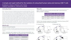 科学海报Isolation of Untouched Human Naive and Memory CD8 T Cells
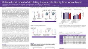 科学海报Unbiased Enrichment of Circulating Tumour Cells Directly from Whole Blood
 沪公网安备31010102008431号