CRISPR/Cas9-mediated heterozygous knockout of the autism gene CHD8 and characterization of its transcriptional networks in cerebral organoids derived from iPS cells.
BACKGROUND CHD8 (chromodomain helicase DNA-binding protein 8),which codes for a member of the CHD family of ATP-dependent chromatin-remodeling factors,is one of the most commonly mutated genes in autism spectrum disorders (ASD) identified in exome-sequencing studies. Loss of function mutations in the gene have also been found in schizophrenia (SZ) and intellectual disabilities and influence cancer cell proliferation. We previously reported an RNA-seq analysis carried out on neural progenitor cells (NPCs) and monolayer neurons derived from induced pluripotent stem (iPS) cells that were heterozygous for CHD8 knockout (KO) alleles generated using CRISPR-Cas9 gene editing. A significant number of ASD and SZ candidate genes were among those that were differentially expressed in a comparison of heterozygous KO lines (CHD8(+/-)) vs isogenic controls (CHD8(+/-)),including the SZ and bipolar disorder (BD) candidate gene TCF4,which was markedly upregulated in CHD8(+/-) neuronal cells. METHODS In the current study,RNA-seq was carried out on CHD8(+/-) and isogenic control (CHD8(+/+)) cerebral organoids,which are 3-dimensional structures derived from iPS cells that model the developing human telencephalon. RESULTS TCF4 expression was,again,significantly upregulated. Pathway analysis carried out on differentially expressed genes (DEGs) revealed an enrichment of genes involved in neurogenesis,neuronal differentiation,forebrain development,Wnt/β-catenin signaling,and axonal guidance,similar to our previous study on NPCs and monolayer neurons. There was also significant overlap in our CHD8(+/-) DEGs with those found in a transcriptome analysis carried out by another group using cerebral organoids derived from a family with idiopathic ASD. Remarkably,the top DEG in our respective studies was the non-coding RNA DLX6-AS1,which was markedly upregulated in both studies; DLX6-AS1 regulates the expression of members of the DLX (distal-less homeobox) gene family. DLX1 was also upregulated in both studies. DLX genes code for transcription factors that play a key role in GABAergic interneuron differentiation. Significant overlap was also found in a transcriptome study carried out by another group using iPS cell-derived neurons from patients with BD,a condition characterized by dysregulated WNT/β-catenin signaling in a subgroup of affected individuals. CONCLUSIONS Overall,the findings show that distinct ASD,SZ,and BD candidate genes converge on common molecular targets-an important consideration for developing novel therapeutics in genetically heterogeneous complex traits.
View Publication
产品类型:
产品号#:
05850
05857
05870
05875
85850
85857
85870
85875
产品名:
mTeSR™1
mTeSR™1
Liu B et al. (MAR 2014)
PLoS ONE 9 3 e90615
Nanog1 in NTERA-2 and recombinant NanogP8 from somatic cancer cells adopt multiple protein conformations and migrate at multiple M.W species
Human Nanog1 is a 305-amino acid (aa) homeodomain-containing transcription factor critical for the pluripotency of embryonic stem (ES) and embryonal carcinoma (EC) cells. Somatic cancer cells predominantly express a retrogene homolog of Nanog1 called NanogP8,which is ˜99% similar to Nanog at the aa level. Although the predicted M.W of Nanog1/NanogP8 is ∼35 kD,both have been reported to migrate,on Western blotting (WB),at apparent molecular masses of 29-80 kD. Whether all these reported protein bands represent authentic Nanog proteins is unclear. Furthermore,detailed biochemical studies on Nanog1/NanogpP8 have been lacking. By combining WB using 8 anti-Nanog1 antibodies,immunoprecipitation,mass spectrometry,and studies using recombinant proteins,here we provide direct evidence that the Nanog1 protein in NTERA-2 EC cells exists as multiple M.W species from ˜22 kD to 100 kD with a major 42 kD band detectable on WB. We then demonstrate that recombinant NanogP8 (rNanogP8) proteins made in bacteria using cDNAs from multiple cancer cells also migrate,on denaturing SDS-PAGE,at ˜28 kD to 180 kD. Interestingly,different anti-Nanog1 antibodies exhibit differential reactivity towards rNanogP8 proteins,which can spontaneously form high M.W protein species. Finally,we show that most long-term cultured cancer cell lines seem to express very low levels of or different endogenous NanogP8 protein that cannot be readily detected by immunoprecipitation. Altogether,the current study reveals unique biochemical properties of Nanog1 in EC cells and NanogP8 in somatic cancer cells.
View Publication
产品类型:
产品号#:
05850
05857
05870
05875
85850
85857
85870
85875
产品名:
mTeSR™1
mTeSR™1
A. Ferrelli et al. (Aug 2025)
HemaSphere 9 8
Mesenchymal stromal cells from JAK2 V617F myeloproliferative neoplasms support healthy and malignant hematopoiesis in a humanized scaffold model in vivo
Myeloproliferative Neoplasms (MPN) are malignancies of hematopoietic stem and progenitor cells (HSPCs) that lead to the overproduction of mature blood cells. These disorders include Essential Thrombocythemia (ET),Polycythemia Vera (PV),and Primary Myelofibrosis (PMF),primarily driven by somatic mutations such as JAK2 V617F . Research indicates that mesenchymal stromal cells (MSCs) support fibrosis in PMF,though their role in ET and PV remains less clear. Furthermore,in vivo studies of ET/PV HSPCs remain a challenge due to low engraftment levels in xenograft models. We employed a 3D scaffold model to create an MPN humanized xenograft mouse model,enabling in vivo functional studies of primary MPN progenitor cells and the supportive role of human MSCs. Using this model,we first demonstrated robust hematopoietic support of healthy (HD) HSPCs by PV and ET MSCs. We then investigated the role of MSCs in sustaining JAK2 V617F mutant cells by using a CRISPR‐Cas9 editing model,along with primary PV and ET HSPCs. Our results showed consistent engraftment of CRISPR‐edited JAK2 V617F mutant HSPCs and PV and ET patient‐derived HSPCs in scaffolds seeded with HD,PV,and ET stroma,providing the first in vivo evidence that PV and ET MSCs can sustain both healthy and MPN‐associated hematopoiesis. Furthermore,HD MSCs were also capable of sustaining PV and ET HSPCs in vivo. Overall,we present the first humanized MPN xenograft model that offers valuable insights into how human BM MSCs interact with JAK2 V617F mutant clones.
View Publication
产品类型:
产品号#:
05150
产品名:
MyeloCult™ H5100
Hidalgo A et al. (JAN 2005)
Blood 105 2 567--75
Enforced fucosylation of neonatal CD34+ cells generates selectin ligands that enhance the initial interactions with microvessels but not homing to bone marrow.
Hematopoietic progenitor/stem cell homing to the bone marrow requires the concerted action of several adhesion molecules. Endothelial P- and E-selectins play an important role in this process,but their ligands on a large subset of neonate-derived human CD34+ cells are absent,leading to a reduced ability to interact with the bone marrow (BM) microvasculature. We report here that this deficiency results from reduced alpha1,3-fucosyltransferase (FucT) expression and activity in these CD34+ cells. Incubation of CD34+ cells with recombinant human FucTVI rapidly corrected the deficiency in nonbinding CD34+ cells and further increased the density of ligands for both P- and E-selectins on all cord blood-derived CD34+ cells. Intravital microscopy studies revealed that these FucTVI-treated CD34+ cells displayed a marked enhancement in their initial interactions with the BM microvasculature,but unexpectedly,homing into the BM was not improved by FucTVI treatment. These data indicate that,although exogenous FucT enzyme activity can rapidly modulate selectin binding avidity of cord blood CD34+ cells,further studies are needed to understand how to translate a positive effect on progenitor cell adhesion in bone marrow microvessels into one that significantly influences migration and lodgement into the parenchyma.
View Publication
A. Wu et al. (Jul 2025)
International Journal of Molecular Sciences 26 13
Identification of a PAK6-Mediated MDM2/p21 Axis That Modulates Survival and Cell Cycle Control of Drug-Resistant Stem/Progenitor Cells in Chronic Myeloid Leukemia
Chronic myeloid leukemia (CML) is a leading example of a malignancy where a molecular targeted therapy revolutionized treatment but has rarely led to cures. Overcoming tyrosine kinase inhibitor (TKI) drug resistance remains a challenge in the treatment of CML. We have recently identified miR-185 as a predictive biomarker where reduced expression in CD34 + treatment-naïve CML cells was associated with TKI resistance. We have also identified PAK6 as a target gene of miR-185 that was upregulated in CD34 + TKI-nonresponder cells. However,its role in regulating TKI resistance remains largely unknown. In this study,we specifically targeted PAK6 in imatinib (IM)-resistant cells and CD34 + stem/progenitor cells from IM-nonresponders using a lentiviral-mediated PAK6 knockdown strategy. Interestingly,the genetic and pharmacological suppression of PAK6 significantly reduced proliferation and increased apoptosis in TKI-resistant cells. Cell survivability was further diminished when IM was combined with PAK6 knockdown. Importantly,PAK6 inhibition in TKI-resistant cells induced cell cycle arrest in the G2-M phase and cellular senescence,accompanied by increased levels of DNA damage-associated senescence markers. Mechanically,we identified a PAK6-mediated MDM2-p21 axis that regulates cell cycle arrest and senescence. Thus,PAK6 plays a critical role in determining alternative cell fates in leukemic cells,and targeting PAK6 may offer a therapeutic strategy to selectively eradicate TKI-resistant cells.
View Publication
产品类型:
产品号#:
04230
产品名:
MethoCult™ H4230
West FD et al. (OCT 2011)
Stem Cells 29 10 1640--1643
Brief report: chimeric pigs produced from induced pluripotent stem cells demonstrate germline transmission and no evidence of tumor formation in young pigs.
The recent development of porcine induced pluripotent stem cells (piPSCs) capable of generating chimeric animals,a feat not previously accomplished with embryonic stem cells or iPSCs in a species outside of rodents,has opened the doors for in-depth study of iPSC tumorigenicity,autologous transplantation,and other key aspects to safely move iPSC therapies to the clinic. The study of iPSC tumorigenicity is critical as previous research in the mouse showed that iPSC-derived chimeras possessed large numbers of tumors,rising significant concerns about the safety of iPSC therapies. Additionally,piPSCs capable of generating germline chimeras could revolutionize the transgenic animal field by enabling complex genetic manipulations (e.g.,knockout or knockin of genes) to produce biomedically important large animal models or improve livestock production. In this study,we demonstrate for the first time in a nonrodent species germline transmission of iPSCs with the live birth of a transgenic piglet that possessed genome integration of the human POU5F1 and NANOG genes. In addition,gross and histological examination of necropsied porcine chimeras at 2,7,and 9 months showed that these animals lacked tumor formation and demonstrated normal development. Tissue samples positive for human POU5F1 DNA showed no C-MYC gene expression,further implicating C-MYC as a cause of tumorigenicity. The development of germline-competent porcine iPSCs that do not produce tumors in young chimeric animals presents an attractive and powerful translational model to study the efficacy and safety of stem cell therapies and perhaps to efficiently produce complex transgenic animals.
View Publication
产品类型:
产品号#:
05850
05857
05870
05875
85850
85857
85870
85875
产品名:
mTeSR™1
mTeSR™1
S. Jaleco et al. (Jul 2003)
The Journal of Immunology 171 61-68
Homeostasis of Naive and Memory CD4 + T Cells: IL-2 and IL-7 Differentially Regulate the Balance Between Proliferation and Fas-Mediated Apoptosis
Cytokines play a crucial role in the maintenance of polyclonal naive and memory T cell populations. It has previously been shown that ex vivo,the IL-7 cytokine induces the proliferation of naive recent thymic emigrants (RTE) isolated from umbilical cord blood but not mature adult-derived naive and memory human CD4(+) T cells. We find that the combination of IL-2 and IL-7 strongly promotes the proliferation of RTE,whereas adult CD4(+) T cells remain relatively unresponsive. Immunological activity is controlled by a balance between proliferation and apoptotic cell death. However,the relative contributions of IL-2 and IL-7 in regulating these processes in the absence of MHC/peptide signals are not known. Following exposure to either IL-2 or IL-7 alone,RTE,as well as mature naive and memory CD4(+) T cells,are rendered only minimally sensitive to Fas-mediated cell death. However,in the presence of the two cytokines,Fas engagement results in a high level of caspase-dependent apoptosis in both RTE as well as naive adult CD4(+) T cells. In contrast,equivalently treated memory CD4(+) T cells are significantly less sensitive to Fas-induced cell death. The increased susceptibility of RTE and naive CD4(+) T cells to Fas-induced apoptosis correlates with a significantly higher IL-2/IL-7-induced Fas expression on these T cell subsets than on memory CD4(+) T cells. Thus,IL-2 and IL-7 regulate homeostasis by modulating the equilibrium between proliferation and apoptotic cell death in RTE and mature naive and memory T cell subsets.
View Publication
产品类型:
产品号#:
15023
15063
产品名:
RosetteSep™人CD8+ T细胞富集抗体混合物
RosetteSep™人CD8+ T细胞富集抗体混合物
Meenhuis A et al. (JUL 2011)
Blood 118 4 916--25
MiR-17/20/93/106 promote hematopoietic cell expansion by targeting sequestosome 1-regulated pathways in mice.
MicroRNAs (miRNAs) are pivotal for regulation of hematopoiesis but their critical targets remain largely unknown. Here,we show that ectopic expression of miR-17,-20,-93 and -106,all AAAGUGC seed-containing miRNAs,increases proliferation,colony outgrowth and replating capacity of myeloid progenitors and results in enhanced P-ERK levels. We found that these miRNAs are endogenously and abundantly expressed in myeloid progenitors and down-regulated in mature neutrophils. Quantitative proteomics identified sequestosome 1 (SQSTM1),an ubiquitin-binding protein and regulator of autophagy-mediated protein degradation,as a major target for these miRNAs in myeloid progenitors. In addition,we found increased expression of Sqstm1 transcripts during CSF3-induced neutrophil differentiation of 32D-CSF3R cells and an inverse correlation of SQSTM1 protein levels and miR-106 expression in AML samples. ShRNA-mediated silencing of Sqstm1 phenocopied the effects of ectopic miR-17/20/93/106 expression in hematopoietic progenitors in vitro and in mice. Further,SQSTM1 binds to the ligand-activated colony-stimulating factor 3 receptor (CSF3R) mainly in the late endosomal compartment,but not in LC3 positive autophagosomes. SQSTM1 regulates CSF3R stability and ligand-induced mitogen-activated protein kinase signaling. We demonstrate that AAAGUGC seed-containing miRNAs promote cell expansion,replating capacity and signaling in hematopoietic cells by interference with SQSTM1-regulated pathways.
View Publication
产品类型:
产品号#:
03231
产品名:
MethoCult™ M3231
X. Liu et al. (Mar 2025)
Stem Cell Research & Therapy 16
Purine metabolism in bone marrow microenvironment inhibits hematopoietic stem cell differentiation under microgravity
Spaceflight and microgravity environments have been shown to cause significant health impairments,including bone loss,immune dysfunction,and hematopoietic disorders. Hematopoietic stem cells (HSCs),as progenitors of the hematopoietic system,are critical for the continuous renewal and regulation of immune cells. Therefore,elucidating the regulatory mechanisms governing HSC fate and differentiation in microgravity environments is of paramount importance. In this study,hindlimb unloading (HU) was employed in mice to simulate microgravity conditions. After 28 days of HU,cells were isolated for analysis. Flow cytometry and colony-forming assays were utilized to assess changes in HSC proliferation and differentiation. Additionally,transcriptomic and untargeted metabolomic sequencing were performed to elucidate alterations in the metabolic pathways of the bone marrow microenvironment and their molecular regulatory effects on HSCs fate. Our findings revealed that 28 days of HU impaired hematopoietic function,leading to multi-organ damage and hematological disorders. The simulated microgravity environment significantly increased the HSCs population in the bone marrow,particularly within the long-term and short-term subtypes,while severely compromising the differentiation capacity of hematopoietic stem/progenitor cells. Transcriptomic analysis of HSCs,combined with metabolomic profiling of bone marrow supernatants,identified 1,631 differentially expressed genes and 58 metabolites with altered abundance. Gene set enrichment analysis indicated that HU suppressed key pathways,including hematopoietic cell lineage and MAPK signaling. Furthermore,integrated analyses revealed that metabolites affected by HU,particularly hypoxanthine enriched in the purine metabolism pathway,were closely associated with hematopoietic cell lineage and MAPK signaling pathways. Molecular docking simulations and in vitro experiments confirmed that hypoxanthine interacts directly with core molecules within these pathways,influencing their expression. These findings demonstrate that hypoxanthine in the bone marrow supernatant acts as a signaling mediator under microgravity,influencing HSCs fate by modulating hematopoietic cell lineage and MAPK signaling pathways. This study offers novel insights into the impact of microgravity on HSC fate and gene expression,underscoring the pivotal role of bone marrow microenvironmental metabolic changes in regulating key signaling pathways that determine hematopoietic destiny. The online version contains supplementary material available at 10.1186/s13287-025-04213-9.
View Publication
产品类型:
产品号#:
03434
03444
产品名:
MethoCult™ GF M3434
MethoCult™ GF M3434
Shi S et al. (SEP 2011)
Journal of Visualized Experiments 55 e3010
A high-throughput automated platform for the development of manufacturing cell lines for protein therapeutics
The fast-growing biopharmaceutical industry demands speedy development of highly efficient and reliable production systems to meet the increasing requirement for drug supplies. The generation of production cell lines has traditionally involved manual operations that are labor-intensive,low-throughput and vulnerable to human errors. We report here an integrated high-throughput and automated platform for development of manufacturing cell lines for the production of protein therapeutics. The combination of BD FACS Aria Cell Sorter,CloneSelect Imager and TECAN Freedom EVO liquid handling system has enabled a high-throughput and more efficient cell line development process. In this operation,production host cells are first transfected with an expression vector carrying the gene of interest (1),followed by the treatment with a selection agent. The stably-transfected cells are then stained with fluorescence-labeled anti-human IgG antibody,and are subsequently subject to flow cytometry analysis (2-4). Highly productive cells are selected based on fluorescence intensity and are isolated by single-cell sorting on a BD FACSAria. Colony formation from single-cell stage was detected microscopically and a series of time-laps digital images are taken by CloneSelect Imager for the documentation of cell line history. After single clones have formed,these clones were screened for productivity by ELISA performed on a TECAN Freedom EVO liquid handling system. Approximately 2,000 - 10,000 clones can be screened per operation cycle with the current system setup. This integrated approach has been used to generate high producing Chinese hamster ovary (CHO) cell lines for the production of therapeutic monoclonal antibody (mAb) as well as their fusion proteins. With the aid of different types of detecting probes,the method can be used for developing other protein therapeutics or be applied to other production host systems. Comparing to the traditional manual procedure,this automated platform demonstrated advantages of significantly increased capacity,ensured clonality,traceability in cell line history with electronic documentation and much reduced opportunity in operator error.
View Publication
产品类型:
产品号#:
30000
产品名:
X. Ma et al. (Jun 2025)
Journal of Experimental & Clinical Cancer Research : CR 44 5
PSMB10 maintains the stemness of chemotherapeutic drug-resistant leukemia cells by inhibiting senescence and cytotoxic T lymphocyte-mediated killing in a ubiquitinated degradation manner
Drug resistance and relapse are still major challenges in acute myeloid leukemia (AML) because of the inability to effectively eradicate leukemia stem cells (LSCs). Senescence induction combined with immune killing may offer promising strategies for LSC eradication. However,whether and how drug-resistant LSCs retain stemness via senescence and immune regulation remains unknown. The immunoproteasome subunit PSMB10 expression levels were analyzed by single-cell RNA-seq data,along with the bioinformatics analysis of publicly available AML datasets,and quantified using RT-qPCR and flow cytometry (FCM) analysis on clinical samples from AML patients. The cellular senescence was evaluated by the assays of cell proliferation,cell cycle,senescence-associated β-galactosidase activity,and senescence-associated secretory phenotype factors. In vitro T-cell killing assay was played to determine immune escape reprogramming of AML cells. FCM was conducted to estimate intracellular drug concentration and cellular apoptosis rates. Human AML xenografts and PSMB10 knockout syngeneic mouse bone marrow transplantation models were utilized to investigate the function of PSMB10. Various techniques were employed for mechanism studies,including Lentivirus transduction or siRNA transfection,western blotting,co-immunoprecipitation assays,luciferase reporter assays,polysome profiling assays,quantitative proteomics,etc. PSMB10 mRNA was significantly upregulated in the surviving nonsenescent LSCs,exhibiting a 13-fold increase compared to senescent LSCs following chemotherapy. The specific high expression of PSMB10 in post-chemotherapy nonsenescent LSCs predicts a poor AML prognosis. The genetic inactivation of PSMB10 resulted in increased senescence and cytotoxic T lymphocyte (CTL) killing,as well as increased intracellular drug concentrations and drug-induced cellular senescence in different types of human AML cells,which also impeded human and murine leukemia initiation and stemness maintenance in vivo with a 19-fold decrease in the frequency of human LSCs and a 7.6-fold decrease of drug-resistant mouse LSCs,while normal hematopoietic cells remained unaffected. Mechanistically,the downregulation of PSMB10 boosted SLC22A16-mediated drug endocytosis and further induced chemotherapy drug-mediated senescence through the RPL6/RPS6-MDM2-P21 pathway in AML cells. Additionally,downregulating PSMB10 also impeded MHC-I protein degradation-induced escape of CTL killing. PSMB10 is a key candidate molecular target for eradicating drug-resistant LSCs via senescence and immune regulation. The online version contains supplementary material available at 10.1186/s13046-025-03420-9.
View Publication


 EasySep™小鼠TIL(CD45)正选试剂盒





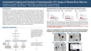 科学海报Automated Imaging and Analysis of Hematopoietic CFU Assays of Mouse Bone Marrow
 沪公网安备31010102008431号