Gene-editing in patient and humanized-mice primary muscle stem cells rescues dysferlin expression in dysferlin-deficient muscular dystrophy
Dystrophy-associated fer-1-like protein (dysferlin) conducts plasma membrane repair. Mutations in the DYSF gene cause a panoply of genetic muscular dystrophies. We targeted a frequent loss-of-function,DYSF exon 44,founder frameshift mutation with mRNA-mediated delivery of SpCas9 in combination with a mutation-specific sgRNA to primary muscle stem cells from two homozygous patients. We observed a consistent >60% exon 44 re-framing,rescuing a full-length and functional dysferlin protein. A new mouse model harboring a humanized Dysf exon 44 with the founder mutation,hEx44mut,recapitulates the patients’ phenotype and an identical re-framing outcome in primary muscle stem cells. Finally,gene-edited murine primary muscle stem-cells are able to regenerate muscle and rescue dysferlin when transplanted back into hEx44mut hosts. These findings are the first to show that a CRISPR-mediated therapy can ameliorate dysferlin deficiency. We suggest that gene-edited primary muscle stem cells could exhibit utility,not only in treating dysferlin deficiency syndromes,but also perhaps other forms of muscular dystrophy. Dysferlin-deficient muscular dystrophy is a devastating and untreatable disease. Using Cas9,the authors restored dysferlin in muscle stem cells from patients ex vivo and show proof-of-concept for autologous cell replacement therapies in a new humanized mouse model.
View Publication
产品类型:
产品号#:
85850
85857
产品名:
mTeSR™1
mTeSR™1
(May 2024)
Nature Microbiology 9 6
Peptostreptococcus anaerobius mediates anti-PD1 therapy resistance and exacerbates colorectal cancer via myeloid-derived suppressor cells in mice
Bacteria such as the oral microbiome member Peptostreptococcus anaerobius can exacerbate colorectal cancer (CRC) development. Little is known regarding whether these immunomodulatory bacteria also affect antitumour immune checkpoint blockade therapy. Here we show that administration of P. anaerobius abolished the efficacy of anti-PD1 therapy in mouse models of CRC. P. anaerobius both induced intratumoral myeloid-derived suppressor cells (MDSCs) and stimulated their immunosuppressive activities to impair effective T cell responses. Mechanistically,P. anaerobius administration activated integrin α2β1–NF-κB signalling in CRC cells to induce secretion of CXCL1 and recruit CXCR2+ MDSCs into tumours. The bacterium also directly activated immunosuppressive activity of intratumoral MDSCs by secreting lytC_22,a protein that bound to the Slamf4 receptor on MDSCs and promoted ARG1 and iNOS expression. Finally,therapeutic targeting of either integrin α2β1 or the Slamf4 receptor were revealed as promising strategies to overcome P. anaerobius-mediated resistance to anti-PD1 therapy in CRC. Interactions between Peptostreptococcus anaerobius and host cells promote recruitment and activation of myeloid-derived suppressor cells,leading to anti-PD1 immune checkpoint inhibitor resistance and exacerbated colorectal cancer in mice.
View Publication
产品类型:
产品号#:
19867
产品名:
EasySep™小鼠MDSC(CD11b+Gr1+)分选试剂盒
(Feb 2024)
Cancer Immunology Research 12 4
High-Specificity CRISPR-Mediated Genome Engineering in Anti-BCMA Allogeneic CAR T Cells Suppresses Allograft Rejection in Preclinical Models
Allogeneic CAR T–cell therapies are being developed for hematologic malignancies. The authors implement a Cas12a chRDNA platform to generate allogeneic immune-cloaked BCMA-specific CAR T cells with resistance to host response–mediated rejection for evaluation in multiple myeloma. AbstractAllogeneic chimeric antigen receptor (CAR) T cell therapies hold the potential to overcome many of the challenges associated with patient-derived (autologous) CAR T cells. Key considerations in the development of allogeneic CAR T cell therapies include prevention of graft-vs-host disease (GvHD) and suppression of allograft rejection. Here,we describe preclinical data supporting the ongoing first-in-human clinical study,the CaMMouflage trial (NCT05722418),evaluating CB-011 in patients with relapsed/refractory multiple myeloma. CB-011 is a hypoimmunogenic,allogeneic anti–B-cell maturation antigen (BCMA) CAR T cell therapy candidate. CB-011 cells feature 4 genomic alterations and were engineered from healthy donor–derived T cells using a Cas12a CRISPR hybrid RNA–DNA (chRDNA) genome-editing technology platform. To address allograft rejection,CAR T cells were engineered to prevent endogenous HLA class I complex expression and overexpress a single-chain polyprotein complex composed of beta-2 microglobulin (B2M) tethered to HLA-E. In addition,T-cell receptor (TCR) expression was disrupted at the TCR alpha constant locus in combination with the site-specific insertion of a humanized BCMA-specific CAR. CB-011 cells exhibited robust plasmablast cytotoxicity in vitro in a mixed lymphocyte reaction in cell cocultures derived from patients with multiple myeloma. In addition,CB-011 cells demonstrated suppressed recognition by and cytotoxicity from HLA-mismatched T cells. CB-011 cells were protected from natural killer cell–mediated cytotoxicity in vitro and in vivo due to endogenous promoter-driven expression of B2M–HLA-E. Potent antitumor efficacy,when combined with an immune-cloaking armoring strategy to dampen allograft rejection,offers optimized therapeutic potential in multiple myeloma. See related Spotlight by Caimi and Melenhorst,p. 385
View Publication
产品类型:
产品号#:
100-0956
10981
17951
100-0695
17951RF
产品名:
ImmunoCult™ XF培养基
ImmunoCult™ XF 人T细胞扩增培养基,500 mL
EasySep™人T细胞分选试剂盒
EasySep™人T细胞分选试剂盒
RoboSep™ 人T细胞分选试剂盒
S. Murty et al. (nov 2020)
Cancer research 80 21 4731--4740
PET Reporter Gene Imaging and Ganciclovir-Mediated Ablation of Chimeric Antigen Receptor T Cells in Solid Tumors.
Imaging strategies to monitor chimeric antigen receptor (CAR) T-cell biodistribution and proliferation harbor the potential to facilitate clinical translation for the treatment of both liquid and solid tumors. In addition,the potential adverse effects of CAR T cells highlight the need for mechanisms to modulate CAR T-cell activity. The herpes simplex virus type 1 thymidine kinase (HSV1-tk) gene has previously been translated as a PET reporter gene for imaging of T-cell trafficking in patients with brain tumor. The HSV1-TK enzyme can act as a suicide gene of transduced cells through treatment with the prodrug ganciclovir. Here we report the molecular engineering,imaging,and ganciclovir-mediated destruction of B7H3 CAR T cells incorporating a mutated version of the HSV1-tk gene (sr39tk) with improved enzymatic activity for ganciclovir. The sr39tk gene did not affect B7H3 CAR T-cell functionality and in vitro and in vivo studies in osteosarcoma models showed no significant effect on B7H3 CAR T-cell antitumor activity. PET/CT imaging with 9-(4-[18F]-fluoro-3-[hydroxymethyl]butyl)guanine ([18F]FHBG) of B7H3-sr39tk CAR T cells in an orthotopic model of osteosarcoma revealed tumor homing and systemic immune expansion. Bioluminescence and PET imaging of B7H3-sr39tk CAR T cells confirmed complete tumor ablation with intraperitoneal ganciclovir administration. This imaging and suicide ablation system can provide insight into CAR T-cell migration and proliferation during clinical trials while serving as a suicide switch to limit potential toxicities. SIGNIFICANCE: This study showcases the only genetically engineered system capable of serving the dual role both as an effective PET imaging reporter and as a suicide switch for CAR T cells.
View Publication
产品类型:
产品号#:
15021
15061
产品名:
RosetteSep™人T细胞富集抗体混合物
RosetteSep™人T细胞富集抗体混合物
Itin PH et al. (NOV 1994)
Endocrinology 135 5 1793--8
Effects of vitamin D metabolites on proliferation and differentiation of cultured human epidermal keratinocytes grown in serum-free or defined culture medium.
We examined the effects of 1,25-dihydroxyvitamin D3 [1,25(OH)2D3],25-hydroxyvitamin D3 (25OHD3),and vitamin D3 on human keratinocyte proliferation and differentiation in a serum-free or defined culture system. Concentrations greater than 10(-8) M 1,25-(OH)2D3 or 10(-7) M 25(OH)2D3 caused marked inhibition of cell growth. Growth inhibition with high doses of 1,25-(OH)2D3 was not stringent,but was mainly exerted in the G1 phase of the cell cycle. Early release from the cell cycle block restored the proliferation of human keratinocytes. The calcium concentration in the medium had no significant effect on the antiproliferative action of 1,25-(OH)2D3,25OHD3,and vitamin D3. We also show that human keratinocyte proliferation is enhanced at doses of 1,25-(OH)2D3 and 25OH2D3 of 10(-9) M or less. Enhanced proliferation of human keratinocytes with physiological concentrations of 1,25-(OH)2D3 could only be shown in fully defined medium that contained no vitamin D3,related sterols,or bovine pituitary extract. Human keratinocyte differentiation was enhanced with higher doses of 1,25-(OH)2D3 when cells were grown in the presence of high calcium concentrations. These studies demonstrate that the lower,physiological concentrations of vitamin D3 metabolites are capable of stimulating the proliferation of epidermal keratinocytes grown under selected conditions that eliminate confounding or unidentified medium culture factors. Vitamin D3 metabolites are shown to exert mitogenic trophic effects in cultured human epithelial cells similar to their established activities in vivo.
View Publication
产品类型:
产品号#:
72412
产品名:
骨化三醇(Calcitriol)
Souness JE et al. ( 1996)
British journal of pharmacology 118 3 649--658
Evidence that cyclic AMP phosphodiesterase inhibitors suppress TNF alpha generation from human monocytes by interacting with a 'low-affinity' phosphodiesterase 4 conformer.
1. We have investigated the inhibitory effects of RP 73401 (piclamilast) and rolipram against human monocyte cyclic AMP-specific phosphodiesterase (PDE4) in relation to their effects on prostaglandin (PG)E2-induced cyclic AMP accumulation and lipopolysaccharide (LPS)-induced TNF alpha production and TNF alpha mRNA expression. 2. PDE4 was found to be the predominant PDE isoenzyme in the cytosolic fraction of human monocytes. Cyclic GMP-inhibited PDE (PDE3) was also detected in the cytosolic and particulate fractions. Reverse transcription polymerase chain reaction (RT-PCR) of human monocyte poly (A+) mRNA revealed amplified products corresponding to PDE4 subtypes A and B of which the former was most highly expressed. A faint band corresponding in size to PDE4D was also observed. 3. RP 73401 was a potent inhibitor of cytosolic PDE4 (IC50: 1.5 +/- 0.6 nM,n = 3). (+/-)-Rolipram (IC50: 313 +/- 6.7 nM,n = 3) was at least 200 fold less potent than RP 73401. R-(-)-rolipram was approximately 3 fold more potent than S-(+)-rolipram against cytosolic PDE4. 4. RP 73401 (IC50: 9.2 +/- 2.1 nM,n = 6) was over 50 fold more potent than (+/-)-rolipram (IC50: 503 +/- 134 nM,n = 6) ) in potentiating PGE2-induced cyclic AMP accumulation. R-(-)-rolipram (IC50: 289 +/- 121 nM,n = 5) was 4.7 fold more potent than its S-(+)-enantiomer (IC50: 1356 +/- 314 nM,n = 5). A strong and highly-significant,linear correlation (r = 0.95,P textless 0.01,n = 13) was observed between the inhibitory potencies of a range of structurally distinct PDE4 inhibitors against monocyte PDE4 and their ED50 values in enhancing monocyte cyclic AMP accumulation. A poorer,though still significant,linear correlation (r = 0.67,P textless 0.01,n = 13) was observed between the potencies of the same compounds in potentiating PGE2-induced monocyte cyclic AMP accumulation and their abilities to displace [3H]-rolipram binding to brain membranes. 5. RP 73401 (IC50: 6.9 +/- 3.3 nM,n = 5) was 71 fold more potent than (+/-)-rolipram (IC50: 490 +/- 260 nM,n = 4) in inhibiting LPS-induced TNF alpha release from monocytes. R-(-)-rolipram (IC50: 397 +/- 178 nM,n = 3) was 5.2-fold more potent than its S-(+)- enantiomer (IC50: 2067 +/- 659 nM,n = 3). As with cyclic AMP,accumulation a closer,linear correlation existed between the potency of structurally distinct compounds in suppressing TNF alpha with PDE4 inhibition (r = 0.93,P textless 0.01,n = 13) than with displacement of [3H]-rolipram binding (r = 0.65,P textless 0.01,n = 13). 6. RP 73401 (IC50: 2 nM) was 180 fold more potent than rolipram (IC50: 360 nM) in suppressing LPS (10 ng ml-1)-induced TNF alpha mRNA. 7. The results demonstrate that RP 73401 is a very potent inhibitor of TNF alpha release from human monocytes suggesting that it may have therapeutic potential in the many pathological conditions associated with over-production of this pro-inflammatory cytokine. Furthermore,PDE inhibitor actions on functional responses are better correlated with inhibition of PDE4 catalytic activity than displacement of [3H]-rolipram from its high-affinity binding site,suggesting that the native PDE4 in human monocytes exists predominantly in a 'low-affinity' state.
View Publication
产品类型:
产品号#:
73382
73384
产品名:
Rolipram
Rolipram
(Apr 2024)
Cell Communication and Signaling : CCS 22 9274
Gut microbiota-derived butyrate restores impaired regulatory T cells in patients with AChR myasthenia gravis via mTOR-mediated autophagy
More than 80% of patients with myasthenia gravis (MG) are positive for anti-acetylcholine receptor (AChR) antibodies. Regulatory T cells (Tregs) suppress overproduction of these antibodies,and patients with AChR antibody-positive MG (AChR MG) exhibit impaired Treg function and reduced Treg numbers. The gut microbiota and their metabolites play a crucial role in maintaining Treg differentiation and function. However,whether impaired Tregs correlate with gut microbiota activity in patients with AChR MG remains unknown. Here,we demonstrate that butyric acid-producing gut bacteria and serum butyric acid level are reduced in patients with AChR MG. Butyrate supplementation effectively enhanced Treg differentiation and their suppressive function of AChR MG. Mechanistically,butyrate activates autophagy of Treg cells by inhibiting the mammalian target of rapamycin. Activation of autophagy increased oxidative phosphorylation and surface expression of cytotoxic T-lymphocyte-associated protein 4 on Treg cells,thereby promoting Treg differentiation and their suppressive function in AChR MG. This observed effect of butyrate was blocked using chloroquine,an autophagy inhibitor,suggesting the vital role of butyrate-activated autophagy in Tregs of patients with AChR MG. We propose that gut bacteria derived butyrate has potential therapeutic efficacy against AChR MG by restoring impaired Tregs.Supplementary InformationThe online version contains supplementary material available at 10.1186/s12964-024-01588-9.
View Publication
产品类型:
产品号#:
18063
18063RF
100-1136
产品名:
EasySep™人CD4+CD127low CD25+调节性T细胞分选试剂盒
EasySep™人CD4+CD127lowCD25+调节性T细胞分离试剂盒
EasySep™人CD4+CD127low CD25+调节性T细胞分选试剂盒
Kortylewski M et al. (DEC 2005)
Nature medicine 11 12 1314--21
Inhibiting Stat3 signaling in the hematopoietic system elicits multicomponent antitumor immunity.
The immune system can act as an extrinsic suppressor of tumors. Therefore,tumor progression depends in part on mechanisms that downmodulate intrinsic immune surveillance. Identifying these inhibitory pathways may provide promising targets to enhance antitumor immunity. Here,we show that Stat3 is constitutively activated in diverse tumor-infiltrating immune cells,and ablating Stat3 in hematopoietic cells triggers an intrinsic immune-surveillance system that inhibits tumor growth and metastasis. We observed a markedly enhanced function of dendritic cells,T cells,natural killer (NK) cells and neutrophils in tumor-bearing mice with Stat3(-/-) hematopoietic cells,and showed that tumor regression requires immune cells. Targeting Stat3 with a small-molecule drug induces T cell- and NK cell-dependent growth inhibition of established tumors otherwise resistant to direct killing by the inhibitor. Our findings show that Stat3 signaling restrains natural tumor immune surveillance and that inhibiting hematopoietic Stat3 in tumor-bearing hosts elicits multicomponent therapeutic antitumor immunity.
View Publication
The endothelial antigen ESAM marks primitive hematopoietic progenitors throughout life in mice.
Although recent advances have enabled hematopoietic stem cells (HSCs) to be enriched to near purity,more information about their characteristics will improve our understanding of their development and stage-related functions. Here,using microarray technology,we identified endothelial cell-selective adhesion molecule (ESAM) as a novel marker for murine HSCs in fetal liver. Esam was expressed at high levels within a Rag1(-) c-kit(Hi) Sca1(+) HSC-enriched fraction,but sharply down-regulated with activation of the Rag1 locus,a valid marker for the most primitive lymphoid progenitors in E14.5 liver. The HSC-enriched fraction could be subdivided into 2 on the basis of ESAM levels. Among endothelial antigens on hematopoietic progenitors,ESAM expression showed intimate correlation with HSC activity. The ESAM(Hi) population was highly enriched for multipotent myeloid-erythroid progenitors and primitive progenitors with lymphopoietic activity,and exclusively reconstituted long-term lymphohematopoiesis in lethally irradiated recipients. Tie2(+) c-kit(+) lymphohematopoietic cells in the E9.5-10.5 aorta-gonad-mesonephros region also expressed high levels of ESAM. Furthermore,ESAM was detected on primitive hematopoietic progenitors in adult bone marrow. Interestingly,ESAM expression in the HSC-enriched fraction was up-regulated in aged mice. We conclude that ESAM marks HSC in murine fetal liver and will facilitate studies of hematopoiesis throughout life.
View Publication
产品类型:
产品号#:
03434
03444
产品名:
MethoCult™ GF M3434
MethoCult™ GF M3434
Hoggatt J et al. (MAY 2009)
Blood 113 22 5444--55
Prostaglandin E2 enhances hematopoietic stem cell homing, survival, and proliferation.
Adult hematopoietic stem cells (HSCs) are routinely used to reconstitute hematopoiesis after myeloablation; however,transplantation efficacy and multilineage reconstitution can be limited by inadequate HSC number,or poor homing,engraftment,or self-renewal. Here we report that mouse and human HSCs express prostaglandin E2 (PGE2) receptors,and that short-term ex vivo exposure of HSCs to PGE2 enhances their homing,survival,and proliferation,resulting in increased long-term repopulating cell (LTRC) and competitive repopulating unit (CRU) frequency. HSCs pulsed with PGE2 are more competitive,as determined by head-to-head comparison in a competitive transplantation model. Enhanced HSC frequency and competitive advantage is stable and maintained upon serial transplantation,with full multilineage reconstitution. PGE2 increases HSC CXCR4 mRNA and surface expression,enhances their migration to SDF-1 in vitro and homing to bone marrow in vivo,and stimulates HSC entry into and progression through cell cycle. In addition,PGE2 enhances HSC survival,associated with an increase in Survivin mRNA and protein expression and reduction in intracellular active caspase-3. Our results define novel mechanisms of action whereby PGE2 enhances HSC function and supports a strategy to use PGE2 to facilitate hematopoietic transplantation.
View Publication
产品类型:
产品号#:
72192
72194
产品名:
前列腺素E2(Prostaglandin E2)
前列腺素E2(Prostaglandin E2)
Charafe-Jauffret E et al. (JAN 2010)
Clinical cancer research : an official journal of the American Association for Cancer Research 16 1 45--55
Aldehyde dehydrogenase 1-positive cancer stem cells mediate metastasis and poor clinical outcome in inflammatory breast cancer.
PURPOSE: To examine the role of cancer stem cells (CSC) in mediating metastasis in inflammatory breast cancer (IBC) and the association of these cells with patient outcome in this aggressive type of breast cancer. EXPERIMENTAL DESIGN: CSCs were isolated from SUM149 and MARY-X,an IBC cell line and primary xenograft,by virtue of increased aldehyde dehydrogenase (ALDH) activity as assessed by the ALDEFLUOR assay. Invasion and metastasis of CSC populations were assessed by in vitro and mouse xenograft assays. Expression of ALDH1 was determined on a retrospective series of 109 IBC patients and this was correlated with histoclinical data. All statistical tests were two sided. Log-rank tests using Kaplan-Meier analysis were used to determine the correlation of ALDH1 expression with development of metastasis and patient outcome. RESULTS: Both in vitro and xenograft assays showed that invasion and metastasis in IBC are mediated by a cellular component that displays ALDH activity. Furthermore,expression of ALDH1 in IBC was an independent predictive factor for early metastasis and decreased survival in this patient population. CONCLUSIONS: These results suggest that the metastatic,aggressive behavior of IBC may be mediated by a CSC component that displays ALDH enzymatic activity. ALDH1 expression represents the first independent prognostic marker to predict metastasis and poor patient outcome in IBC. The results illustrate how stem cell research can translate into clinical practice in the IBC field.
View Publication


 EasySep™小鼠TIL(CD45)正选试剂盒





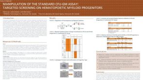 科学海报Manipulation of the Standard CFU-GM Assay Targeted Screening of Hematopoietic Myeloid Progenitors
 沪公网安备31010102008431号