
 EasySep™小鼠TIL(CD45)正选试剂盒


搜索结果: 'methocult media formulations for mouse hematopoietic cells serum containing'
-

产品类型:
产品号#:
73322
产品名:
BIO-Acetoxime
-
产品类型:
产品号#:
17684
17684RF
产品名:
EasySep™ PE正选试剂盒 II
RoboSep™ PE正选试剂盒 II
-
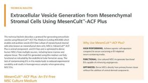 技术公告Extracellular Vesicle Generation from Mesenchymal Stromal Cells Using MesenCult™-ACF Plus
产品类型:
细胞类型:
内皮细胞,间充质基质细胞,间充质干祖细胞
产品号#:
08000
05445
05448
产品名:
MesenCult™ -ACF Plus培养基
MesenCult™-ACF Plus培养试剂盒
发布日期: 12/01/2023 -
产品类型:
产品号#:
34411
34415
34421
34425
34450
34460
85850
85857
产品名:
AggreWell™ 400 24孔板,1个
AggreWell™400 24孔板,5个
AggreWell™ 400 6孔板,1个
AggreWell™ 400 6孔板,5个
AggreWell™400 24孔板启动套装
AggreWell™ 400 6孔板启动套装
mTeSR™1
mTeSR™1
-
产品类型:
产品号#:
01702
产品名:
ALDEFLUOR™检测缓冲液
-
产品类型:
产品号#:
04434
04444
产品名:
MethoCult™ H4434 Classic
MethoCult™ H4434 Classic
-
产品类型:
产品号#:
73782
73784
产品名:
R848
R848
-
产品类型:
产品号#:
05700
05701
05702
05715
产品名:
NeuroCult™ 基础培养基(小鼠和大鼠)
NeuroCult™ 扩增添加物(小鼠和大鼠)
NeuroCult™扩增试剂盒(小鼠和大鼠)
NeuroCult™成年中枢神经系统(CNS)组织酶解试剂盒(小鼠和大鼠)
-
产品类型:
产品号#:
05150
05350
产品名:
MyeloCult™ H5100
-
产品类型:
产品号#:
09600
09650
产品名:
StemSpan™ SFEM
StemSpan™ SFEM
-
产品类型:
产品号#:
09600
09650
产品名:
StemSpan™ SFEM
StemSpan™ SFEM


 沪公网安备31010102008431号