Zhang Z et al. (JAN 2006)
Human molecular genetics 15 2 337--46
Palmitoyl-protein thioesterase-1 deficiency mediates the activation of the unfolded protein response and neuronal apoptosis in INCL.
Numerous proteins undergo modification by palmitic acid (S-acylation) for their biological functions including signal transduction,vesicular transport and maintenance of cellular architecture. Although palmitoylation is an essential modification,these proteins must also undergo depalmitoylation for their degradation by lysosomal proteases. Palmitoyl-protein thioesterase-1 (PPT1),a lysosomal enzyme,cleaves thioester linkages in S-acylated proteins and removes palmitate residues facilitating the degradation of these proteins. Thus,inactivating mutations in the PPT1 gene cause infantile neuronal ceroid lipofuscinosis (INCL),a devastating neurodegenerative storage disorder of childhood. Although rapidly progressing brain atrophy is the most dramatic pathological manifestation of INCL,the molecular mechanism(s) remains unclear. Using PPT1-knockout (PPT1-KO) mice that mimic human INCL,we report here that the endoplasmic reticulum (ER) in the brain cells of these mice is structurally abnormal. Further,we demonstrate that the level of growth-associated protein-43 (GAP-43),a palmitoylated neuronal protein,is elevated in the brains of PPT1-KO mice. Moreover,forced expression of GAP-43 in PPT1-deficient cells results in the abnormal accumulation of this protein in the ER. Consistent with these results,we found evidence for the activation of unfolded protein response (UPR) marked by elevated levels of phosphorylated translation initiation factor,eIF2alpha,increased expression of chaperone proteins such as glucose-regulated protein-78 and activation of caspase-12,a cysteine proteinase in the ER,mediating caspase-3 activation and apoptosis. Our results,for the first time,link PPT1 deficiency with the activation of UPR,apoptosis and neurodegeneration in INCL and identify potential targets for therapeutic intervention in this uniformly fatal disease.
View Publication
产品类型:
产品号#:
05700
05701
05702
产品名:
NeuroCult™ 基础培养基(小鼠和大鼠)
NeuroCult™ 扩增添加物(小鼠和大鼠)
NeuroCult™扩增试剂盒(小鼠和大鼠)
Li J-M et al. (FEB 2007)
Molecular endocrinology (Baltimore,Md.) 21 2 499--511
Angiotensin II-induced neural differentiation via angiotensin II type 2 (AT2) receptor-MMS2 cascade involving interaction between AT2 receptor-interacting protein and Src homology 2 domain-containing protein-tyrosine phosphatase 1.
Angiotensin II (Ang II) type 2 (AT2) receptors are abundantly expressed not only in the fetal brain where they probably contribute to brain development,but also in pathological conditions to protect the brain against stroke; however,the detailed mechanisms are unclear. Here,we demonstrated that AT2 receptor signaling induced neural differentiation via an increase in MMS2,one of the ubiquitin-conjugating enzyme variants. The AT2 receptor,MMS2,Src homology 2 domain-containing protein-tyrosine phosphatase 1 (SHP-1),and newly cloned AT2 receptor-interacting protein (ATIP) were highly expressed in fetal rat neurons and declined after birth. Ang II induced MMS2 expression in a dose-dependent manner,reaching a peak after 4 h of stimulation,and this effect was enhanced with AT1 receptor blocker,valsartan,but inhibited by AT2 receptor blocker PD123319. Moreover,we observed that an AT2 receptor agonist,CGP42112A,alone enhanced MMS2 expression. Neurons treated with small interfering RNA of MMS2 failed to exhibit neurite outgrowth and synapse formation. Moreover,the increase in AT2 receptor-induced MMS2 mRNA expression was enhanced by overexpression of ATIP but inhibited by small interfering RNA of SHP-1 and overexpression of catalytically dominant-negative SHP-1 or a tyrosine phosphatase inhibitor,sodium orthovanadate. After AT2 receptor stimulation,ATIP and SHP-1 were translocated into the nucleus after formation of their complex. Furthermore,increased MMS2 expression mediates the inhibitor of DNA binding 1 proteolysis and promotes DNA repair. These results provide a new insight into the contribution of AT2 receptor stimulation to neural differentiation via transactivation of MMS2 expression involving the association of ATIP and SHP-1.
View Publication
产品类型:
产品号#:
05700
05703
05704
产品名:
NeuroCult™ 基础培养基(小鼠和大鼠)
NeuroCult™ 分化添加物(小鼠和大鼠)
NeuroCult™ 分化试剂盒(小鼠和大鼠)
Walker TL et al. (APR 2007)
The Journal of neuroscience : the official journal of the Society for Neuroscience 27 14 3734--42
The doublecortin-expressing population in the developing and adult brain contains multipotential precursors in addition to neuronal-lineage cells.
Doublecortin (DCX) has recently been promulgated as a selective marker of cells committed to the neuronal lineage in both the developing and the adult brain. To explore the potential of DCX-positive (DCX+) cells more stringently,these cells were isolated by flow cytometry from the brains of transgenic mice expressing green fluorescent protein under the control of the DCX promoter in embryonic,early postnatal,and adult animals. It was found that virtually all of the cells (99.9%) expressing high levels of DCX (DCX(high)) in the embryonic brain coexpressed the neuronal marker betaIII-tubulin and that this population contained no stem-like cells as demonstrated by lack of neurosphere formation in vitro. However,the DCX+ population from the early postnatal brain and the adult subventricular zone and hippocampus,which expressed low levels of DCX (DCX(low)),was enriched for neurosphere-forming cells,with only a small subpopulation of these cells coexpressing the neuronal markers betaIII-tubulin or microtubule-associated protein 2. Similarly,the DCX(low) population from embryonic day 14 (E14) brain contained neurosphere-forming cells. Only the postnatal cerebellum and adult olfactory bulb contained some DCX(high) cells,which were shown to be similar to the E14 DCX(high) cells in that they had no stem cell activity. Electrophysiological studies confirmed the heterogeneous nature of DCX+ cells,with some cells displaying characteristics of immature or mature neurons,whereas others showed no neuronal characteristics whatsoever. These results indicate that DCX(high) cells,regardless of location,are restricted to the neuronal lineage or are bone fide neurons,whereas some DCX(low) cells retain their multipotentiality.
View Publication
产品类型:
产品号#:
05700
05701
05702
产品名:
NeuroCult™ 基础培养基(小鼠和大鼠)
NeuroCult™ 扩增添加物(小鼠和大鼠)
NeuroCult™扩增试剂盒(小鼠和大鼠)
Binder LI et al. (SEP 1984)
Proceedings of the National Academy of Sciences of the United States of America 81 17 5613--7
Heterogeneity of microtubule-associated protein 2 during rat brain development.
The electrophoretic pattern of the large microtubule-associated protein,MAP2,changes during rat brain development. Immunoblots of NaDodSO4 extracts obtained from the cerebral cortex,cerebellum,and thalamus at 10-15 days after birth reveal only a single electrophoretic species when probed with any of three MAP2 monoclonal antibodies. By contrast,adult MAP2 contains two immunoreactive species,MAP2a and MAP2b. The single band of MAP2 from immature brain electrophoretically comigrates with adult MAP2b. Between postnatal days 17 and 18,immature MAP2 simultaneously resolves into two species in both the cerebellum and cerebral cortex. Immunoblots of NaDodSO4 extracts from spinal cord demonstrate the adult complement of MAP2 by day 10,indicating that MAP2 does not change coordinately throughout the entire central nervous system. In vitro cAMP-dependent phosphorylation of immature MAP2 causes a band split reminiscent of that seen during brain development in vivo. The possibility that the developmentally regulated changes observed in MAP2 during brain maturation are due to timed phosphorylation events is discussed.
View Publication
Brigidi GS et al. (SEP 2015)
Nature communications 6 8200
Activity-regulated trafficking of the palmitoyl-acyl transferase DHHC5.
Synaptic plasticity is mediated by the dynamic localization of proteins to and from synapses. This is controlled,in part,through activity-induced palmitoylation of synaptic proteins. Here we report that the ability of the palmitoyl-acyl transferase,DHHC5,to palmitoylate substrates in an activity-dependent manner is dependent on changes in its subcellular localization. Under basal conditions,DHHC5 is bound to PSD-95 and Fyn kinase,and is stabilized at the synaptic membrane through Fyn-mediated phosphorylation of a tyrosine residue within the endocytic motif of DHHC5. In contrast,DHHC5's substrate,δ-catenin,is highly localized to dendritic shafts,resulting in the segregation of the enzyme/substrate pair. Neuronal activity disrupts DHHC5/PSD-95/Fyn kinase complexes,enhancing DHHC5 endocytosis,its translocation to dendritic shafts and its association with δ-catenin. Following DHHC5-mediated palmitoylation of δ-catenin,DHHC5 and δ-catenin are trafficked together back into spines where δ-catenin increases cadherin stabilization and recruitment of AMPA receptors to the synaptic membrane.
View Publication
产品类型:
产品号#:
05711
100-1281
产品名:
NeuroCult™ SM1 神经添加物
NeuroCult™ SM1 神经添加物
Dobie FA and Craig AM (JUL 2011)
The Journal of neuroscience : the official journal of the Society for Neuroscience 31 29 10481--93
Inhibitory synapse dynamics: coordinated presynaptic and postsynaptic mobility and the major contribution of recycled vesicles to new synapse formation.
Dynamics of GABAergic synaptic components have been studied previously over milliseconds to minutes,revealing mobility of postsynaptic scaffolds and receptors. Here we image inhibitory synapses containing fluorescently tagged postsynaptic scaffold Gephyrin,together with presynaptic vesicular GABA transporter (VGAT) or postsynaptic GABA(A) receptor γ2 subunit (GABA(A)Rγ2),over seconds to days in cultured rat hippocampal neurons,revealing modes of inhibitory synapse formation and remodeling. Entire synapses were mobile,translocating rapidly within a confined region and exhibiting greater nonstochastic motion over multihour periods. Presynaptic and postsynaptic components moved in unison,maintaining close apposition while translocating distances of several micrometers. An observed flux in the density of synaptic puncta partially resulted from the apparent merging and splitting of preexisting clusters. De novo formation of inhibitory synapses was observed,marked by the appearance of stably apposed Gephyrin and VGAT clusters at sites previously lacking either component. Coclustering of GABA(A)Rγ2 supports the identification of such new clusters as synapses. Nascent synapse formation occurred by gradual accumulation of components over several hours,with VGAT clustering preceding that of Gephyrin and GABA(A)Rγ2. Comparing VGAT labeling by active uptake of a luminal domain antibody with post hoc immunocytochemistry indicated that recycling vesicles from preexisting boutons significantly contribute to vesicle pools at the majority of new inhibitory synapses. Although new synapses formed primarily on dendrite shafts,some also formed on dendritic protrusions,without apparent interconversion. Altogether,the long-term imaging of GABAergic presynaptic and postsynaptic components reveals complex dynamics and perpetual remodeling with implications for mechanisms of assembly and synaptic integration.
View Publication
产品类型:
产品号#:
05711
100-1281
产品名:
NeuroCult™ SM1 神经添加物
NeuroCult™ SM1 神经添加物
Zhang Z and Alexanian AR (MAY 2014)
Journal of tissue engineering and regenerative medicine 8 5 407--413
The neural plasticity of early-passage human bone marrow-derived mesenchymal stem cells and their modulation with chromatin-modifying agents.
Mesenchymal stem cells (MSCs) in their immature state express a variety of genes of the three germ layers at relatively low or moderate levels that might explain their phenomenal plasticity. Numerous recent studies have demonstrated that under the appropriate conditions in vitro and in vivo the expression of different sets of these genes can be upregulated,turning MSCs into variety of cell lineages of mesodermal,ectodermal and endodermal origin. While transdifferentiation of MSCs is still controversial,these unique properties make MSCs an ideal autologous source of easily reprogrammable cells. Recently,using the approach of cell reprogramming by biological active compounds that interfere with chromatin structure and function,as well as with specific signalling pathways that promote neural fate commitment,we have been able to generate neural-like cells from human bone marrow (BM)-derived MSCs (hMSCs). However,the efficiency of neural transformation of hMSCs induced by this approach gradually declined with passaging. To elucidate the mechanisms that underlie the higher plasticity of early-passage hMSCs,comparative analysis of the expression levels of several pluripotent and neural genes was conducted for early- and late-passage hMSCs. The results demonstrated that early-passage hMSCs expressed the majority of these genes at low and moderate levels that gradually declined at late passages. Neural induction further increased the expression of some of these genes in hMSCs,accompanied by morphological changes into neural-like cells. We concluded that low and moderate expression of several pluripotent and neural genes in early-passage hMSCs could explain their higher plasticity and pliability for neural induction. Copyright textcopyright 2012 John Wiley & Sons,Ltd.
View Publication
产品类型:
产品号#:
05850
05857
05870
05875
05710
85850
85857
85870
85875
产品名:
mTeSR™1
mTeSR™1
Ostrakhovitch EA et al. (DEC 2012)
Archives of biochemistry and biophysics 528 1 21--31
Directed differentiation of embryonic P19 cells and neural stem cells into neural lineage on conducting PEDOT-PEG and ITO glass substrates.
Differentiation of pluripotent and lineage restricted stem cells such as neural stem cells (NSCs) was studied on conducting substrates of various nature without perturbation of the genome with exogenous genetic material or chemical stimuli. Primary mouse adult neural stem cells (NSCs) and P19 pluripotent embryonal (P19 EC) carcinoma cells were used. Expression levels of neuronal markers β-III-tubulin and neurofilament were evaluated by immunochemistry and flow cytometry. It was shown that the ability of the substrate to induce differentiation directly correlated with its conductivity. Conducting substrates (conducting oxides or doped pi-conjugated organic polymers) with different morphology,structure,and conductivity mechanisms all promoted differentiation of NSC and P19 cells into neuronal lineage to a similar degree without use of additional factors such as poly-L-ornithine coating or retinoic acid,as verified by their morphology and upregulation of the neuronal markers but not astrocyte marker GFAP. However,substrates with low conductance below ca. 10(-4) S cm(-2) did not show this ability. Morphology of differentiating cells was visualized by atomic force microscopy. NSCs cells increased β-III-tubulin expression by 95% and P19 cells by over 30%. Our results suggest that the substrate conductivity is a key factor governing the cell fate. Differentiation of P19 cells into neuronal lineage on conducting substrates was attributed to downregualtion of Akt signaling pathway and increase in expression of dual oxidase 1 (DUOX 1).
View Publication
产品类型:
产品号#:
05700
05701
05702
05703
05704
05715
产品名:
NeuroCult™ 基础培养基(小鼠和大鼠)
NeuroCult™ 扩增添加物(小鼠和大鼠)
NeuroCult™扩增试剂盒(小鼠和大鼠)
NeuroCult™ 分化添加物(小鼠和大鼠)
NeuroCult™ 分化试剂盒(小鼠和大鼠)
NeuroCult™成年中枢神经系统(CNS)组织酶解试剂盒(小鼠和大鼠)
Belkind-Gerson J et al. (JAN 2013)
Neurogastroenterology and motility : the official journal of the European Gastrointestinal Motility Society 25 1 61--9.e7
Nestin-expressing cells in the gut give rise to enteric neurons and glial cells.
BACKGROUND Neuronal stem cells (NSCs) are promising for neurointestinal disease therapy. Although NSCs have been isolated from intestinal musclularis,their presence in mucosa has not been well described. Mucosa-derived NSCs are accessible endoscopically and could be used autologously. Brain-derived Nestin-positive NSCs are important in endogenous repair and plasticity. The aim was to isolate and characterize mucosa-derived NSCs,determine their relationship to Nestin-expressing cells and to demonstrate their capacity to produce neuroglial networks in vitro and in vivo. METHODS Neurospheres were generated from periventricular brain,colonic muscularis (Musc),and mucosa-submucosa (MSM) of mice expressing green fluorescent protein (GFP) controlled by the Nestin promoter (Nestin-GFP). Neuronal stem cells were also grown as adherent colonies from intestinal mucosal organoids. Their differentiation potential was assessed using immunohistochemistry using glial and neuronal markers. Brain and gut-derived neurospheres were transplanted into explants of chick embryonic aneural hindgut to determine their fate. KEY RESULTS Musc- and MSM-derived neurospheres expressed Nestin and gave rise to cells of neuronal,glial,and mesenchymal lineage. Although Nestin expression in tissue was mostly limited to glia co-labelled with glial fibrillary acid protein (GFAP),neurosphere-derived neurons and glia both expressed Nestin in vitro,suggesting that Nestin+/GFAP+ glial cells may give rise to new neurons. Moreover,following transplantation into aneural colon,brain- and gut-derived NSCs were able to differentiate into neurons. CONCLUSIONS & INFERENCES Nestin-expressing intestinal NSCs cells give rise to neurospheres,differentiate into neuronal,glial,and mesenchymal lineages in vitro,generate neurons in vivo and can be isolated from mucosa. Further studies are needed for exploring their potential for treating neuropathies.
View Publication
产品类型:
产品号#:
05700
05701
05702
05703
05704
05715
产品名:
NeuroCult™ 基础培养基(小鼠和大鼠)
NeuroCult™ 扩增添加物(小鼠和大鼠)
NeuroCult™扩增试剂盒(小鼠和大鼠)
NeuroCult™ 分化添加物(小鼠和大鼠)
NeuroCult™ 分化试剂盒(小鼠和大鼠)
NeuroCult™成年中枢神经系统(CNS)组织酶解试剂盒(小鼠和大鼠)
Gerardo Valadez J et al. (JAN 2013)
Cancer letters 328 2 297--306
Identification of Hedgehog pathway responsive glioblastomas by isocitrate dehydrogenase mutation.
The Hedgehog (Hh) pathway regulates the growth of a subset of adult gliomas and better definition of Hh-responsive subtypes could enhance the clinical utility of monitoring and targeting this pathway in patients. Somatic mutations of the isocitrate dehydrogenase (IDH) genes occur frequently in WHO grades II and III gliomas and WHO grade IV secondary glioblastomas. Hh pathway activation in WHO grades II and III gliomas suggests that it might also be operational in glioblastomas that developed from lower-grade lesions. To evaluate this possibility and to better define the molecular and histopathological glioma subtypes that are Hh-responsive,IDH genes were sequenced in adult glioma specimens assayed for an operant Hh pathway. The proportions of grades II-IV specimens with IDH mutations correlated with the proportions that expressed elevated levels of the Hh gene target PTCH1. Indices of an operational Hh pathway were measured in all primary cultures and xenografts derived from IDH-mutant glioma specimens,including IDH-mutant glioblastomas. In contrast,the Hh pathway was not operational in glioblastomas that lacked IDH mutation or history of antecedent lower-grade disease. IDH mutation is not required for an operant pathway however,as significant Hh pathway modulation was also measured in grade III gliomas with wild-type IDH sequences. These results indicate that the Hh pathway is operational in grades II and III gliomas and glioblastomas with molecular or histopathological evidence for evolvement from lower-grade gliomas. Lastly,these findings suggest that gliomas sharing this molecularly defined route of progression arise in Hh-responsive cell types.
View Publication
产品类型:
产品号#:
05751
产品名:
NeuroCult™ NS-A 扩增试剂盒(人)
Maynard KR and Stein E (NOV 2012)
The Journal of neuroscience : the official journal of the Society for Neuroscience 32 47 16637--50
DSCAM contributes to dendrite arborization and spine formation in the developing cerebral cortex.
Down syndrome cell adhesion molecule,or DSCAM,has been implicated in many neurodevelopmental processes including axon guidance,dendrite arborization,and synapse formation. Here we show that DSCAM plays an important role in regulating the morphogenesis of cortical pyramidal neurons in the mouse. We report that DSCAM expression is developmentally regulated and localizes to synaptic plasma membranes during a time of robust cortical dendrite arborization and spine formation. Analysis of mice that carry a spontaneous mutation in DSCAM (DSCAM(del17)) revealed gross morphological changes in brain size and shape in addition to subtle changes in cortical organization,volume,and lamination. Early postnatal mutant mice displayed a transient decrease in cortical thickness,but these reductions could not be attributed to changes in neuron production or cell death. DSCAM(del17) mutants showed temporary impairments in the branching of layer V pyramidal neuron dendrites at P10 and P17 that recovered to normal by adulthood. Defects in DSCAM(del17) dendrite branching correlated with a temporal increase in apical branch spine density and lasting changes in spine morphology. At P15 and P42,mutant mice displayed a decrease in the percentage of large,stable spines and an increase in the percentage of small,immature spines. Together,our findings suggest that DSCAM contributes to pyramidal neuron morphogenesis by regulating dendrite arborization and spine formation during cortical circuit development.
View Publication

 EasySep™小鼠TIL(CD45)正选试剂盒


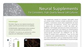 产品手册神经添加物用于培养高一致性、高品质的神经细胞
 沪公网安备31010102008431号