(Apr 2024)
Frontiers in Cell and Developmental Biology 12 5
Forskolin induces FXR expression and enhances maturation of iPSC-derived hepatocyte-like cells
The generation of iPSC-derived hepatocyte-like cells (HLCs) is a powerful tool for studying liver diseases,their therapy as well as drug development. iPSC-derived disease models benefit from their diverse origin of patients,enabling the study of disease-associated mutations and,when considering more than one iPSC line to reflect a more diverse genetic background compared to immortalized cell lines. Unfortunately,the use of iPSC-derived HLCs is limited due to their lack of maturity and a rather fetal phenotype. Commercial kits and complicated 3D-protocols are cost- and time-intensive and hardly useable for smaller working groups. In this study,we optimized our previously published protocol by fine-tuning the initial cell number,exchanging antibiotics and basal medium composition and introducing the small molecule forskolin during the HLC maturation step. We thereby contribute to the liver research field by providing a simple,cost- and time-effective 2D differentiation protocol. We generate functional HLCs with significantly increased HLC hallmark gene (ALB,HNF4?,and CYP3A4) and protein (ALB) expression,as well as significantly elevated inducible CYP3A4 activity. Graphical Abstract
View Publication
产品类型:
产品号#:
100-0276
100-1130
产品名:
mTeSR™ Plus
mTeSR™ Plus
Lee Y-KK et al. (JAN 2016)
International journal of cardiology 203 964--971
Efficient attenuation of Friedreich's ataxia (FRDA) cardiomyopathy by modulation of iron homeostasis-human induced pluripotent stem cell (hiPSC) as a drug screening platform for FRDA.
BACKGROUND Friedreich's ataxia (FRDA),a recessive neurodegenerative disorder commonly associated with hypertrophic cardiomyopathy,is caused by silencing of the frataxin (FXN) gene encoding the mitochondrial protein involved in iron-sulfur cluster biosynthesis. METHODS Application of our previously established FRDA human induced pluripotent stem cell (hiPSC) derived cardiomyocytes model as a platform to assess the efficacy of treatment with either the antioxidant coenzyme Q10 analog,idebenone (IDE) or the iron chelator,deferiprone (DFP),which are both under clinical trial. RESULTS DFP was able to more significantly suppress synthesis of reactive oxygen species (ROS) than IDE at the dosages of 25 $\$ and 10nM respectively which agreed with the reduced rate of intracellular accumulation of iron by DFP treatment from 25 to 50 $\$ With regard to cardiac electrical-contraction (EC) coupling function,decay velocity of calcium handling kinetics in FRDA-hiPSC-cardiomyocytes was significantly improved by DFP treatment but not by IDE. Further mechanistic studies revealed that DFP also modulated iron induced mitochondrial stress as reflected by mitochondria network disorganization and decline level of respiratory chain protein,succinate dehydrogenase (CxII) and cytochrome c oxidase (COXIV). In addition,iron-response protein (IRP-1) regulatory loop was overridden by DFP as reflected by resumed level of ferritin (FTH) back to basal level and the attenuated transferrin receptor (TSFR) mRNA level suppression thereby reducing further iron uptake. CONCLUSIONS DFP modulated iron homeostasis in FRDA-hiPSC-cardiomyocytes and effectively relieved stress-stimulation related to cardiomyopathy. The resuming of redox condition led to the significantly improved cardiac prime events,cardiac electrical-coupling during contraction.
View Publication
Wellington M et al. (DEC 2003)
Infection and immunity 71 12 7228--31
Enhanced phagocytosis of Candida species mediated by opsonization with a recombinant human antibody single-chain variable fragment.
Specific antibody opsonization significantly enhances the level of phagocytosis of Candida in the absence of complement. Furthermore,we have described a system using a recombinant human antibody single-chain variable fragment that allows a comparative study of phagocytosis of multiple Candida species opsonized via a common antigen.
View Publication
MiRNA-Mediated Regulation of the SWI/SNF Chromatin Remodeling Complex Controls Pluripotency and Endodermal Differentiation in Human ESCs.
MicroRNAs and chromatin remodeling complexes represent powerful epigenetic mechanisms that regulate the pluripotent state. miR-302 is a strong inducer of pluripotency,which is characterized by a distinct chromatin architecture. This suggests that miR-302 regulates global chromatin structure; however,a direct relationship between miR-302 and chromatin remodelers has not been established. Here,we provide data to show that miR-302 regulates Brg1 chromatin remodeling complex composition in human embryonic stem cells (hESCs) through direct repression of the BAF53a and BAF170 subunits. With the subsequent overexpression of BAF170 in hESCs,we show that miR-302's inhibition of BAF170 protein levels can affect the expression of genes involved in cell proliferation. Furthermore,miR-302-mediated repression of BAF170 regulates pluripotency by positively influencing mesendodermal differentiation. Overexpression of BAF170 in hESCs led to biased differentiation toward the ectoderm lineage during EB formation and severely hindered directed definitive endoderm differentiation. Taken together,these data uncover a direct regulatory relationship between miR-302 and the Brg1 chromatin remodeling complex that controls gene expression and cell fate decisions in hESCs and suggests that similar mechanisms are at play during early human development.
View Publication
产品类型:
产品号#:
05110
产品名:
STEMdiff™定型内胚层检测试剂盒
Dí et al. (DEC 2010)
Cardiovascular research 88 3 502--11
Endothelial progenitor cells undergo an endothelial-to-mesenchymal transition-like process mediated by TGFbetaRI.
AIMS: Endothelial progenitor cells (EPC) have been shown to repair pulmonary endothelium,although they can also migrate into the arterial intima and differentiate into smooth muscle-like (mesenchymal) cells contributing to intimal hyperplasia. The molecular mechanisms by which this process proceeds have not been fully elucidated. Here,we study whether genes involved in the endothelial-to-mesenchymal transition (EnMT) may contribute to the mesenchymal phenotype acquisition of EPC and we evaluate whether transforming growth factor β1 (TGFβ1) is involved in this process. METHODS AND RESULTS: Our results show that co-culture of EPC with smooth muscle cells (SMC) increases the expression of the mesenchymal cell markers α-smooth muscle actin,sm22-α,and myocardin,and decreases the expression of the endothelial cell marker CD31. In the same conditions,we also observed a concomitant increase in the gene expression of the EnMT-related transcription factors: slug,snail,zeb1,and endothelin-1. This indicates that mesenchymal phenotype acquisition occurred through an EnMT-like process. Inhibition of TGFβ receptor I (TGFβRI) downregulated snail gene expression,blocked the EnMT,and facilitated the differentiation of EPC to the endothelial cell lineage. Furthermore,TGFβRI inhibition decreased migration of EPC stimulated by SMC without affecting their functionality and adhesion capacity. CONCLUSION: These results indicate that EPC may differentiate into SMC-like cells through an EnMT-like process and that TGFβI plays an important role in the fate of EPC.
View Publication
CXCR7 Mediates Neural Progenitor Cells Migration to CXCL12 Independent of CXCR4
Neural progenitor cell (NPC) migration is an essential process for brain development,adult neurogenesis,and neuroregeneration after brain injury. Stromal cell-derived factor-1 (SDF-1,CXCL12) and its traditional receptor CXCR4 are well known to regulate NPC migration. However,the discovery of CXCR7,a newly identified CXCL12 receptor,adds to the dynamics of the existing CXCL12/CXCR4 pair. Antagonists for either CXCR4 or CXCR7 blocked CXCL12-mediated NPC migration in a transwell chemotaxis assay,suggesting that both receptors are required for CXCL12 action. We derived NPC cultures from Cxcr4 knockout (KO) mice and used transwell and stripe assays to determine the cell migration. NPCs derived from Cxcr4 KO mice polarized and migrated in response to CXCL12 gradient,suggesting that CXCR7 could serve as an independent migration receptor. Furthermore,Cxcr4 KO NPCs transplanted into the adult mouse striatum migrated in response to the adjacent injection of CXCL12,an effect that was blocked by a CXCR7 antagonist,suggesting that CXCR7 also mediates NPC migration in vivo. Molecular mechanism studies revealed that CXCR7 interact with Rac1 in the leading edge of the polarized NPCs in the absence of CXCR4. Both CXCR7 and Rac1 are required for extracellular signal-regulated kinases (ERK) 1/2 activation and subsequent NPC migration,indicating that CXCR7 could serve as a functional receptor in CXCL12-mediated NPC migration independent of CXCR4. Together these results reveal an essential role of CXCR7 for CXCL12-mediated NPC migration that will be important to understand neurogenesis during development and in adulthood.
View Publication
产品类型:
产品号#:
05700
05701
05702
产品名:
NeuroCult™ 基础培养基(小鼠和大鼠)
NeuroCult™ 扩增添加物(小鼠和大鼠)
NeuroCult™扩增试剂盒(小鼠和大鼠)
Raynaud CM et al. (JAN 2013)
PLoS ONE 8 1 e54524
Human Embryonic Stem Cell Derived Mesenchymal Progenitors Express Cardiac Markers but Do Not Form Contractile Cardiomyocytes
Mesenchymal progenitors or stromal cells have shown promise as a therapeutic strategy for a range of diseases including heart failure. In this context,we explored the growth and differentiation potential of mesenchymal progenitors (MPs) derived in vitro from human embryonic stem cells (hESCs). Similar to MPs isolated from bone marrow,hESC derived MPs (hESC-MPs) efficiently differentiated into archetypical mesenchymal derivatives such as chondrocytes and adipocytes. Upon treatment with 5-Azacytidine or TGF-β1,hESC-MPs modified their morphology and up-regulated expression of key cardiac transcription factors such as NKX2-5,MEF2C,HAND2 and MYOCD. Nevertheless,NKX2-5+ hESC-MP derivatives did not form contractile cardiomyocytes,raising questions concerning the suitability of these cells as a platform for cardiomyocyte replacement therapy. Gene profiling experiments revealed that,although hESC-MP derived cells expressed a suite of cardiac related genes,they lacked the complete repertoire of genes associated with bona fide cardiomyocytes. Our results suggest that whilst agents such as TGF-β1 and 5-Azacytidine can induce expression of cardiac related genes,but treated cells retain a mesenchymal like phenotype.
View Publication
产品类型:
产品号#:
05850
05857
05870
05875
07913
07930
07931
07940
07955
07956
07959
07954
85850
85857
85870
85875
100-1061
07952
产品名:
Dispase(5 U/mL)
CryoStor® CS10
CryoStor® CS10
CryoStor® CS10
CryoStor® CS10
CryoStor® CS10
mTeSR™1
mTeSR™1
CryoStor® CS10
CryoStor® CS10
Ungrin MD et al. (JAN 2008)
PloS one 3 2 e1565
Reproducible, ultra high-throughput formation of multicellular organization from single cell suspension-derived human embryonic stem cell aggregates.
BACKGROUND Human embryonic stem cells (hESC) should enable novel insights into early human development and provide a renewable source of cells for regenerative medicine. However,because the three-dimensional hESC aggregates [embryoid bodies (hEB)] typically employed to reveal hESC developmental potential are heterogeneous and exhibit disorganized differentiation,progress in hESC technology development has been hindered. METHODOLOGY/PRINCIPAL FINDINGS Using a centrifugal forced-aggregation strategy in combination with a novel centrifugal-extraction approach as a foundation,we demonstrated that hESC input composition and inductive environment could be manipulated to form large numbers of well-defined aggregates exhibiting multi-lineage differentiation and substantially improved self-organization from single-cell suspensions. These aggregates exhibited coordinated bi-domain structures including contiguous regions of extraembryonic endoderm- and epiblast-like tissue. A silicon wafer-based microfabrication technology was used to generate surfaces that permit the production of hundreds to thousands of hEB per cm(2). CONCLUSIONS/SIGNIFICANCE The mechanisms of early human embryogenesis are poorly understood. We report an ultra high throughput (UHTP) approach for generating spatially and temporally synchronised hEB. Aggregates generated in this manner exhibited aspects of peri-implantation tissue-level morphogenesis. These results should advance fundamental studies into early human developmental processes,enable high-throughput screening strategies to identify conditions that specify hESC-derived cells and tissues,and accelerate the pre-clinical evaluation of hESC-derived cells.
View Publication
产品类型:
产品号#:
72302
72304
72307
72308
27845
27945
27840
27865
27940
27965
100-1044
产品名:
Y-27632(二盐酸盐)
Y-27632(二盐酸盐)
Y-27632(二盐酸盐)
Y-27632(二盐酸盐)
Y-27632(二盐酸盐)
Breems DA et al. (JUL 1994)
Leukemia 8 7 1095--104
Frequency analysis of human primitive haematopoietic stem cell subsets using a cobblestone area forming cell assay.
Stroma-dependent long-term bone marrow cultures (LTBMC) assay the ability of primitive haematopoietic stem cells (HSC) for long-term production of clonable progenitors. We have developed a limiting dilution type LTBMC assay allowing frequency analysis of transiently repopulating HSC and long-term culture initiating cells (LTC-IC) without the necessity to replate large numbers of wells. Normal or 5-FU-treated Ficoll bone marrow cells (BMC),or BMCs sorted on CD34 or HLA-DR expression,or Rh123 retention,(input range 40-70,000 CFU-GM/BFU-E/10(5) cells) were plated at limiting dilution on unirradiated adherent layers formed by a novel murine preadipose cell line (FBMD-1). The percentage of wells with at least one phase-dark haematopoietic clone (cobblestone area,CA) beneath the stromal layer was weekly determined for at least 8 weeks,and CA-forming cell (CAFC) frequencies were calculated using Poisson statistics. Parallel LTBMCs of the same samples were weekly assessed for supernate CFU-GM/BFU-E production. Weekly addition of rhIL-3 with rhG-CSF supported a high average clonogenic output per CA and dramatically increased CA size,but did not significantly alter the apparent CAFC frequency. The generation of CFU-GM per CA was constant over a period of 6 weeks with weekly means of eight normal BM samples,ranging between 5-16. At week 6 the mean CAFC frequency was 29 (1 SEM,8.8)/10(5). Early appearing CAFC were highly sensitive to 5-FU,and were contained over the full Rh123 and HLA-DR fluorescence profile of CD34pos cells,whereas CAFC week 5-8 were predominantly contained in the CD34pos Rh123dull HLA-DRlow fraction in agreement with previously reported LTC-IC characteristics. In conclusion,the CAFC assay enumerates LTC-IC using a direct visual endpoint and allows study of LTC-IC heterogeneity with respect to progenitor cell generation per stem cell clone in various haematologic diseases.
View Publication
产品类型:
产品号#:
05150
05350
产品名:
MyeloCult™ H5100
Araoka T et al. (JAN 2014)
PloS one 9 1 e84881
Efficient and rapid induction of human iPSCs/ESCs into nephrogenic intermediate mesoderm using small molecule-based differentiation methods.
The first step in developing regenerative medicine approaches to treat renal diseases using pluripotent stem cells must be the generation of intermediate mesoderm (IM),an embryonic germ layer that gives rise to kidneys. In order to achieve this goal,establishing an efficient,stable and low-cost method for differentiating IM cells using small molecules is required. In this study,we identified two retinoids,AM580 and TTNPB,as potent IM inducers by high-throughput chemical screening,and established rapid (five days) and efficient (80% induction rate) IM differentiation from human iPSCs using only two small molecules: a Wnt pathway activator,CHIR99021,combined with either AM580 or TTNPB. The resulting human IM cells showed the ability to differentiate into multiple cell types that constitute adult kidneys,and to form renal tubule-like structures. These small molecule differentiation methods can bypass the mesendoderm step,directly inducing IM cells by activating Wnt,retinoic acid (RA),and bone morphogenetic protein (BMP) pathways. Such methods are powerful tools for studying kidney development and may potentially provide cell sources to generate renal lineage cells for regenerative therapy.
View Publication
The longevity of organisms is maintained by stem cells. If an organism loses the ability to maintain a balance between quiescence and differentiation in the stem/progenitor cell compartment due to aging and/or stress,this may result in death or age-associated diseases,including cancer. Ewing sarcoma is the most lethal bone tumor in young patients and arises from primitive stem cells. Here,we demonstrated that endogenous Ewing sarcoma gene (Ews) is indispensable for stem cell quiescence,and that the ablation of Ews promotes the early onset of senescence in hematopoietic stem progenitor cells. The phenotypic and functional changes in Ews-deficient stem cells were accompanied by an increase in senescence-associated β-galactosidase staining and a marked induction of p16(INK4a) compared with wild-type counterparts. With its relevance to cancer and possibly aging,EWS is likely to play a significant role in maintaining the functional capacity of stem cells and may provide further insight into the complexity of Ewing sarcoma in the context of stem cells.
View Publication

 EasySep™小鼠TIL(CD45)正选试剂盒


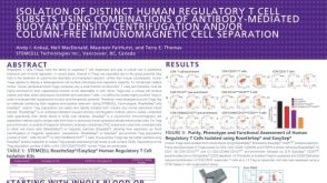 科学海报Isolation of Distinct Human Regulatory T Cell Subsets Using Combinations of Antibody Mediated Buoyant Density Centrifugation
 沪公网安备31010102008431号